anatomy exam 2
 Call Kai
 Learn
Learn Practice Test
 Spaced Repetition
 Match
Match Flashcards
 Knowt Play
1/156

There's no tags or description
Looks like no tags are added yet.
Name | Mastery | Learn | Test | Matching | Spaced | Call with Kai | Chat |
|---|
No analytics yet
Send a link to your students to track their progress
157 Terms
bones consist of
-bone tissues (compact + spongy)
-other connective tissues: fat, hemopoietic tissue, blood vessels, nerves, + hyaline cartilage
main components of bone
extracellular matric (ecm): calcified structure providing rigidity
bone cells: osteoblasts, osteoclasts, and osteocytes
macroscopic view of bones
compact (cortical) bone: dense outer layer
spongy (cancellous bone): inner layer w/ trabeculae for structural support
function of bones
• Mechanical Support: Provides a structural framework for the body and
attachment points for muscles.
• Protection: Shields vital organs such as the brain, heart, and lungs.
• Mineral Homeostasis: Serves as a reservoir for essential minerals like calcium
and phosphorus, releasing them as needed to maintain balance.
• Hematopoiesis: Produces blood cells in the red bone marrow.
bone matrix
-Organic (1/3 of bone mass) AKA Osteoid:
• Type I collagen: Provides tensile strength and flexibility.
• Ground substance: Contains proteoglycans and glycoproteins.
- Inorganic (2/3 of bone mass):
• Hydroxyapatite crystals (calcium phosphate): Ensure compressional strength and durability
osteoid + mineralized bone
Unmineralized organic matrix (collagen + ground substance).
Mineralized bone = Osteoid + Inorganic minerals (hydroxyapatite crystals)
osteoblasts
build new bone tissue (bone formation)
formed from osteoprogenitor cells
produce bone matrix + increase bone density of bones
secretes osteoid
osteocytes
mature bone cells that maintain the bone matrix and signal for repairs
-majority of bone cells in the adult skeleton are these!!!
maintain bone matrix + communicate w other cells. phenotype change that is not reversible.
osteoclasts
break down bone tissue (bone resorption) to regulate calcium levels
-least amount out of the three
-similar to macrophages. breaking down material + reduce bone density. derived form monocytes that are blood cells.
-have multiple nuclei
too much can lead to osteoporosis
osteoblast origin, function, characteristics, phenotype change
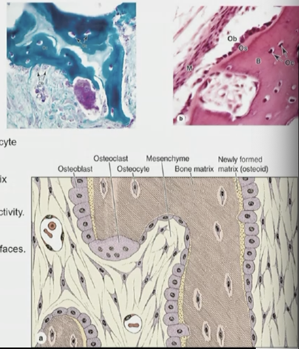
Origin: Mesenchyme→ Osteoprogenitor cells→ Osteoblasts → Osteocyte
Functions: Produce the organic and unmineralized component of bone matrix
(osteoid); Promote calcification through enzymes (alkaline phosphatase) activity.
Characteristics:
• Cuboidal in shape and arranged in linear formations on bone surfaces.
Phenotype Change:
• Into osteoprogenitor cells
• Into osteocytes (not reversible to osteoblast)

osteocyte cell origins + location + features + lifespan
Origins: Derived from osteoblasts trapped within the bone matrix.
Location: Reside in lacunae within the mineralized matrix.
Key Features:
• Possess dendritic processes extending through canaliculi for
communication→ What type of cell-to-cell junction? gap junctions
• Form a connected network with other osteocytes and surface lining cells.
Functions:
• Regulate bone remodeling by signaling osteoblasts and osteoclasts.
• Secrete growth factors to maintain bone tissue.
Lifespan: long lifespan, Highly viable and maintain bone health over time.

Gap Junctions in Bone Tissue
• Cell Communication: Gap junctions allow osteocytes to communicate with each other and with osteoblasts. This communication is crucial for coordinating bone remodeling and repair.
• Nutrient and Signal Exchange: Through gap junctions, small molecules like ions, nutrients, and signaling molecules (e.g., calcium and cAMP) can pass between cells, maintaining bone health and responding to mechanical stress.
• Regulation of Bone Remodeling: Osteocytes act as mechanosensors, and gap junctions enable them to transmit mechanical signals to osteoblasts and osteoclasts, influencing bone formation and resorption

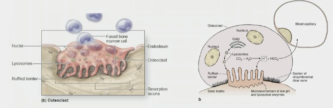
Osteoclast cells structure + origin, functions + key structures
Structure and Origin:
• Large, multinucleated cells derived from hematopoietic progenitor cells in the bone marrow.
• Found on bone surfaces, especially in areas of active resorption.
Functions:
• Release Ca²⁺ into the bloodstream by breaking down bone matrix for calcium homeostasis.
• Remove old or damaged bone to facilitate remodeling and repair.
Key Features:
• Reside in Howship lacunae (resorption bays) to produce hydrochloric acid that break down bone matrix. (acidic environment) enzymes break down hydroxyapatite crystals + release calcium + phosphate.
• Active cells have a ruffled border to increase surface area for efficient resorption.
Types of Bone
Primary (woven) bone
Secondary (lamellar) bone
primary (woven) bone
• Immature (not fully mienralized)bone with irregular/random collagen fiber arrangement.
• Rapidly formed, typically during fetal development or fracture repair.
• Less organized, less mineralized and weaker than mature bone
occurs during: initial stages of development, early phases of fracture repair
transition: gradually replaced by lamellar bone during remodeling

secondary (lamellar) bone:
• Mature (mineralized state) bone with parallel, organized w concentric lamellae collagen fibers in a lamellar structure.
• Replaces woven bone during bone remodeling.
• Stronger and more resilient
structure includes: osteons (haversian systems) (small units of compact bone), lacunae + canaliculi (Lacunae house osteocytes, and canaliculi allow communication and nutrient exchange), perforating (volkmann’s) canals (allow blood vessels + nerves to run through diff osteons + make their way to spongy part of bone; link central canals with the periosteum and medullary cavity)
osteons (haversian systems)
smallest units of the bone
within these units, there is space that has blood vessels + nerves
Units of compact bone with concentric lamellae (parallel layers) surrounding a central canal.
types of secondary bone tissue
cortical vs trabecular bone
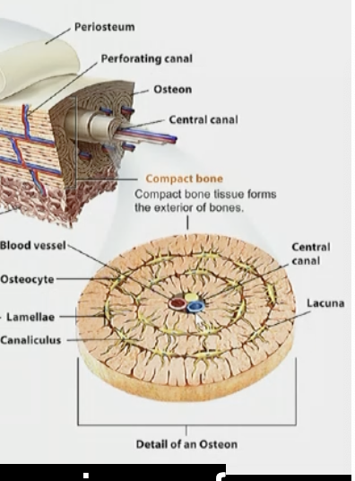
Cortical (compact) bone
Dense and forms the outer layer of bones. many osteons present
comprised of concentric rings of collegen + lamellar
• Provides structural support and protection against physical stress.
-central canal that has blood vessels + nerves
-periosteum, cellular part that has osteoporogenic cells that become osteoblasts + outer fibrous layer

trabecular (spongy aka cancellous) bone
Porous and found inside bones. still has concentric rings of collagen (lamellar structure)
comprised of concentric rings of collegen + lamellar
• Houses bone marrow and supports metabolic functions such as mineral exchange.
-no periosteum. one layer of cells predominantly osteoblasts
provides nutrients + oxygen: bone marrow/ blood vessels + blood cells
periosteum
Functions:
layer covering the compact bone w/ fibrous outer layer that is dense connective tissue + the cellular level inside w osteoporogenic cells + bunch of nerves/blood vessels
-Supports fracture healing and bone repair.
- Covers and protects the entire skeleton.
Structure:
• Outer Layer: Dense CT, vascularized, and innervated fibrous connective tissue.
• Inner Layer: Cellular layer housing; osteoprogenitor cells for bone growth and repair.
Collagen Arrangement:
• Collagen fibers run parallel to the bone surface.
• Exception: Sharpey’s fibers; Type 1 (perforating fibers) attach to cellular layer to external laminar
binding periosteum to the bone → Penetrate the bone at an angle
endosteum
-found in spongy bone/travecula
Composition:
• A single layer of osteoprogenitor cells and osteoblasts.
• Contains a minimal amount of connective tissue.
Structure:
• Much thinner than the periosteum, consisting of only one cell layer.
Functions:
• Facilitates endosteal bone formation.
• Serves as the interface between the bone and bone marrow.
cartilage
• Chondrocytes embedded in a flexible, specialized extracellular matrix.
• Primarily composed of Type II collagen.
• Provides support, cushioning, and flexibility in joints
bone
• Osteocytes embedded in a mineralized, rigid extracellular matrix.
• Primarily composed of Type I collagen.
• Provides structural support, protection, and calcium storage.
similarities btwn cartilage and bone
• Both are connective tissues.
• Both have a two-layered outer covering: perichondrium in cartilage and periosteum in bone
exceptions for fibrocartilage/articular cartilage
bone remodeling
Resorption Phase:
• Osteoclasts break down old osteons, forming tunnel-like cavities.
Formation Phase:
• Tunnels are filled by osteoprogenitor cells and blood vessels, derived from the periosteum and endosteum.
• Osteoblasts secrete osteoid, forming concentric lamellae.
• Osteocytes become trapped, completing the new osteon structure.
Significance:
• Maintains bone strength and adapts to mechanical stress. (work out, heavy weights; increasing bone density)
• Regulates calcium and phosphate homeostasis + constant process
osteogenesis (formation of bone)
-endochondral ossification (within cartilage)
-intramembranous ossification

-endochondral ossification (within cartilage)
• Begins with mesenchyme cells forming a cartilaginous model.
• Cartilage is gradually replaced by bone tissue during development.
• Examples: Long bones like the thigh, shin, femur and humerus

-intramembranous ossification
• Direct differentiation of mesenchyme cells into osteoblasts produce more matrix + through mineralization becomes flat/irregular bones.
• Forms bone without a cartilaginous intermediate.
• Examples: Flat bones like the skull and clavicle.
The frontal bone develops through _________ bone formation.

endochondral bone formation
-fetal hyaline bone all cartilage (has perichondrium for blood supply)
blood within the cartilage creates chemical changes causing cells to become osteoprogenitor → osteoblast.
-perichondrium changes into periosteum.
-osteoblasts make a bone collar (deposit osteoid around the cartilage)
-chondrocytes enlarge (hypertrophy) + cartilage matrix begins to calcify. calcification blocks nutrient diffusion → undergo apoptosis. lacunae becomes empty, leaving calcified cartilage scaffold.
-bone replaces cartilage (osteoblasts lay osteod onto scaffold + osteoclasts break down bone creating medullary (marrow) cavity.
-secondary ossification centers form + ephiphyseal/growth plate allows bone to grow in length until it closes.
BASIC EXPLANATION: Cartilage → blood vessels arrive → osteoblasts form → bone collar → cartilage calcifies → chondrocytes die → osteoblasts replace cartilage with bone → growth plate remains for lengthening.

intramembranous bone formation
Mesenchymal Cell Differentiation:
• Mesenchymal cells cluster and differentiate into osteoblasts (no hyaline cartilage
involved)
Matrix Deposition:
• Osteoblasts secrete osteoid, forming small spicules and trabeculae.
Recruitment and Growth:
• More mesenchymal cells are recruited and differentiate into osteoblasts.
• Appositional growth adds additional layers of matrix.
Formation of Spongy Bone:
• Spicules merge to form trabeculae, creating spongy bone.
Transition to Lamellar Bone:
• Woven bone, which is initially formed, is eventually replaced by lamellar bone, which is organized into compact and spongy bone layers.
Interstitial Growth
• Occurs within the cartilage at the epiphyseal plates.
• Responsible for increasing the length of long bones during development.
• Driven by chondrocyte proliferation and subsequent cartilage ossification.

Appositional Growth
• Involves the addition of new bone tissue to the surface of existing bone.
• Increases the diameter and thickness of bones (adds layers on periphery of bones.)
-osteoclasts open up space in medulla cavity (to not make it too dense or heavy)
• Occurs throughout life as part of bone remodeling.
• Mediated by osteoblast activity on the periosteum

Interstitial Bone Growth Zones
From epiphysis → diaphysis:
Resting: inactive chondrocytes
Proliferation: chondrocytes divide → columns
Hypertrophic: chondrocytes enlarge
Calcification: cartilage calcifies → chondrocytes die
Ossification: osteoblasts replace cartilage with bone
regulation of bone metabolism
The skeleton stores 99% of the body's calcium and releases it as needed to maintain stable blood calcium levels.
• Bone calcium is exchanged with blood calcium to regulate vital functions like muscle contraction, nerve signaling, and blood clotting.
How?
• Cancellous Bone Role:
• Most calcium exchange occurs in cancellous (spongy) bone due to its large surface area and vascularity.
• Mediating Cells:
• Osteoclasts break down bone to release calcium.
• Osteoblasts deposit calcium into the bone matrix.
• Hormonal Regulation:
• Parathyroid Hormone (PTH): Stimulates osteoclast (indirectly by inhibiting osteoblasts) activity to increase blood calcium levels.
• Calcitonin (Thyroid gland): Inhibits osteoclasts (directly) to reduce calcium release. osteoblast activity becomes higher than osteoclasts + can absorb more calcium into the bone.
• Vitamin D: Enhances calcium absorption from the gut/small intestine and its deposition in bone
osteoporosis (bone MA)
is a condition characterized by reduced bone mass and deterioration of bone, increasing the risk of fractures.
• Causes:
• Imbalance between bone resorption (osteoclast activity) and bone formation (osteoblast activity).
• Hormonal changes (e.g., menopause leading to decreased estrogen levels).
• Nutritional deficiencies (e.g., calcium or vitamin D).
• Effects:
• Common fracture sites: Spine, hips, and wrists.
• Loss of bone density primarily affects cancellous bone
rickets (bone MA)
is a condition in children caused by defective calcification of growing bones, leading to soft and weakened bones.
Causes:
• Vitamin D Deficiency:
• Most common cause; impairs calcium absorption from the small intestine.
• Calcium Deficiency:
• Directly impacts bone mineralization.
Symptoms:
• Bowed legs (genu varum).
• Delayed growth and development.
• Pain in the spine, pelvis, and legs.
• Weakness and muscle cramps.
MA: Similar Condition in adults called Osteomalacia.
gigantism
• Cause: Excess growth hormone secretion during childhood before the epiphyseal plates close.
• Effect: Abnormally tall stature due to excessive growth of long bones.
dwarfism
• Cause: Insufficient growth hormone secretion (pituitary dwarfism) or genetic disorders like achondroplasia (defective receptors).
• Effect: Short stature due to impaired growth of long bones.
acromegaly
In adults, an increase in gH causes acromegaly, a disease in which the bones (mainly the long ones) become very thick.
Nervous tissue composed of…
neurons: signal-transmitting cells/communication
neuroglia: Non-conducting cells that support, protect, and nourish neurons
subdivisions of nervous tissue
central nervous system (CNS): brain and spinal cord; main processing/control center
peripheral nervous system (PNS): cranial, spinal, and autonomic nerves, alone with their ganglia; info to and from CNS

neurons
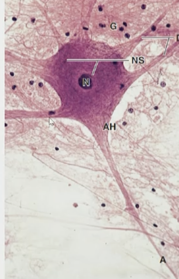
Fundamental units responsible for communication within the nervous system
Composed of:
• A cell body (soma); houses nucleus + other organelles NOT INCLUDING dendrites + axons
• Processes: dendrites (numerous, short, receive signals to soma/toward cell body) and axons (long, transmit signals away cell body or soma)
neurons characteristics
Specialized for Signal Transmission
• Designed to transmit electrical impulses across long distances in the body.
Longevity
• Neurons are non-dividing cells, typically lasting a lifetime.
Limited Neurogenesis in Specific Regions
• Some brain areas, such as the hippocampus, short term to long term mems (FYI), contain neural stem cells capable of generating new neurons to replace damaged ones

ganglia/ganglion
collection of nerve cell bodies in the peripheral nervous system (PNS); supported by satellite cells
sensory + autonomic ganglia
nuclei
a collection of nerve cell bodies in the central nervous system (CNS)
gray matter
formed by neuron cell bodies, dendrites, interneurons (multipolar) within the cns
outside regions of the brain + in some deeper parts within the brain
-involved in processing + integrating info
white matter
formed by nerve fibers (axon tracts) within the CNS/ also has myelin sheath (insulating cover) to speed up signal
-surrounds gray matter
-composed of myelinated axons forming ascending (sensory) and descending (motor) tracts
-contains glial cells
-facilitates communication btwn diff spinal levels + the brain regions.
functional classification of neurons
sensory neurons
motor neurons
interneurons
sensory neurons
Sending impulses from sensory receptors to the central nervous system (CNS).
afferent (input transmission)

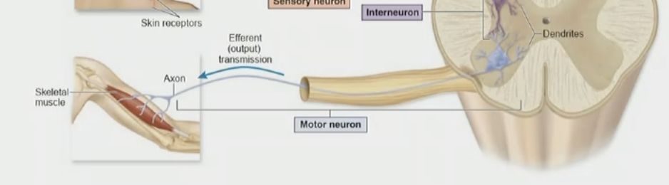
motor neurons
Carry signals away from the CNS or ganglia to muscles and glands (effector cells).
efferent (output) transmission
somatic → voluntary skeletal muscle
autonomic → involuntary organs/glands (sympathetic/parasympathetic)
interneurons
Serve as connectors, enabling communication between sensory and motor neurons; account for 99.9% of all neurons.
neuron basics: cell body (soma)
Perikaryon (Soma): Another name for the cell body of a neuron.
Key Components:
• Nucleus: Contains the genetic material.
• Nissl Bodies: Stacks of rough endoplasmic reticulum (ER) with ribosomes for protein synthesis.
• Axon Hillock: Pyramid-shaped region where the axon originates; lacks Golgi apparatus and Nissl bodies.
• Neurofibrils: Cytoskeletal components aiding in intracellular transport and structural support.
Neuron Basics: Classification by Number of Processes
Multipolar Neurons
• Found in motor neurons and interneurons (most common).
• Have multiple dendrites and a single axon.
Bipolar Neurons
• Located in the retina, olfactory epithelium, and vestibulocochlear nerve (least common).
• Have one dendrite and one axon extending from the cell body.
(Pseudo) Unipolar Neurons
• Found in sensory neurons.
• Cell body located in ganglia near the central nervous system.
• Single process divides into peripheral and central branches.

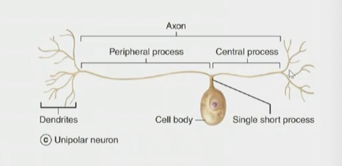
unipolar neurons
• Have a single process.
• During development, axon and dendrite fuse into one fiber.
• The entire process is anatomically referred to as the axon.
• The segment carrying signals to the cell body is the physiologic dendrite or peripheral process (longer part).
• The segment carrying signals away is the central process (similar to axon).
synapse
• Neurons communicate. the intercellular junction between a neuron and:
• Another neuron
• A muscle cell (neuromuscular junction)
• A gland cell (neuroglandular junction)

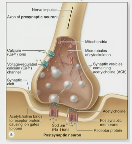
characteristics of synapse
Includes:
• Pre-synaptic bouton (sending side; neurotransmitter vesicles; terminal axonal ending)
• Synaptic cleft (narrow intercellular space; chemical signal)
• Post-synaptic membrane (dendrite; receiving side; contains receptors)
Chemical = neurotransmitter crosses a gap
Electrical = electricity passes directly through gap junctions
types of synapes:
• Inhibitory synapses: Reduce activity in the target neuron.
• Excitatory synapses: Increase activity in the target neuron
Neurotransmitters:
• Multiple types depending on the neuron and synapse function.
inhibitory synapses
reduce activity in the target neuron
excitatory synapses
Increase activity in the target neuron.
membrane potential “difference”
the electrical potential difference across the cell membrane.
• At rest, the membrane potential/inside is negative
(approximately -85 mV).
• It reflects the balance of ion flow, primarily K⁺ and Na⁺ outside the cell more + concentration + abundant.
• Intracellular K⁺ concentration is higher than extracellular, while for Na⁺, the reverse is true.
• Resting potential is maintained by ion channels and the Na⁺/K⁺ pump.
Rest → threshold → Na⁺ IN → K⁺ OUT → return to rest
connective tissue around peripheral nerves
Endoneurium → ONE axon
Perineurium → ONE fascicle/bundle/packet
Epineurium → ENTIRE nerve; outermost layer including blood vessels. structural support + nutrients.

threshold potential
voltage that must be reached to trigger an action potential, usually around −55 mV.
action potential
transient change in membrane potential involving depolarization and repolarization
rapid electrical signal traveling down the axon.
Local Potential: sub-threshhold changes in membrane potential caused by stimuli
Depolarization: Na⁺ enters → membrane becomes more positive
Repolarization: sodium channels close; K⁺ leaves → becomes negative again
Hyperpolarization/afterpotential: briefly becomes too negative
Returns to resting potential (70mv)
propagation: action potential travels along the axon, ensuring signal transmission

neuropil
Dense network of unmyelinated axons, dendrites, and glial cells’ processes.
• Fill the space between cell bodies in gray matter.
glial (supportive cells)
Oligodendrocytes
Astrocytes
Microglia
Ependymal Cells

glial cell: oligodendrocytes
Provide myelin sheaths to insulate around neurons in the CNS.
speeds up rxns by allowing electrical signals to travel faster!!!!
-one of these can wrap around multiple neurons.

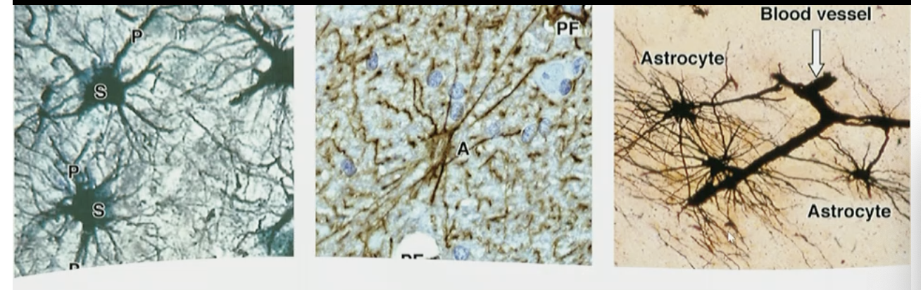
astrocytes
Act as intermediaries between capillaries and neurons; regulate nutrient exchange and support/form the blood-brain barrier
-found in hippocampus
-activate stem cells to become neurons within that region/regeneration
-maintain brain homeostasis/memory processing
-contain filal fibrillary acidic protein (GFAP), repairing neurons
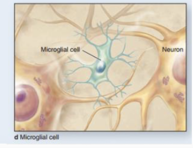
microglia
similar to macrophages; but not derived from blood or monocytes; derived from MESENCHYME
Function as immune cells in the CNS by removing debris and pathogens
-active phenotype: respond to injury or infection, phagocytize debris
-inactive phenotype: survey the environment, maintain homeostasis

glial cells:
Ependymal Cells
modified epithelial cells that cover the central canal within the spinal cord
Line ventricles in the brain and central canal of the spinal cord; form the choroid plexus and produce cerebrospinal fluid (CSF).
-choroid plexus (specialized structures where ependymal cells produce + secrete CSF; found in each ventricle in the brain

meninges
3 protective layers surrounding the brain and spinal cord (CNS):
Dura → Arachnoid → Pia
Dura mater: tough outer layer; Dense irregular CT
Arachnoid mater: web-like middle layer; Loose CT w/ collagen + retiuclar fibers
Pia mater: thin inner layer directly touching CNS; loose CT
CSF is found in the subarachnoid space between arachnoid and pia.
Mnemonic: DAP = Dura, Arachnoid, Pia
nodes of ranvier
gaps in myelin → allow rapid conduction

Schwann Cells (neurolemmocytes)
only in pns
Myelinated Fibers:
•wraps around ONLY ONE axon multiple times forming myelin sheaths.
Unmyelinated Fibers:
-envelop multiple axons w/o forming myelin sheath. structural support SLOWER
• Aid in nerve regeneration
structural comparison:
-myelinated fibers show compact insulation
-unmyelinated fibers LACK insulation
peripheral nervous system
Structure:
• Includes nerves, ganglia, and nerve endings.
-every nerve fiber enclosed by neurolemma or sheath of Schwann.
Nerve Processes:
• Can be myelinated (with insulating myelin sheaths) or unmyelinated.
Specializations:
• Axonal and dendritic processes facilitate communication between neurons and target cells.
satellite cells
• Surround neuron cell bodies in dorsal (posterior) root ganglia.
• Regulate the microenvironment (ion balance and metabolic support).
space around the meninges
• Epidural Space: Found between the dura mater and the vertebrae, containing fat and blood vessels.
• Subdural Space: Between the dura mater and arachnoid mater.
• Subarachnoid Space: Between the arachnoid and pia mater, contains cerebrospinal
fluid (CSF); moves more
(spina bifida)
-neural tube defect; is not fully closed
-can be surgically closed; if not, space remains exposed to outside environment. meninges could be infected + cause meningitis.
severe case of spina bifida (myelomeningocele)
It is the most severe form of spina bifida where the spinal cord and meninges protrude through an opening in the spine.
Clinical Signs:
• Protruding sac on the back, often with exposed nervous tissue.
• Potential for leakage of cerebrospinal fluid (CSF).
• High risk of infection such as meningitis.
Complications (FYI):
• Paralysis or motor deficits below the lesion.
• Bowel and bladder dysfunction.
• Hydrocephalus due to disrupted CSF flow
cerebral cortex
grey matter of cerebrum (where cell bodies r)
Motor areas → control voluntary movement/frontal lobe
(houses prefrontal cortex; high level executive functions)
Sensory areas → receive/process sensory/touch/temp information/parietal lobe
Association areas → thinking, memory, language, interpretation/temporal lobe

precentral gyrus
MOTOR
Located in frontal lobe
Contains primary motor cortex
Controls voluntary skeletal muscle movement, muscle contractions, direction of movement
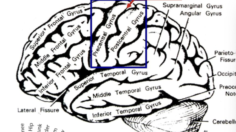
postcentral gyri
SENSORY
Located in parietal lobe
Contains primary somatosensory cortex
Receives touch, pressure, pain, temperature, etc
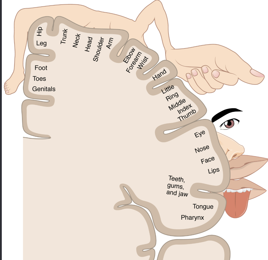
somatotopic organization
Neurons in the brain are arranged in a somatotopic map, reflecting the organization of the body.
different areas of the cortex correspond to specific body parts.
Example: one cortical area controls the hand, another controls the leg.
brainstem
midbrain: controls visual + auditory reflexes
pons: relays signals btwn cerebrum + cerebellus
medulla: regulates vital autonomic functions like breathing, heart rate, blood pressure
Especially involved in:
- Breathing
- Heart rate/blood pressure
- Consciousness/arousal
- Reflexes
- Cranial nerve functions
brainstem functional centers
• Houses cranial nerve nuclei (for eye movements, facial sensation, hearing reflexes, swallowing, etc.).
• Contains autonomic centers regulating breathing, heart rate, and blood pressure.
brainstem pathway for nerve fibers
• Ascending tracts carry sensory input to the cortex.
• Descending tracts transmit motor commands for voluntary movement.
• Links the cerebrum, cerebellum, and spinal cord
clinical relevance of brainstem
Damage may cause sensory and motor deficits, cranial nerve dysfunction, loss of consciousness, or disruption of vital functions.
structure of spinal nerves
• Cervical Nerves: 8 pairs.
• Thoracic Nerves: 12 pairs.
• Lumbar Nerves: 5 pairs.
• Sacral Nerves: 5 pairs.
• Coccygeal Nerves: 1 pair
Formation of Spinal Nerves
Spinal nerves are formed by the union of:
• Dorsal rootlets (posterior): Carry sensory input to the spinal cord.
• Ventral rootlets (anterior): Carry motor output from the spinal cord.
dorsal + ventral → spinal nerve
DAVE:
Dorsal = Afferent
Ventral = Efferent
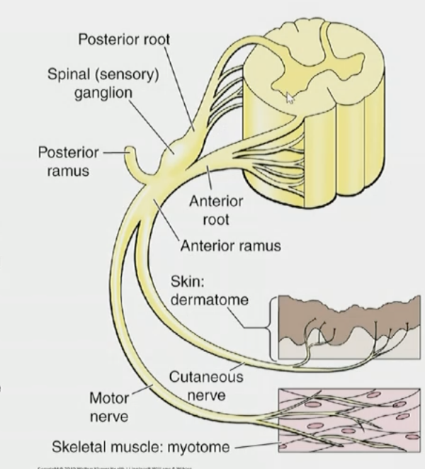
Neuron Cell Body Locations for spinal nerve + cord
Sensory (Afferent) Neurons:
• Cell bodies are located in the dorsal/posterior root ganglia.
Motor (Efferent) Neurons:
• Cell bodies are located in the ventral/anterior horn gray matter of the spinal cord.
spinal reflex
involuntary movements to protect
fast, automatic response that can be processed by the spinal cord without waiting for the brain.

knee jerk reflex
Tap patellar tendon → quadriceps stretches → sensory neuron through dorsal root → spinal cord → motor neuron → quadriceps contracts → leg kicks
At the same time, the hamstrings are inhibited/relax so they don't oppose the quadriceps. This is called reciprocal inhibition.
babinski reflex
the big toe dorsiflexes (curls upwards) + the other toes fan outward in response to stroking the sole of the foot
-normal in children up to 2 years old
-disappears as the nervous system matures
What is the connection between Babinski Reflex and Corticospinal tract development?
Babies: Babinski is normal because the corticospinal tract isn't fully developed/myelinated yet.
Immature corticospinal tract → less inhibition → Babinski present
Mature corticospinal tract → inhibition develops → Babinski disappears
⭐ If Babinski returns in an adult, it can indicate UMN/corticospinal tract damage.
upper motor neurons (UMNs)
• Originate in the precentral gyrus/primary motor cortex of the cerebral cortex.
• Carry signals down to the spinal cord.
axons travel down through brain in the corticospinal tract
-most fibers cross in medulla
-synapses w/ lower motor neuron
in relaxed state: inhibiting lower motor neurons to relax target muscle
lower motor neurons (LMNs)
• Located in the anterior (ventral) horn of the spinal cord.
-axon exits through ventral root → spinal nerve → peripheral nerve
-synapses with skeletal muscle at the neuromuscular junction
• muscle contracts
ex. glute muscles
Clinical Note about corticospinal (descending) tract
• Damage to the corticospinal tract can result in motor deficits:
• Above the decussation: Contralateral weakness.
• Below the decussation: Ipsilateral weakness
voluntary motor pathway
Precentral gyrus (UMN) → lateral corticospinal tract → crosses in medulla → spinal cord → ventral horn (LMN) → ventral root → spinal nerve → skeletal muscle to produce movement
UMN Defect
-spastic paralysis
-no muscle atrophy
-hyperreflexia
-babinski reflex present
LMN Defect
-falccid paralysis
-significant atrophy (no contraction)
-hypoeflexia
-babinski reflex not present
basal ganglia
group of neural cell bodies that play a critical role in motor function by interacting with cerebral cortex.
Initiate/select appropriate movements
Suppress unwanted movements
Regulate movement
Make movements smooth/appropriate
They don't directly command the muscle—they modify motor activity through feedback to the cortex.
functions of basal ganglia
1. Initiation of Voluntary Movements: The basal ganglia help start intended movements and suppress unintended movements.
2. Postural Adjustments: They control adjustments needed for maintaining balance and posture during voluntary movements.