NPB 101: Lecture 22 - Cardiovascular Physiology 5: Capillaries, Veins, & Regulation
 Call Kai
 Learn
 Practice Test
 Spaced Repetition
 Match
 Flashcards
 Knowt Play
1/18

Earn XP
Description and Tags
Learning Objectives: Relate capillary structure and blood-flow velocity to the exchange of substances between blood and tissues. Distinguish diffusion from bulk flow and predict how changes in hydrostatic and osmotic forces affect fluid movement. Explain how lymphatic drainage maintains fluid balance and how impaired drainage contributes to edema. Explain how veins function as blood-volume reservoirs and how venous tone, valves, skeletal muscle activity, and respiration affect venous return. Trace the baroreceptor reflex from a change in arterial pressure through sensory signaling and autonomic responses. Predict coordinated changes in heart rate, contractility, arteriolar resistance, and venous return following disturbances such as standing suddenly or losing blood.
19 Terms
Capillaries
The smallest and most numerous blood vessels in the human body.
Designed for exchange
~8 µm diameter (small size)
Thin, porous walls
Single layer of endothelial cells
Connection between cells forms small pores
Water soluble substances are exchanges between plasma and interstitial fluid
Densely distributed
Most cells are 100 µm of a capillary
Large total SA
Low velocity of flow
Lipid soluble substances are exchanged across capillary wall
Proteins generally do not cross wall unless moved by vesicular transport
Do cells cross through the capillary walls?
Cells are generally too large to cross capillary walls
Exception: leukocytes (white blood cells) to fight infection

Capillary Exchange
The movement of gases, nutrients, fluids, and metabolic wastes between the blood inside capillaries and the surrounding interstitial fluid of body tissues.
Mechanisms:
Diffusion
Bulk flow
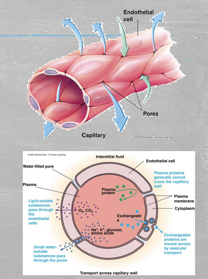
Diffusion
Substances in the blood mov independently of one another down their concentration gradients.

Bulk flow
The organized movement of a fluid and its dissolved substances together in the same direction, driven by a pressure or temperature gradient rather than by concentration differences.
Maintains fluid balance between blood and interstitial fluid
Capillary pores permit fluid, but not proteins or cells
What happens to bulk flow if you lose a liter of blood?
More absorption; decreased flow to tissues.
Want to maintain blood pressure
What if you replace the volume with saline?
More filtration; increased flow to tissues

Filtration
Flow into tissues.
Increases with high blood pressure

Absorption
Flow into plasma.
Increases with high plasma proteins

Lymphatics
A vital network of tissues, organs, and vessels that acts as the body’s drainage, waste management, and immune defense system. Helps maintain fluid balance.
Components:
Lymph
A fluid derived from interstitial fluid
Lymph capillaries
Lymph vessels
Contain valves
Lymph nodes
Lymph
A clear, watery fluid that leaks out of blood capillaries into the spaces around the body’s cells and circulates through the lymphatic system.
Veins and Venules
The two parts of the venous system that carry deoxygenated blood back to the heart.
Structure:
Less smooth muscle than arteries
More elastin than arteries

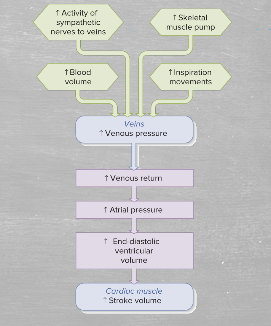
Venous return
The volume of blood entering each atrium per minute
Depends on many factors
Venous capacity
Volume of blood the veins can accommodate
Depends on distensibility of the venous walls and any externally applied force
Sympathetic activity (Venous Return)
Sympathetic stimulation produces vasoconstriction to increase venous pressure and enhance venous return.

Skeletal muscle activity (Venous Return)
Contraction of skeletal muscles compresses veins and increases venous pressure which counteracts the effect of gravity

Venous valves (Venous Return)
Located within the lumen of large veins and prevent the backflow of venous blood.
Why don’t arteries have valves?
Arteries don’t need valves because large pressure from the left ventricle drives bulk flow

Respiratory activity (Venous Return)
Pressure within the chest cavity transiently decreases during respiration. This increases the pressure gradient between the veins in the lower extremities and the chest.
Cardiac suction (Venous Return)
During ventricular contraction, atrial pressure transiently falls below 0 mmHg. This increases the venous pressure gradient and sucks venous blood in the atria.
Baroreceptor Reflex
A fast-acting homeostatic negative-feedback loop that the body uses to maintain stable arterial blood pressure.
Autonomically regulates cardiac output and total peripheral resistance
Responds to changes in arterial blood pressure by altering firing rate
Signaling alters the ratio of activity in the parasympathetic and sympathetic neurons of the cardiovascular control centers
Anatomy:
Aortic arch baroreceptor
Carotid sinus baroreceptor
Detects change in posture
Important to keep blood flow to brain
Pulmonary vessels, walls of heart

Baroreceptor
Specialized mechanoreceptors located in blood vessel and heart walls that detect changes in stretch or tension causes by blood pressure fluctuations.
Sensitive to:
Arterial pressure
Pulse pressure
Firing rate of afferent neuron increases with pressure
Baroreceptor firing leads to adjusting ANS output through the cardiovascular center
What happens to baroreceptor firing when standing quickly after lying down?
B: Decreased firing
due to low blood pressure
ANS Control of Mean Arterial Blood Pressure
