US patient care and prep
 Call Kai
 Learn
 Practice Test
 Spaced Repetition
 Match
 Flashcards
 Knowt Play
1/38

There's no tags or description
Looks like no tags are added yet.
39 Terms
What types of patients may be referred for an ultrasound examination?
Emergency patients.
GP referral patients.
Follow-up scan patients.
Inpatients.
What information might be included in a patient's clinical presentation?
Pain.
Swelling.
Known cancer.
Laboratory results.
What information should be included on an ultrasound request card?
Detailed clinical information.
Medical history.
Symptoms.
Test results.
Why is it important to review a patient's previous medical history and imaging?
Previous reports and scans can provide useful information about the patient's condition and help inform the current investigation.
Why should sonographers ask patients additional questions about their symptoms?
Information may be missing from the request card. Asking appropriate questions helps provide a better understanding of the patient's condition and informs the investigation.
What questions might a sonographer ask when taking a patient's medical history? (name a few only)
Are you experiencing pain?
How long have you had the pain?
Where exactly is the pain?
Can you point to the pain?
Does the pain radiate anywhere?
Have you experienced any previous problems with the area?
Are you taking any medication, and what is it for?
Have you had previous surgery or a biopsy?
Has a lump increased in size?
Does the pain occur at a particular time?
What four characteristics of solid organs can be assessed using ultrasound?
Size.
Shape.
Texture.
Blood flow.
What is Doppler ultrasound used to assess?
Blood flow patterns within blood vessels, including the presence and direction of blood flow.

What happens to sound waves reflected from moving objects, such as blood cells? What is this known as?
Their frequency changes. This is known as the Doppler shift.
What is the Doppler shift?
The difference between the frequency of the sound waves emitted by the transducer and the frequency of the sound waves received back.
What is the purpose of calculating the Doppler shift and Doppler angle?
They allow the determination of blood circulation patterns.
In colour Doppler, what do red and blue represent?
Red: Blood moving towards the transducer.
Blue: Blood moving away from the transducer.

What are examples of fluid-filled structures that can be examined using ultrasound?
Gallbladder.
Bladder.
Cysts.
Blood vessels.

What procedures can ultrasound help guide?
Biopsies.
Aspirations.
Drainage procedures.
Fertility tests.
What are three major limitations of ultrasound?
Bowel gas.
Obesity.
Bone.
Why does bowel gas limit ultrasound imaging?
Intestinal gas produces intense acoustic shadowing, which can obscure structures located beyond the gas.
Why can obesity make ultrasound examinations more difficult?
Overlying adipose tissue can prevent the ultrasound waves from penetrating deeply enough to visualise the region of interest.
How does bone appear on an ultrasound image?
Bone appears very bright white, known as hyperechoic.
Why does bone produce acoustic shadowing?
Bone creates a significant acoustic impedance mismatch, causing it to reflect sound waves strongly. Sound waves cannot pass through the bone, so structures deep to it appear dark.
What type of waves does ultrasound use?
High-frequency sound waves.
What happens to ultrasound waves when they travel through the body?
They can be:
Reflected.
Refracted.
Absorbed.
How is an ultrasound image produced?
The transducer emits sound waves into the body. Some waves are reflected back towards the transducer, and these returning echoes are used to produce the ultrasound image.
What does hyperechoic mean?
A structure that reflects a large amount of sound and appears bright or white on the ultrasound image.
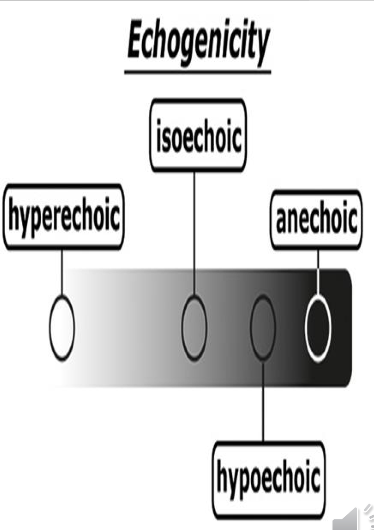
What does hypoechoic mean?
A structure that reflects fewer sound waves and appears darker on the ultrasound image.
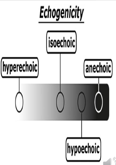
What does anechoic mean?
A structure that produces no internal echoes and appears completely black on the ultrasound image.

What does isoechoic mean?
A structure that has the same echogenicity (brightness) as the surrounding tissue.

How do tissue density and the speed of sound through tissue affect ultrasound image quality?
They influence how sound waves interact with tissues and therefore affect the echoes received and the resulting image.
Why does bone generally appear hyperechoic?
Its high density produces strong reflection of sound waves, creating a bright appearance.
Why does fluid appear anechoic?
Fluid reflects no sound waves, so it appears black on the ultrasound image.
What is the difference between homogeneous and heterogeneous tissue?
Homogeneous: Tissue has a uniform, consistent appearance.
Heterogeneous: Tissue has an uneven, varied appearance.

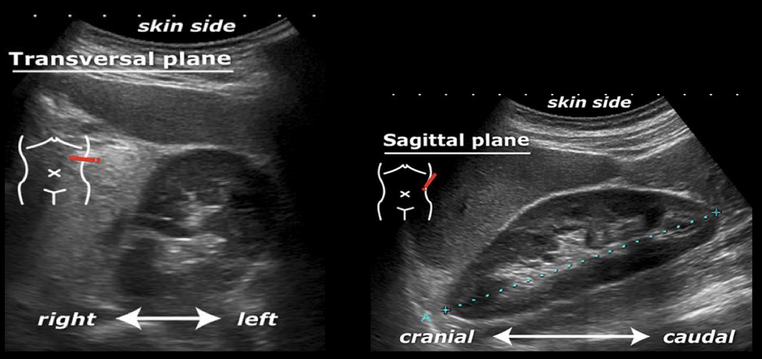
What is B-mode ultrasound?
Brightness mode ultrasound, which produces real-time 2D sectional images showing anatomical structures.
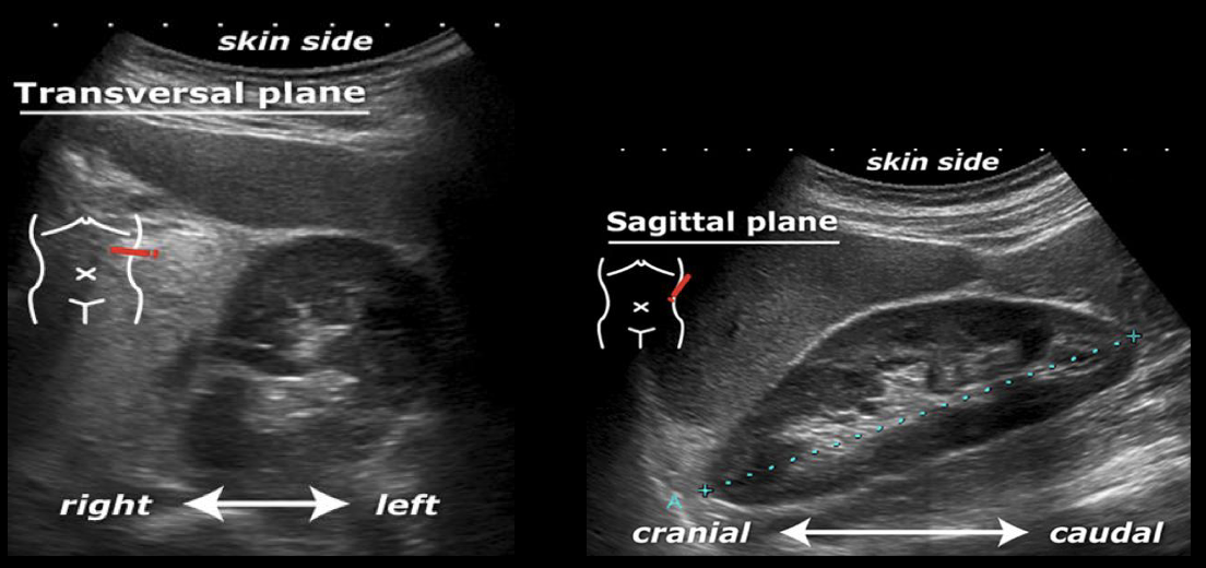
What information does B-mode ultrasound provide?
Real-time 2D images.
Structural information.
Measurements.
Sectional images.

What two scanning planes are commonly used in B-mode ultrasound?
Sagittal plane.
Transverse plane.
What is colour Doppler ultrasound used to assess?
Presence of blood flow.
Direction of blood flow.
Nature of blood flow (arterial or venous).
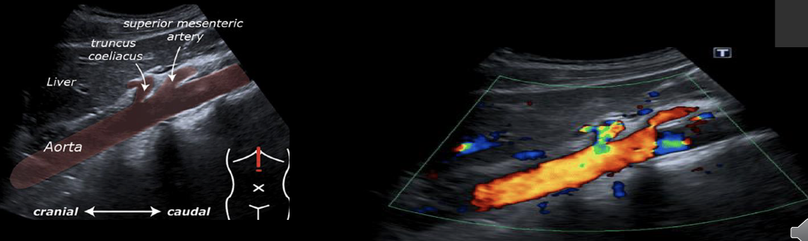
What is pulsed Doppler ultrasound?
A Doppler technique that samples blood flow from a particular point within a particular blood vessel.

What can pulsed Doppler help differentiate?
Arteries from veins.
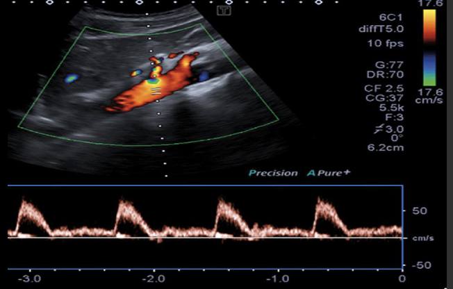
What can pulsed Doppler measure to assess blood vessels?
It can measure the maximum velocity of blood flow and help calculate the percentage of stenosis when systolic velocity is raised above a recommended baseline.
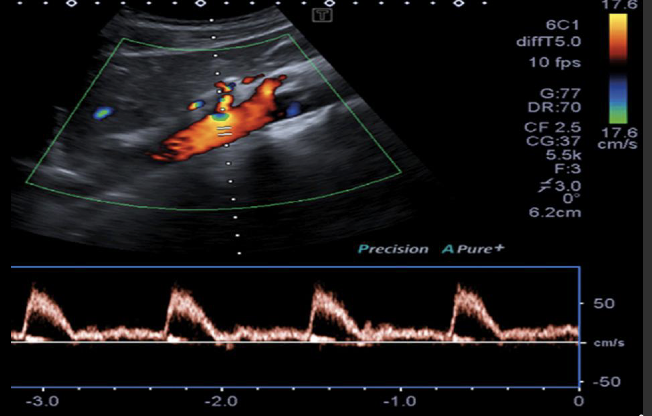
What is 3D ultrasound mainly used for?
Obstetric examinations, particularly prenatal anatomical assessment.

What other uses does 3D ultrasound have besides obstetrics?
Some gynaecological assessments.