Advanced Chest Pathology + IC
 Call Kai
 Learn
 Practice Test
 Spaced Repetition
 Match
 Flashcards
 Knowt Play
1/95

There's no tags or description
Looks like no tags are added yet.
96 Terms
What is an infection that inflames the air sacs in one or both lungs?
Pneumonia
With pneumonia, the air sacs may fill with what?
Fluid or pus
Infection that inflames the air sacs in one or both lungs; the air sacs may fill with fluid or pus
Can bacterial or viral pneumonia be treated with antibiotics?
ONLY bacterial

What pathology is shown in this X-ray?
Viral Pneumonia
Hyper inflated lungs with diffuse interstitial marking; more severe cases - focal areas of atelectasis are present

What pathology is shown in this X-ray?
RSV
What is COPD?
Inefficient exchange of respiratory gases causing difficulty to breath
What is the technical classification of Bronchitis with COPD?
Additive
What is the technical classification of Emphysema with COPD?
Subtractive (extra air)

What pathology is shown on this X-ray?
COPD
Disease that causes thick, sticky mucus to form in the lungs, pancreas and other organs
Inherited condition (genetic)
White, fuzzy patches all over lung fields

What is the pathology shown in this X-ray?
Cystic Fibrosis
Latent TB: Person carries the bacteria but is symptom free and not contagious
Active TB: The bacteria is multiplying in the body faster than the immune system can combat, resulting in a dangerous infection that is also contagious
What type of TB occurs when a person carries the bacteria but is symptom free and not contagious?
Latent
What type of TB occurs when the bacteria is multiplying in the body faster than the immune system can combat, resulting in a dangerous infection that is also contagious?
Active TB
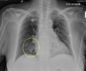
What pathology is shown in this X-ray?
TB

What pathology is shown in this X-ray?
TB

What pathology is shown in this X-ray?
TB
Hyperlucent area with absence of pulmonary markings

What pathology is shown in this X-ray?
Pneumothorax
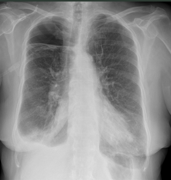
What pathology is shown in this X-ray?
Pneumothorax

What pathology is shown in this X-ray?
Pneumothorax
Accumulation of fluid in the pleural space
Blunting of the costophrenic angles with upward concave border of the fluid level

What pathology is shown in this X-ray?
Pleural Effusion
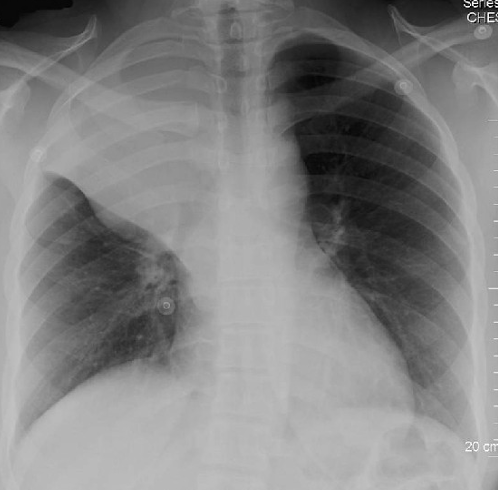
What pathology is shown in this X-ray?
Atelectasis

What pathology is shown in this X-ray?
Atelectasis
Enlarged heart
Very large heart, widened in appearance from typical

What pathology is shown in this X-ray?
Cardiomegaly

What pathology is shown in this X-ray?
Congestive Heart Failure
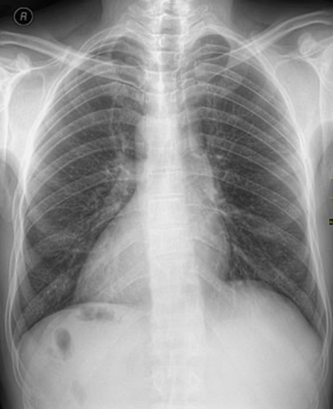
What pathology is shown in this X-ray?
Dextrocardia situs inversus

What pathology is shown in this X-ray?
Pulmonary Embolism (westermark’s sign)

What pathology is shown in this X-ray?
Pulmonary embolism (Hampton’s hump)
What are the two most common conditions associated with COPD?
Chronic bronchitis and emphysema