Pathophysiology Exam II: Hematogical, neurological, and respiratory System
 Call Kai
 Learn
 Practice Test
 Spaced Repetition
 Match
 Flashcards
 Knowt Play
 Podcast
1/103

There's no tags or description
Looks like no tags are added yet.
104 Terms
Hematopoiesis
Production of all blood cells, self-renewing
Erythropoiesis
production of red blood cells
Leukopoiesis
production of white blood cells
Thrombopoiesis
production of platelets
Hemostasis
stoppage of bleeding and formations of clots
Cell type: RBC
Primary function: Oxygen Delivery.
If low: Fatigue, pallor
Cell type: WBC
Primary Function: Fights infections
if low: Infection risk
Platelets
Primary Function: Stops bleeding
If low: Bleeding risk
Leukemia
cancer of white blood cells
Acute Lymphoblastic Leukemia
ACUTE
Most common childhood leukemia
Increased lymphoblasts
Symptoms:
Fatigue
Bruising
Recurrent infections
Major types of WBC
Neutrophil, Eosinophil, Basophil, Monocyte, Lymphocyte
Hemostasis Steps
vascular spasm, platelet plug, coagulation
Acute Myelogenous Leukemia (AML)
ACUTE
Hallmark: Auer rods
Priority concerns:
Infection
Bleeding
Petechiae
Bleeding gums
Chronic Lymphocytic Leukemia (CLL)
CHRONIC
Hallmark: Smudge cells
Older adults, progression is slow
Chronic Myelogenous Leukemia (CML)
Hallmark: Philadelphia chromosome
Common findings:
Splenomegaly
Left upper quadrant discomfort
Fatigue
Lymphoma
malignant tumor of lymph nodes and lymph tissue
Hodgkin's lymphoma
distinguished from other lymphomas by the presence of large, cancerous lymphocytes known as Reed-Sternberg cells
Anemia
A condition in which the blood is deficient in red blood cells, in hemoglobin, or in total volume.
iron deficency anemia
anemia caused by inadequate iron intake
Risk factors: Heavy menstrual periods, Chronic blood loss
Findings: fatique, pallor, microcytic RBCS, Pica
Vitamin B12 Deficiency Anemia
produced by inadequate intake of B12 in diet
Health impact: Neurological symptoms, numbness, tinging, balance changes
Non-Hodgin's Lymphoma
a group of more than 20 different types of lymphomas that occur in older adults and do not show Reed-Sternberg cells

Thrombocytopenia
low platelet count
Causes
Easy bruising
Petechiae
Bleeding Gums
Prolonged Bleeding
Sickle Cell Disease
Genetic disorder in which red blood cells have abnormal hemoglobin molecules and take on an abnormal shape.
Results: RBC sickiling, Vascular Occlusion, tissue ischemia
CNS
central nervous system; brain and spinal cord
PNS
Peripheral nervous system; nerves outside CNS.
Parkinson disease
degeneration of dopamine-producing neurons in the basal ganglia, occurring in later life and leading to tremors, weakness of muscles, and slowness of movement.
Clasic findings: resting tremor, rigidity, bradykinesia, shuffling gait
carbidopa-levodopa
A medication used to treat Parkinson's disease. Improves movement, reduces tremors, improves walkings
Multiple Sclerosis (MS)
disease of the central nervous system characterized by the demyelination (deterioration of the myelin sheath) of nerve fibers, with episodes of neurologic dysfunction (exacerbation) followed by recovery (remission)
Findings: weakness, unillateral deficits, blurred vision, fatigue
Multiple Sclerosis clinical point
Fevers can worsen symptoms because heat impairs conduction through damaged myelin
Auer rods
AML
Smudge cells
CLL
Reed-Sternberg cells
Hodgkin lymphoma
Philadelphia chromosome
CML
B12 deficiency
Neurologic symptoms
Thrombocytopenia
Bleeding
Parkinson
↓ dopamine
MS
Demyelination
TIA
Future stroke risk
Meningitis
Fever + headache + neck stiffness
Seizure care
Airway & safety
Early ICP sign
Decreased LOC
Fixed dilated pupil
Emergency
Surfactant
Reduces surface tension
Epiglottitis
Drooling
Pneumothorax
Unilateral decreased breath sounds
COPD
Airflow obstruction
Emphysema
Loss of recoil
TB
Cough + weight loss + night sweats
PE
Sudden chest pain + dyspnea
CN III
Pupil response
CN III/IV/VI
Eye movements
CN IX/X
Swallowing/gag reflex
CN XI
Shoulder shrug
Carbidopa-levodopa
Improves Parkinson symptoms
Antiseizure medications
Never abruptly stop
Normal pH range
7.35-7.45
Normal PaCO₂ range
35-45 mmHg
Normal HCO₃ range
22-26 mEq/L
Respiratory Acidosis lab values
pH ↓, CO₂ ↑
Respiratory Acidosis common causes
COPD, Hypoventilation
Respiratory Alkalosis lab values
pH ↑, CO₂ ↓
Respiratory Alkalosis common cause
Hyperventilation
Transient Ischemic Attack (TIA)
coined Mini-stoke (but not a stroke)
brief episode of loss of blood flow to the brain, usually caused by a partial occlusion that results in temporary neurologic deficit (impairment); often precedes a CVA
Strong warming sign of future stroke
Temporary neurologic symptoms
Symptoms resolve completely
stroke
a medical emergency that occurs when blood flow to the brain is cut off or severely reduced, depriving brain cells of oxygen and causing them to die within minutes
Ischemic Stroke: Accounts for about 85% to 87% of all cases and happens when a blood clot or plaque blocks an artery supplying the brain.
Hemorrhagic Stroke: Caused by a weakened blood vessel that leaks or ruptures, putting dangerous pressure on brain tissue.
Stroke Symptoms
Sudden weakness or numbness of the face, arm or leg, especially on one side of the body
•Sudden dizziness, loss of balance or coordination, or trouble walking
•Sudden confusion, trouble speaking or understanding
•Sudden trouble seeing •Sudden severe headache
Clinical priorities of a stroke
Time of symptom onset, neurologic assessment, rapid diagnostic testing
Meningitis
CNS infection
inflammation of the meninges
Menigitis symptoms
fever, stiff neck, neuro symptoms, headache, backache; bulging fontanels in infant.
Nochal Rigidity=Cant touch chin to chest (sign of meningitis)
Big risk factor for college age students living close together (Dorms)
encephaliitis
CNS infection, inflammation of the brain
Encephalitis symptoms
headache, fever, cerebral dysfunction, disordered thought patterns, and, often, seizures
Seizures
Sudden, uncontrolled electrical disturbances in the brain.
Priority during seizure
Protect patient from injury
Priority after seizure
Airway, side-laying positions, assess oxygenation
(NEVER stop anti-epileptic medication abruptly)
Increased intracranial pressure (ICP)
Elevated pressure within the skull affecting brain function.
First indicator: decrease level of consciousness, Fixed dialated pupil, pupillary changes
Priority: Notify Provider Immediately
ICP interventions
Elevate HOB ~30 degrees
Maintain neck alignment
Avoid excessive coughing
Prevent Valsalva maneuver
Allow rest periods
Traumatic Brain Injury (TBI)
A traumatic insult to the brain capable of producing physical, intellectual, emotional, social, and vocational changes.
Treatment for TBI
Hypertonic Saline
Pulls fluid out of swollen brain tissue
reduces cerebral edema
Lowers ICP
Spinal Cord Injury
the type of paralysis is determined by the level of the vertebra closest to the injury

CN III (3)
Name: Oculomotor
Function: Pupil constriction, eye movement
Assessment: PERRLA, Light Response
FIndings: Nonreactive pupil = possible CN III Dysfunction
CN III, IV, VI (3,4,6)
Names (in order): oculomotor, trochlear, abducens
Control: Extra-ocular eye movements
Assessment: Six cardinal fields of gaze
Remembers: eyes move together with CN III, IV, VI
CN IX, X (9,10)
Names: glossopharyngeal and vagus
Function: swallowing, gag reflex
Assessments: degree of difficulty when swallowing, or coughing while drinking
Risk: Aspiration
CN XI (11)
Name: Accessory
Function: Shoulder Shrug, Head turn against resistance
Assessment: shrug shoulders
Epileptic vs. non-epileptic seizures
Epileptic seizures are caused by sudden, abnormal electrical discharges in the brain, while non-epileptic seizures (often called functional or psychogenic seizures) look similar but show normal brain wave activity on an EEG (caused by psychological factors like stress, trauma, or emotional processing, or physical mimics like low blood sugar, heart arrhythmias, or fainting (syncope))
Alzheimer's Disease
a progressive and irreversible brain disorder characterized by gradual deterioration of memory, reasoning, language, and, finally, physical functioning
Vascular Dementia
form of dementia caused by a stroke or other restriction of the flow of blood to the brain
Delirium
an acutely disturbed state of mind that occurs in fever, intoxication, and other disorders and is characterized by restlessness, illusions, and incoherence of thought and speech.
Major parts of the brain
Cerebrum, Cerebellum, Brainstem, Limbic system

glial cells
cells in the nervous system that support, nourish, and protect neurons
Major regions of the spinal cords and (spinal nerves)
Cervical, thoracic, lumbar, Sacral

Upper Respiratory system
Immune system, filtration, Humidity
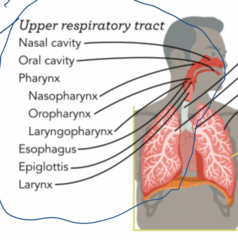
Lower respiratory tract
Air and gas exchange

Pneumonia
An inflammation of lung tissue, where the alveoli in the affected areas fill w/fluid
Signs: fever, cough, crackles (air moving through alveolar fluid)
Surfactant
chemical produced in the lungs to maintain the surface tension of the alveoli and keep them from collapsing
Epiglottitis
A disease in which the epiglottis becomes inflamed and enlarged and may cause an upper airway obstruction.
Concerning findings (Medical emergency!!! usually in children): Drooling, Difficulty swallowing
Priority: Airway Management
Pneumothorax
air in the pleural cavity caused by a puncture of the lung or chest wall
Common findings: Sudden dyspnea, chest pain, unilateral decrease breathing sounds
COPD (chronic obstructive pulmonary disease)
is a progressive, long-term lung condition that makes it hard to breathe by blocking airflow and damaging lung tissue
Findings: Prolonged expiration, air trapping, diminished breath sounds
Emphysema
a condition in which the air sacs of the lungs are damaged and enlarged, causing breathlessness.
WHY: loss of alveolar elastic recoil
Results: air trapping, hyperinflation
Tuberculosis (TB)
infectious disease caused by Mycobacterium tuberculosis; lungs usually are involved, but any organ in the body may be affected
SYMPTOMS: persistent cough, weight loss, night sweats, hemoptysis (coughing up blood or mucus from the lungs)
AIRBORNE PRECAUTIONS
Hypoxemia
Low oxygen level in the arterial blood
hypoxemia vs hypoxia
hypoxemia is low oxygen in the arterial blood and hypoxia is low oxygen in the tissues; hypoxia may be cause by hypoxemia or may be due to cardiac or other problems, including toxins