DMN (Lesson 86): Response to injury and common disorders of the liver
 Call Kai
 Learn
 Practice Test
 Spaced Repetition
 Match
 Flashcards
 Knowt Play
1/34

There's no tags or description
Looks like no tags are added yet.
35 Terms
Portal tracts
reside peripherally at the angles of the hexagon and contain three structures: 1) bile duct, 2) portal vein, 3) hepatic artery
Hepatic plates
one-cell-thick layers of hepatocytes that extend radially from the central vein and are separated by blood-filled hepatic sinusoids which have discontinuous endothelial cells
Patterns of hepatocellular necrosis: Random or embolic pattern
Multifocal areas of necrosis scattered randomly throughout the liver
Indicates a hematogenous route of entry (septicemia)
Typical of many blood-borne infectious agents including, viruses, bacteria, and protozoa
Image of Random hepatic necrosis. Multiple pale, yellow, foci of necrosis (Sawdust liver), cow. Cause: bacteria

Patterns of hepatocellular necrosis: Zonal
Specific areas of necrosis that are restricted to a particular portion of the hepatic lobule
Occurs with various specific types of liver damage such as hypoxic injury and hepatotoxicities
Centrilobular hepatic degeneration and necrosis
Occurs with hypoxic injury because Zone 3 receives the least oxygenated blood
Also occurs with hepatotoxicity that requires mixed function oxidases which are present in Zone 3
Mixed function oxidases function in the metabolism of drugs and toxins
Cytochrome P450 enzymes function in the liver?
convert drugs or chemicals into toxic metabolites
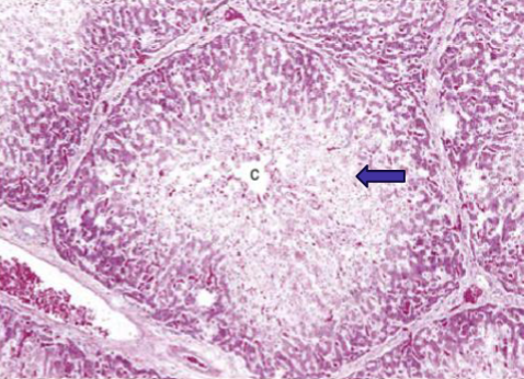
Centrilobular necrosis: Circumferential zone of hepatocellular necrosis around the central vein(Zone 3-arrow)
Midzonal necrosis
Sometimes reported in pigs and horses with aflatoxicosis

Periportal necrosis
Occurs following exposure to toxins such as phosphorus

General considerations of the liver
liver has a considerable functional reserve and good regenerative capacity(quickly in a matter of days to weeks)
Approximately 75% of the hepatic parenchyma can be removed without significant impairment of function: Clinical signs of hepatic failure are not expressed until functional reserve is lost
Consequences of hepatic failure
❖ Icterus (hyperbilirubinemia )
❖ Hepatic encephalopathy ( due to hyperammonemia)
❖ Vascular and hemodynamic alterations (e.g acquired portosystemic shunts)
❖ Cutaneous manifestations (hepatocutaneous syndrome and photosensitization)
❖ Immunologic manifestations due to reduced Kupffer cell function
❖ Coagulopathy resulting in bleeding tendencies
❖ Ascites secondary to hypoalbuminemia and/or portal hypertension
❖ Hypoglycemia
Consequences of hepatic failure: Icterus (hyperbilirubinemia)
Increased concentrations of conjugated or unconjugated bilirubin in blood
Yellow discoloration of tissues
More evident in visible mucus membranes, sclera, blood vessels
Hepatocellular regeneration: Regeneration after hepatic injury
Occurs rapidly if some hepatocytes survive in a lobule and the lobular stroma persists
Necrotic foci are quickly replaced by cellular division of adjacent hepatocytes
However, if whole lobules are destroyed, healing is by scarring
Hepatic fibrosis = an increase in the amount of collagen within the liver.
Occurs as a response to chronic injury and repeated injury.
Stellate cells within the space of Disse produce collagen
Disrupts the exchange between hepatocytes and plasma → diminished hepatic synthetic, catabolic, and excretory functions
Biliary hyperplasia
Proliferation of newly formed biliary ducts within the portal tracts and periportal regions
Predominantly occurs with diseases that cause biliary inflammation, obstruction, and cholestasis
Biliary hyperplasia indicates long-standing (chronic) hepatic injury
End stage liver(cirrhosis)
Repeated injury and regeneration cycles promote the formation of fibrosis and nodular regeneration.
Diffuse process in the liver characterized by severe fibrosis and replacement of normal hepatic lobules with nodular regeneration and abnormal vasculature
pathogenesis of end stage liver(cirrhosis)
Chronic hepatotoxicity
Chronic hepatitis and/or cholangiohepatitis
Chronic biliary obstructive disease
Chronic degenerative liver disease
Acute hepatitis
Characterized by inflammation, hepatocellular necrosis, and apoptosis
Results in an embolic pattern which is randomly multifocal
Can result in death from acute liver failure if severe.
Cause of acute hepatitis
Occurs with systemic infections
• Viruses (herpesvirus)
• Bacteria (Clostridium piliforme, lepto)
• Fungi (blastomycosis, histoplasmosis)
• Protozoan (Toxoplasma gondii, Neospora caninum)
Chronic hepatitis
Characterized by fibrosis, mononuclear inflammatory cells, hepatocellular loss, nodular regeneration, and structural remodeling of the hepatic lobule and vasculature
Progressive loss of function may lead to hepatic failure and end-stage liver
Chronic hepatitis occurs with any chronic persistent antigenic stimulus
Chronic infections
Immune mediated disease
Metabolic disease
Long-term exposure to toxins or drugs
Nonspecific Reactive Hepatitis
Diffuse process in the liver that is a response to systemic inflammation
Most commonly stemming from gastrointestinal disease
Characterized by mild mononuclear inflammatory infiltrates in the portal tracts, less commonly around central veins
Cholangitis
Inflammation of the biliary ducts; may be either intrahepatic or extrahepatic
Chronic cholangitis may lead to cholestasis and icterus (hyperbilirubinemia )
Neutrophilic cholangitis (infectious): acute or chronic
Caused by ascending bacterial infection
Rupture of an infected intrahepatic bile duct may lead to hepatic abscess
Bacterial culture most often reveals: E. coli, Enterococcus, Bacteroides, Streptococcus, or Clostridium
Lymphocytic cholangitis (immune mediated)
A chronic, immune-mediated biliary disease of cat
Cholangiohepatitis
Inflammation of the biliary ducts and extension of inflammation into the surrounding liver parenchyma
Cause of cholangiohepatitis
Caused by an extension of bacterial cholangitis into the surrounding periportal hepatic parenchyma → more severe disease
Causes greater elevation of serum ALT due to damage of more hepatocytes
Liver neoplasia can arise from
Hepatocytes, Bile duct epithelial cells
Mesenchymal elements (e.g connective tissue and blood vessels)
Metastatic (neoplasm from another organ)
hepatocellular adenoma: Benign neoplasm of hepatocytes
Commonly described in young ruminants
Usually single, variably sized, brown masses
Compress adjacent parenchyma
Hepatocellular carcinoma: Malignant neoplasms of hepatocytes
Not common in all domestic species → Reported in dogs and ruminants, especially sheep
Described as massive(single mass), nodular and diffuse
Most common presentation of hepatocellular carcinoma
Solitary well demarcated mass involving entire lobe

Cholangiocellular adenoma: single, firm, discrete, white mass
Mass comprises tubules with well differentiated cuboidal epithelium separated by minimal stroma
Most common in cats
Cholangiocellular carcinoma
Common in all species
Large single mass or multiple masses
Gray-tan unencapsulated masses → Often metastatic to other tissues
Fatty liver: hepatocellular lipid accumulation
Within hepatocytes, free fatty acids are esterified to triglycerides, or converted to cholesterol or phospholipids
Triglycerides can be complexed with apoproteins to form low-density lipoproteins which are released into the plasma as an energy source
Lipidosis occurs when hepatocyte triglyceride accumulation exceeds the rate of lipoprotein release