MSK/Paed IMAGE REC
 Call Kai
 Learn
 Practice Test
 Spaced Repetition
 Match
 Flashcards
 Knowt Play
1/22

There's no tags or description
Looks like no tags are added yet.
23 Terms

Describe the image
Transverse plane of knee shows a 25 mm echogenic foreign body penetrating from subcutaneous to muscle layer. Assess the foreign body's size, orientation and full depth of penetration from the skin surface into the underlying tissues, identifying the specific muscle involved and its proximity to anatomical landmarks (e.g. ankle joint). Use a thick gel layer or stand-off pad to improve visualisation of superficial foreign bodies. Assess adjacent fascia, muscles, the interosseous membrane, vessels, tendons and nerves for involvement or injury. Use colour Doppler to assess for surrounding hyperaemia and vascular abnormalities. If punctured - haematoma. Extend exam by looking for collections


Describe the image
In DDH assessment, the acetabular roof does not appear sharp, suggesting acetabular dysplasia. The alpha angle is less than 60°. Assess femoral head coverage: normal coverage should be at least 58%, while coverage below 50% is significantly dysplastic.


Describe the image


Describe the image
The left kidney demonstrates diffuse increased cortical echogenicity with poor/loss of corticomedullary differentiation, suggesting diffuse renal parenchymal disease. The renal pelvis and calyces appear prominent, suggesting hydronephrosis. Extend the examination to measure the renal pelvic AP diameter at the PUJ and identify the point of ureteric tapering. Follow the ureter distally towards the VUJ, assess bladder distension and consider prone positioning with a high-frequency linear transducer. Likely diagnosis: Autosomal recessive polycystic kidney disease (ARPKD) – characterised by diffusely echogenic kidneys with poor corticomedullary differentiation; numerous tiny cysts may be below ultrasound resolution.

Describe the image
The APL and EPB tendons (first extensor compartment) demonstrate thickening of the right tendon and tendon sheath compared to the left, with associated hyperaemia on colour Doppler, suggestive of tenosynovitis associated with inflammatory arthritis (potentially related to SLE). Assess surrounding soft tissues for increased echogenicity, vascularity and oedema. Check for focal probe tenderness.
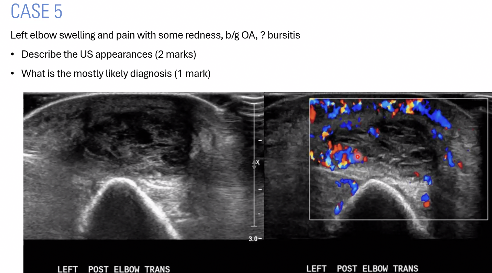
Describe the image
The olecranon bursa demonstrates an ill-defined, complex fluid collection containing mixed cystic and solid components, with thick septations and intervening cystic spaces. There is associated increased echogenicity, dermal thickening and hyperaemia of the surrounding tissues on colour Doppler, suggestive of complex olecranon bursitis. Assess for probe tenderness and compressibility. Internal vascularity should also be assessed.
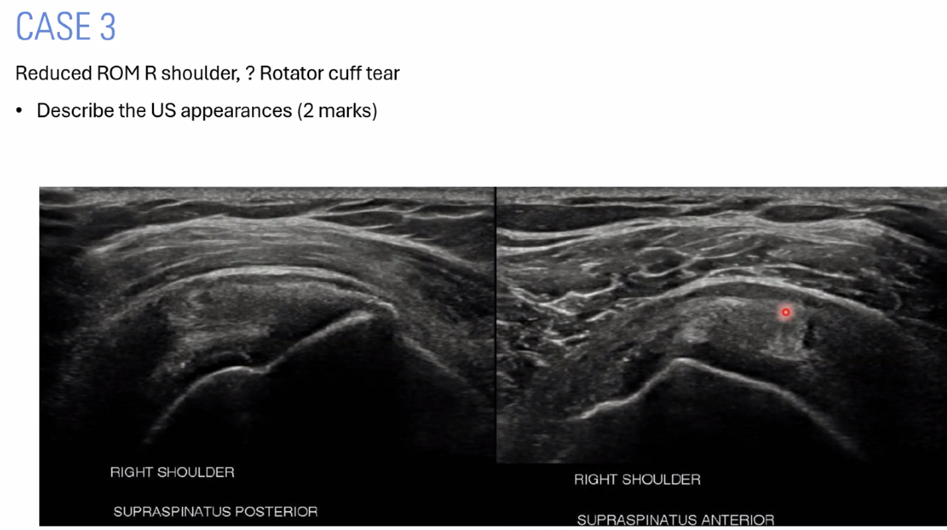
Describe the image
The supraspinatus tendon appears thickened and heterogeneous at its proximal insertion, with some loss of the normal fibrillar architecture. However, tendon continuity is maintained, with no evidence of a tear. There is associated mild thickening of the subacromial-subdeltoid bursa. Findings are suggestive of supraspinatus tendinopathy with mild bursitis.
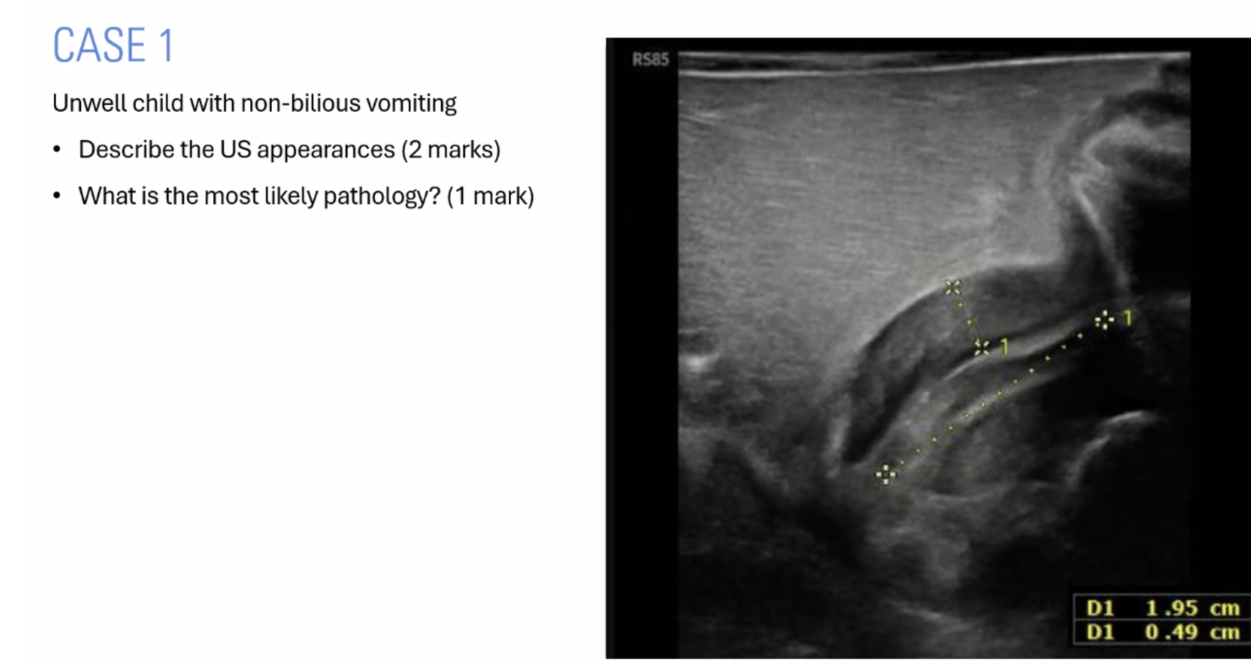
Describe the image
The pylorus demonstrates muscular wall thickening measuring approximately 4.9 mm (>3 mm) and an elongated pyloric canal measuring 1.95 cm (>17 mm), with narrowing/near-obliteration of the lumen. Findings are consistent with hypertrophic pyloric stenosis.
Vomit appearance | Think |
|---|---|
Non-bilious (no green bile) | HPS |
Bilious (green) | Intestinal obstruction distal to ampulla; consider malrotation/volvulus |
Bilious vomit is green because it contains bile, which is a digestive fluid produced by the liver.
Bile contains pigments called bile pigments, particularly bilirubin (yellow) and biliverdin (green).
When bile enters the duodenum, it mixes with the intestinal contents.
If there is an obstruction and bile refluxes back into the stomach, it can be vomited, giving the vomit a characteristic green colour.


Describe the image
The bowel demonstrates a classic target/donut appearance, with concentric hypoechoic and echogenic layers and echogenic mesenteric fat within the intussuscepted segment, suggestive of intussusception. Assess internal vascularity and measure the AP/transverse diameter. Determine the bowel segments involved (small vs large bowel) by following the colon from the caecum to the hepatic flexure and assessing the ileocaecal junction, commonly located in the RLQ. Look for a potential lead point, interloop fluid, free fluid, abnormal lymph nodes and complications such as abscess. Extend the examination by sweeping through the abdomen to determine the full extent of the intussusception and look for lead points.
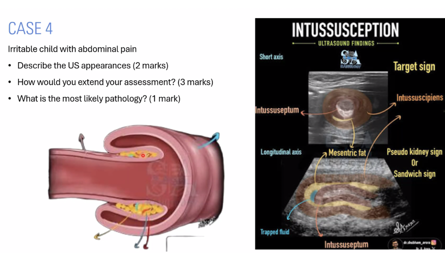

Describe the image
The uterus and vagina appear markedly distended with homogeneous low-level internal echoes, consistent with retained menstrual blood within the uterine cavity and vaginal vault (haematometrocolpos). The endometrial cavity and myometrial boundaries are poorly delineated due to the distension. The vagina appears as a large fluid-filled structure posterior to the urinary bladder.

Describe the image
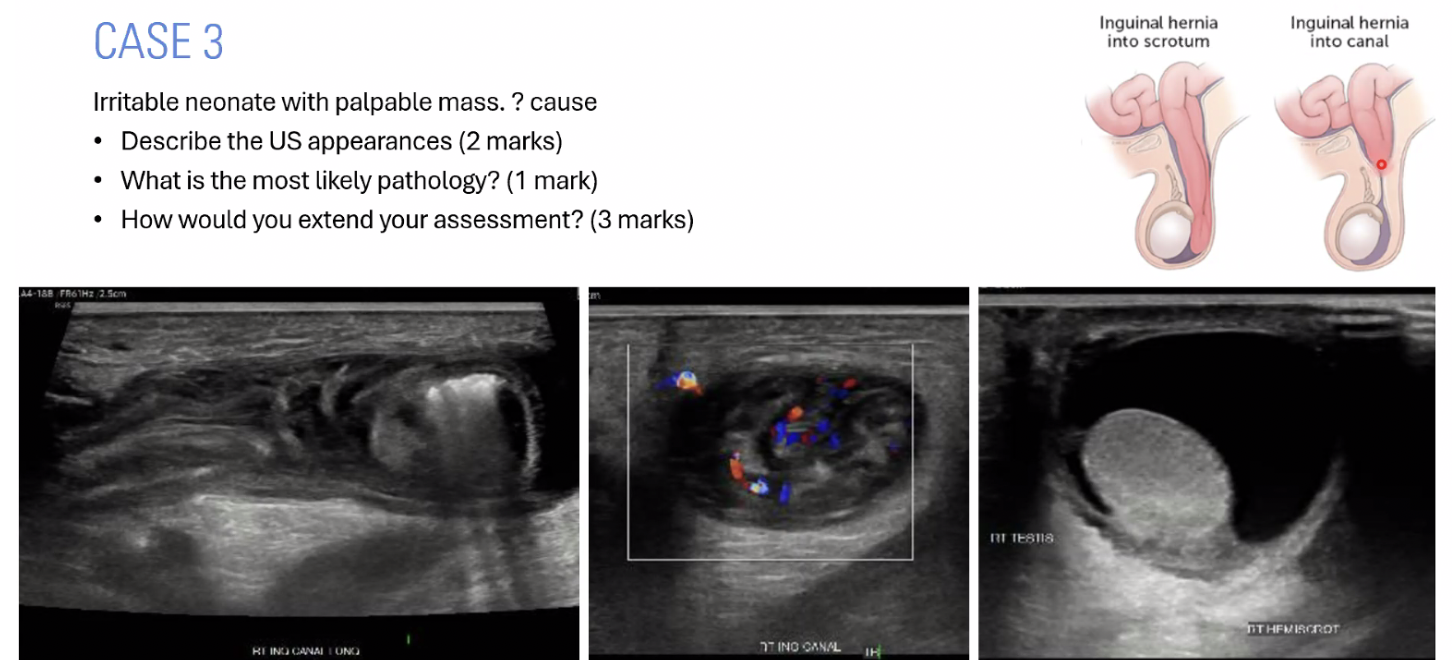
Inguinoscrotal Hernia & Communicating Hydrocele
Inguinoscrotal hernia: Abdominal contents (usually bowel or omentum) pass through the inguinal canal and may extend into the scrotum alongside the testis. Bowel involvement may demonstrate peristalsis and internal echogenic foci with dirty shadowing due to gas.
Communicating hydrocele: Fluid tracks from the peritoneal cavity through a patent processus vaginalis into the scrotum. A large hydrocele may make the testis difficult to visualise.
Image findings:The right inguinal canal demonstrates a distended canal containing a well-defined, heterogeneous structure with internal echogenic foci and dirty shadowing, suggestive of bowel-containing inguinal hernia. The hernia extends towards the right hemiscrotum, consistent with an inguinoscrotal hernia. An associated right hydrocele is also demonstrated. Adjacent lymph nodes maintain their echogenic fatty hila.
Types of Inguinal Hernia
Indirect inguinal hernia: Passes through the deep inguinal ring, lateral to the inferior epigastric vessels, following the pathway of the processus vaginalis. Most common in neonates and children due to a patent processus vaginalis.
Direct inguinal hernia: Passes through a weakness in the posterior wall of the inguinal canal within Hesselbach's triangle, medial to the inferior epigastric vessels. Usually acquired and more common in older adults.
Key Ultrasound Assessment
Measure the hernia sac and neck.
Identify the contents (bowel, omentum or fluid).
Assess reducibility and peristalsis.
Assess vascularity of the herniated contents using colour Doppler.
Look for signs of incarceration or strangulation (particularly reduced/absent vascularity).
Assess for mass effect on the testis and document any associated hydrocele.

Describe the image
Supraspinatus Full-Thickness Tear (FTT): The supraspinatus tendon demonstrates loss of its normal fibrillar architecture, with absent tendon fibres at the expected location and fluid occupying the tendon defect, suggestive of a full-thickness tear. There is associated tendon volume loss and fluid at the rotator interval. The deltoid muscle may be seen dipping into the defect at the bursal surface, which can become more apparent with probe pressure. Assess the extent of the tear and consider the clinical history, particularly whether there is a history of trauma.

Describe the image
The ovary demonstrates a well-defined, thin-walled, cystic, ovoid lesion containing homogeneous low-level internal echoes with a characteristic ground-glass appearance, suggestive of an endometrioma (chocolate cyst). Surrounding ovarian tissue and follicles may be identified, helping confirm ovarian origin. Measure the lesion in three planes and assess for associated deep infiltrating endometriosis (DIE) elsewhere in the pelvis. Distinguish the ground-glass appearance from reticular or honeycomb patterns.

Describe the image
A well-defined, encapsulated, heterogeneous mixed solid/cystic lesion is demonstrated within the subcutaneous tissues of the anterior abdominal wall at the midline umbilicus. There is a defect in the linea alba with continuity into the abdominal cavity, suggestive of an umbilical hernia containing fat and possibly bowel. Measure the hernia sac and neck/defect diameter, and assess the contents and reducibility.

Describe the image
Within the muscle belly of the left vastus medialis, there is a well-defined, homogeneous, hyperechoic solid lesion demonstrating no internal vascularity on colour Doppler, suggestive of an intramuscular lipoma. Differentials include focal fatty atrophy or post-traumatic scarring. Assess the surrounding fascial planes and check for focal probe tenderness.

Describe the image
The Achilles tendon demonstrates heterogeneous echogenicity, loss of the normal fibrillar architecture and fibre discontinuity, consistent with an Achilles tendon tear/rupture. Assess the location and extent of the tear, including the myotendinous junction, and measure the tendon gap in neutral and full plantarflexion to help guide conservative versus surgical management (a gap >10 mm may influence management). Assess the peritenon and plantaris tendon, as well as the adjacent calf muscle myotendinous junction, for associated injury. Perform a Thompson's test as part of the clinical assessment.
How would you extend the examination if there is a full Achilles tendon rupture?
Extend the examination to assess the plantaris tendon, gastrocnemius and soleus muscles for associated injury, as well as the surrounding peritenon. Measure the Achilles tendon gap in neutral and plantarflexion to assess tendon apposition. Assess the deep calf veins, including the posterior tibial, peroneal and popliteal veins, for DVT using compression and Doppler assessment.
Exam pearl: Don't forget DVT! Reduced mobility following an Achilles rupture and subsequent immobilisation increase the risk of venous thromboembolism.

Describe the image
The right kidney is enlarged, measuring approximately 10.3 cm, and demonstrates diffuse increased echogenicity with loss of corticomedullary differentiation. Innumerable small microcystic spaces are seen diffusely throughout the renal parenchyma, with associated posterior acoustic enhancement. Appearances are suggestive of autosomal recessive polycystic kidney disease (ARPKD). Note that the kidney is markedly enlarged for a 1-day-old neonate.

Describe the image
The uterus is enlarged, with marked distension of the endometrial cavity and cervical canal by fluid containing low-level internal echoes, extending into the vaginal cavity. Appearances are consistent with haematometrocolpos (retained menstrual blood within the uterus and vagina). In a patient presenting with primary amenorrhoea, consider an outflow obstruction such as an imperforate hymen or, less commonly, a transverse vaginal septum. Early diagnosis is important to prevent complications such as retrograde menstruation and associated endometriosis.

Describe the image
The olecranon bursa demonstrates a well-defined, heterogeneous complex fluid collection with internal echogenic debris, associated peripheral and mild internal hyperaemia on colour Doppler. The surrounding subcutaneous tissues appear echogenic with oedema and fat induration. Findings are suggestive of complex olecranon bursitis; consider an infective process depending on clinical history, particularly following trauma. Assess and document probe tenderness and compressibility. Obtain three-dimensional measurements to calculate collection volume for potential drainage and comparison on follow-up imaging.
Describe the image
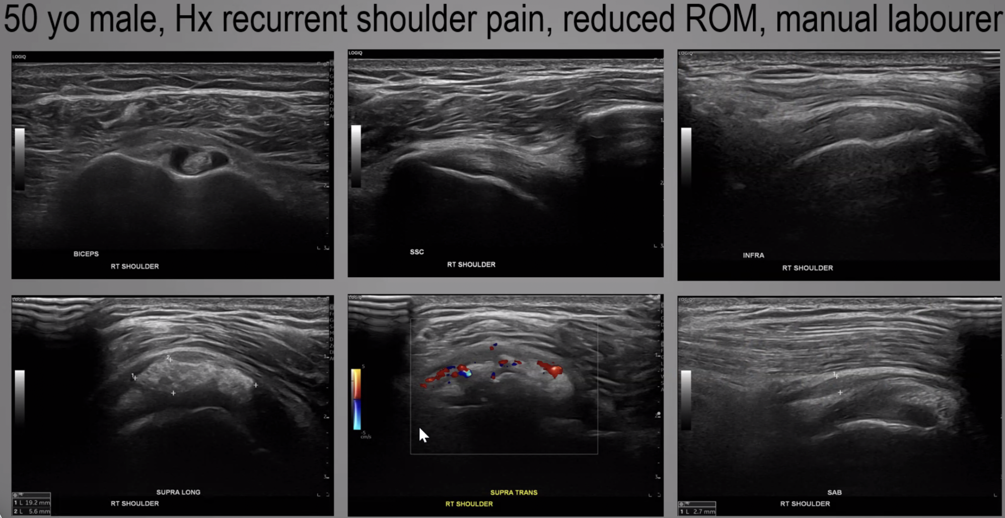
The long head of biceps tendon demonstrates a small amount of simple sheath fluid, with the tendon itself appearing unremarkable. The subscapularis and infraspinatus tendons appear normal. The supraspinatus demonstrates an ill-defined echogenic intratendinous calcific focus with increased surrounding vascularity, suggestive of soft calcific tendinopathy in the resorptive phase (Stage III). The subacromial-subdeltoid (SASD) bursa appears thickened and distended with fluid (measuring 2.7 mm), consistent with bursitis. Overall appearances are suggestive of supraspinatus calcific tendinopathy with associated SASD bursitis.

Describe the image
The bowel demonstrates multiple concentric rings with a characteristic target/doughnut (bullseye) appearance, representing telescoping of a proximal bowel segment into a distal segment, consistent with intussusception. Normal bowel typically contains luminal gas producing dirty shadowing; absence of this may help identify the intussuscepted segment. Extend the examination to determine the bowel segments involved (small-to-small, small-to-large or large-to-large) by following the colon and identifying the ileocaecal junction. Assess the length and diameter of the intussusception, internal vascularity and peristalsis, and look for a potential pathological lead point (e.g. lymph nodes or diverticula). Assess for free/interloop fluid and complications. Consider intermittent intussusception if the finding resolves during scanning.

Describe the image
The right flexor pollicis longus (FPL) tendon demonstrates abrupt fibre discontinuity and loss of the normal fibrillar architecture, with a visible retracted tendon stump, consistent with a complete tendon laceration. An adjacent heterogeneous hypoechoic fluid collection with internal echoes is seen within the tendon gap, likely representing a haematoma. There is also associated disruption of the A4 pulley.