knee, part 1
 Call Kai
 Learn
 Practice Test
 Spaced Repetition
 Match
 Flashcards
 Knowt Play
1/29

There's no tags or description
Looks like no tags are added yet.
30 Terms
distal femur landmarks
medial epicondyle
lateral epicondyle
medal condyle
lateral condyle
condyles
contact (joint articulation)
epicondyles
attachment (ligaments and muscles)
medial epicondyle
attachment site for the medial collateral ligament (MCL)
lateral epicondyle
attachment site for the lateral collateral ligament (LCL)
medial condyle
articulates with the medial condyle of the tibia
lateral condyle
articulates with the lateral condyle of the tibia
function of CONDYLES (distal femur)
articulate with the tibial plateau and help distribute body weight during standing, walking, and running
articulate with menisci
patella
sesamoid bone that articulates with the patellar surface of the femur
tibial plateau
broad, relatively flat superior surface of the tibia that articulates with the medial and lateral femoral condyles of the femur
medial and lateral
medial tibial plateau
larger and generally more concave; articulates with medial femoral condyle
lateral tibial plateau
smaller and generally flatter/less concave; articulates with lateral femoral condyle
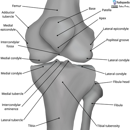
important tibial plateau structures
medial and lateral tibial condyles
intercondylar eminence
intercondylar tubercles
intercondylar areas

distal tibial and fibula landmarks
medial/lateral condyles
tibial tuberosity
intercondylar eminence
soleal line
medial/lateral condyles: DISTAL TIBFIB
articulate with femoral condyles; weight-bearing
tibial tuberosity function
patellar ligament attachment; knee extension
knee joint properties
asymmetrical due to Q-angle of the hip + more weight-bearing on medial compartment
weight-bearing and shock absorption require fibrocartilage (menisci)
contains tibiofemoral joint and patellofemoral joint
tibiofemoral joint
medial and lateral femoral condyles articulate with the corresponding medial and lateral tibial condyles (tibial plateau)
patellofemoral joint
posterior articular surface of the patella articulates with the patellar surface (trochlear groove) of the femur
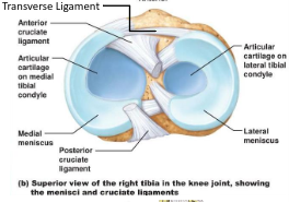
medial meniscus
C-shaped fibrocartilage in knee joint; less mobile, more firmly attached to the MCL
injured more frequently

lateral meniscus
more circular, more mobile fibrocartilage in knee joint
meniscus functions
shock absorption, distribute compressive forces, increase stability, reduce friction
help absorb shock and improve joint stability

coronary ligaments
connect the outer edges of the menisci to the tibial plateau, and anchor menisci to the tibia while allowing some movement during knee motion

transverse ligament
connects the anterior horns of the medial and lateral menisci, and helps maintain their relative positioning and coordinate movement during knee motion
internal ligaments - cruciate ligaments
ACL
PCL

ACL (anterior cruciate ligament)
prevents anterior translation of tibia relative to femur
helps prevent hyperextension
resists excessive rotation

PCL (posterior cruciate ligament)
prevents posterior translation of tibia relative to femur; stronger than ACL
ACL injury
common with pivoting, cutting, or sudden deceleration
PCL injury
common with posterior force to tibia (“dashboard injury”)
positive Lachman’s test
indicates excessive anterior translation of the tibia, suggesting ACL injury