Fatty Acid Storage & Release
 Call Kai
 Learn
 Practice Test
 Spaced Repetition
 Match
 Flashcards
 Knowt Play
1/10

There's no tags or description
Looks like no tags are added yet.
11 Terms
CD-36 and FABP work together to transport Long-chain fatty acids:
Sequential steps involved in the uptake of long-chain fatty acids by cells:
Release of fatty acids from albumin
Binding to CD36 which can accommodate up to two fatty acids at a time
Guidance of the fatty acid through the CD36 and be exposed to the plasma membrane surface
Exit of the fatty acid from CD36 to the outer leaflet of the phospholipid bilayer; transmembrane translocation (‘flip-flop’) of single fatty acids
Binding to the interior of FABP that is anchored by binding to the intracellular part of CD36
Diffusion into the soluble cytoplasm of the fatty acid–FABP complex toward sites of intracellular fatty acid metabolism.

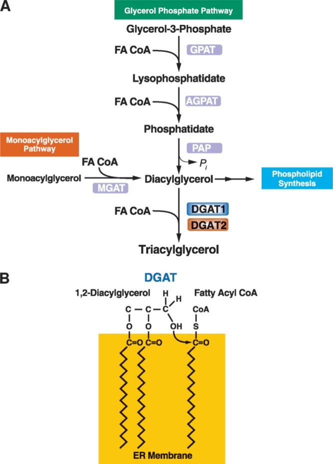
The starting point for TAG synthesis is derived from one of two routes:
Glycerol-3-phosphate
Monoacylglycerol
Triacylglycerol Synthesis
Glycerol + glucose → G-3-P (via GlyK)
G-3-P → LPA (via GPATs)
LPA → PA (via AGPATs)
PA → sn-1,2-DG (via lipins)
sn-1,2-DG → TG (via DGATs)
or
2-MG → sn-1,2-DG (via MGATs)
sn-1,2-DG → TG (via DGATs)
Triacylglycerol storage: Lipid Droplets
Lipid droplets (LD) are key components of many cells
Storage of TAG, cholesteryl esters, fat-soluble vitamins
LD synthesis occurs at the membrane shared by the ER and cytosol
LD have a phospholipid monolayer

Lipid droplet synthesis:
Newly synthesized TAG ‘nucleates’ the ER membrane until a critical mass is reached
The critical mass of TAG is extruded into the cytosol in a phosphospholipid (PL) monolayer
The PL monolayer is subsequently pinched off from the ER to release an LD (that may fuse with other LDs in the cytosol)
To use TAG stored within LD:
Intracellular lipases will associate with LDs to hydrolyze the TAG to release fatty acids
Much of what is known about the process comes from work with white adipose tissue and the liver
The processes in WAT and liver differ, and similarly in other tissues additional components appear to be involved in some other tissues (thus you cannot generalize)
TAG hydrolysis within the heart and skeletal muscle is comparable to that in WAT
TAG hydrolysis within white adipose tissue:
Three enzymes are involved:
Adipose triglyceride lipase (ATGL): cleaves the first fatty acid from TG, producing diacylglycerol (DAG) and a free fatty acid (FFA).
Hormone-sensitive lipase (HSL): mobilizes stored fat by hydrolyzing diacylglycerols (DAG) and TG into free fatty acids and glycerol - regulated by hormones.
Monoglyceride lipase (MGL)
The process is tightly regulated:
Insulin or excess intracellular acyl-CoA suppress lipolysis, while stress hormones stimulate lipolysis

Adipose triglyceride lipase (ATGL)
cleaves the first fatty acid from TG, producing diacylglycerol (DAG) and a free fatty acid (FFA)
Hormone-sensitive lipase (HSL)
mobilizes stored fat by hydrolyzing diacylglycerols (DAG) and TG into free fatty acids and glycerol - regulated by hormones
TAG hydrolysis within the liver:
While ATGL, HSL, and MGL are present in the liver, the majority of TAG hydrolysis is via carboxylesterase-1 (CES1) – (also known as triacylglycerol hydrolase (TGH)
CES1 alone can yield three fatty acids and glycerol
Fate of released FA:
The FA may be ‘activated’ to be used by other pathways or for oxidation
The FA may be used for intracellular signaling and gene regulation
The FA may be released from cells to be carried by albumin
Up to eight FA are carried per albumin