ana path final case study
 Call Kai
 Learn
 Practice Test
 Spaced Repetition
 Match
 Flashcards
 Knowt Play
1/26

There's no tags or description
Looks like no tags are added yet.
27 Terms

Case study 1
Background info (Part 1):
Mr Grocott is a 56-year-old man with a history of cigarette smoking. A CT scan of his liver demonstrated a tumour. He underwent fine-needle aspiration (FNA) of the liver lesion with rapid on-site evaluation (ROSE). The smears were examined in the lab, and a final report identifying adenocarcinoma was issued to the treating doctors.
Mr Grocott has a history of malignant melanoma diagnosed approximately six months ago. Representative images from the liver FNA are shown in Figures 1A and 1B.
Question 1A: Provide a detailed, systematic cytological description of the cells observed in Figures 1A and 1B
Cells are arranged in cohesive, 3D clusters, with occasional individual cells
Cells are predominantly round to oval, with some variation in size & shape
Nuclei are enlarged, variably sized & often eccentrically positioned, with an increased N:C ratio
Nuclei exhibit irregular nuclear contours, coarse chromatin, hyperchromasia & prominent nucleoli
Cytoplasm is moderate to abundant, pale-staining & frequently vacuolated, with variably defined cell borders

Case study 1
Background info (Part 1):
Mr Grocott is a 56-year-old man with a history of cigarette smoking. A CT scan of his liver demonstrated a tumour. He underwent fine-needle aspiration (FNA) of the liver lesion with rapid on-site evaluation (ROSE). The smears were examined in the lab, and a final report identifying adenocarcinoma was issued to the treating doctors.
Mr Grocott has a history of malignant melanoma diagnosed approximately six months ago. Representative images from the liver FNA are shown in Figures 1A and 1B.
Question 1B: Analyse the cytological features observed in Figures 1A and 1B and explain how they support the reported diagnosis. For each feature discussed, explain how it contributes to the diagnostic interpretation
The presence of cohesive, 3D clusters supports glandular differentiation, which is characteristic of adenocarcinoma
Nuclei positioned away from the centre of the cells (eccentric nuclei) support glandular differentiation, consistent with adenocarcinoma
Clear cytoplasmic vacuoles may indicate mucin production, supporting the glandular origin of the tumour
Enlarged, conspicuous nucleoli indicate increased cellular activity & support the presence of a neoplastic process
Nuclear pleomorphism, irregular nuclear contours & hyperchromasia include abnormal nuclear morphology, supporting malignancy
Case study 1
Background info (Part 1):
Mr Grocott is a 56-year-old man with a history of cigarette smoking. A CT scan of his liver demonstrated a tumour. He underwent fine-needle aspiration (FNA) of the liver lesion with rapid on-site evaluation (ROSE). The smears were examined in the lab, and a final report identifying adenocarcinoma was issued to the treating doctors.
Mr Grocott has a history of malignant melanoma diagnosed approximately six months ago.
Question 1C: Compare fine-needle aspiration with an exfoliative cytology specimen. Discuss the collection, preparation and staining of each specimen, including how different staining methods contribute to cytological evaluation
FNA is a procedure in which a fine needle & syringe are used to directly aspirate cells from the target lesion, such as the liver in this case
Exfoliative cytology, such as sputum cytology, examines cells that are naturally shed from an epithelial surface into a body fluid or secretion rather than being directly aspirated from a lesion
In FNA, the aspirated material is expelled onto glass slides & prepared as smears for cytological assessment. ROSE may also be performed to assess specimen adequacy while the procedure is still underway
In sputum cytology, selected areas of the sputum specimen, particularly suspicious or blood-stained areas, are smeared directly onto slides for examination
Pap-stained smears are prepared by immediate wet fixation in 95% ethanol, which helps prevent air-drying artefact & preserves nuclear morphology well
Diff-Quik smears are usually air-dried & then methanol-fixed, making them suitable for rapid assessment & Romanowsky-type staining
Pap staining is especially useful because it provides excellent nuclear detail, allowing clear assessment of chromatin, nuclear membranes & nucleoli
Diff-Quik staining is useful because it enhances cytoplasmic detail & can make the N:C ratio easier to appreciate
Pap staining is generally better for assessing crowded cell groups & nuclear features, whereas Diff-Quik is more helpful for cytoplasmic features & rapid preliminary assessment
Therefore, FNA & exfoliative cytology differ in collection method & specimen type, but both rely on appropriate smear preparation & staining to allow accurate cytological interpretation


Case study 1
Background info (Part 2):
Mr Grocott is referred to a surgeon for removal of the liver lesion. He undergoes surgery, and part of his liver is removed and sent to the histopathology lab.
A macroscopic image of the liver and tumour before formalin fixation is shown in Figure 2A.
Describe the complete workflow required to prepare sections from the excised liver specimen for microscopic evaluation. Begin with receipt of the specimen in the lab and continue through to the production of stained sections
The excised liver specimen is received in the histopathology lab. Patient identification, specimen labelling & the request form are checked to ensure accurate identification & prevent specimen mix-ups
A unique lab accession number is assigned, and the specimen details are entered into the Lab Info System (LIS). The specimen is assessed to determine whether additional investigations or special handling are required
The liver specimen is placed in 10% neutral buffered formalin (NBF), containing approximately 4% formaldehyde, to preserve cellular & tissue morphology
Formaldehyde forms cross-links between proteins, stabilising tissue structures & preventing autolysis, putrefaction & cellular degradation
A minimum fixative-to-tissue volume ratio of approximately 10:1 is used. Large specimens may be sliced to improve formalin penetration & ensure adequate fixation throughout the tissue
The liver specimen undergoes macroscopic examination (grossing/cut-up), during which its size, weight, shape, colour & overall appearance are noted
The tumour is identified & described, including its size, location, appearance & relationship to surrounding liver tissue & surgical margins
Representative samples of the tumour, adjacent normal liver tissue & relevant surgical margins are selected & placed into appropriately labelled tissue cassettes for processing
Fixed tissue undergoes dehydration using graded concentrations of ethanol, progressing towards absolute alcohol, to gradually remove water while minimising tissue shrinkage & distortion
Alcohol is replaced by a clearing agent, aka xylene, which is miscible with both ethanol & paraffin wax, allowing subsequent wax infiltration
The clearing agent is replaced with molten paraffin wax, which penetrates the tissue & provides structural support for embedding & sectioning
Processed tissue is carefully oriented within an embedding mould to ensure that representative tissue structures & diagnostically important areas are available during microtomy
Molten paraffin wax is added to the mould & allowed to solidify on a cold plate, producing a firm, correctly labelled tissue block suitable for sectioning
The paraffin block is cooled & trimmed using a rotary microtome to expose the tissue surface & prepare it for representative sectioning
Thin tissue sections, approximately 3-4 µm thick, are cut using a sharp microtome blade, producing ribbons of consecutive sections
Sections are floated on a warm water bath (approx. 46-49°C) to remove wrinkles, collected onto labelled glass slides & dried to ensure adhesion before staining
Paraffin wax is removed using xylene, & the sections are rehydrated through alcohol concentrations into water to prepare them for aqueous staining reagents
Haematoxylin, following oxidation to haematein & combination with an aluminium mordant, stains nuclei blue-purple. Differentiation may be required, followed by bluing to enhance nuclear contrast
Eosin, an acidic dye, stains positively charged tissue components, including cytoplasm, muscle & CT, in varying shades of pink & red
Stained sections are dehydrated through ascending alcohol concentrations, cleared using xylene & coverslipped with DPX. Slides undergo quality checks before being sent to the pathologist for microscopic examination

Case study 1
Background info (Part 3):
Upon examination of the tumour sections, the pathologist identifies a neoplastic/malignant process and determines that immunohistochemistry is required. An H&E-stained microscopic image of the neoplastic process is shown in Figure 3A.
In addition, the pathologist observes an abnormal area adjacent to the tumour showing increased collagen and distortion of the trabecular pattern. Special stains are performed to further characterise this area.
An H&E-stained microscopic image of the area is shown in Figure 3B.
Question 3A: Select an appropriate special stain to highlight one or both of the abnormal features shown in Figure 3B. Outline the steps & underlying principles of the selected staining method
Masson’s trichrome stain is selected to demonstrate the increased collagen deposition in the liver tissue adjacent to the tumour, allowing collagen to be distinguished from surrounding tissue components
Tissue sections are dewaxed using xylene & rehydrated through descending alcohol concentrations into water, allowing aqueous staining reagents to penetrate the tissue
Secondary fixation using Bouin’s fluid enhances tissue acidophilia & increases the affinity of tissue components for anionic (acid) dyes, counteracting the reducing staining affinity associated with formalin fixation
Weigert’s iron haematoxylin is applied to stain nuclei blue-black. Its iron mordant produces an acid-resistant nuclear stain that withstands subsequent acidic staining solutions
Masson’s trichrome operates through dye permeation, using anionic dyes of different molecular sizes that penetrate & are retained by tissue components according to their porosity & dye molecular size
The smaller red acid dye, Brilliant Crocein, initially penetrates & stains both collagen & cytoplasmic components, including muscle, red
Phosphotungstic acid (PTA) acts as a differentiating agent, removing the red dye from collagen while allowing it to remain within cytoplasm & muscle
The larger blue acid dye, aniline blue, subsequently penetrates & stains collagen fibres blue, while cytoplasm & muscle retain their red staining
Acetic acid is used to maintain acidic staining conditions & improve differentiation. Sections are then dehydrated, cleared with xylene & coverslipped using DPX
The resulting stain demonstrates collagen fibres in blue, cytoplasm & muscle in red, & nuclei in blue-black, allowing assessment of increased collagen deposition & fibrosis in the liver tissue

Case study 1
Background info (Part 3):
Upon examination of the tumour sections, the pathologist identifies a neoplastic/malignant process and determines that immunohistochemistry is required.
In addition, the pathologist observes an abnormal area adjacent to the tumour showing increased collagen and distortion of the trabecular pattern. Special stains are performed to further characterise this area.
Question 3B: Explain why immunohistochemistry is an appropriate ancillary investigation for this patient
Mr Grocott has a history of malignant melanoma diagnosed approx. 6 months earlier & has subsequently developed a tumour in the liver, raising the possibility of metastatic disease
Although the liver FNA identified adenocarcinoma, further investigation is required to determine whether the liver tumour represents a primary malignancy or a secondary (metastatic) tumour
Immunohistochemistry (IHC) uses specific antibodies to detect antigens expressed by tumour cells, providing additional info about their cellular differentiation & tissue of origin
An appropriate IHC antibody panel can help distinguish metastatic malignant melanoma from adenocarcinoma originating elsewhere, including a possible lung primary, by identifying characteristic patterns of pos & neg staining
The IHC findings are interpreted alongside tumour morphology & clinical history to establish the most likely tissue origin, support an accurate diagnosis & guide subsequent clinical management
Case study 1
Background info (Part 3):
Upon examination of the tumour sections, the pathologist identifies a neoplastic/malignant process and determines that immunohistochemistry is required.
In addition, the pathologist observes an abnormal area adjacent to the tumour showing increased collagen and distortion of the trabecular pattern. Special stains are performed to further characterise this area.
Question 3C: Outline the major steps involved in immunohistochemistry and briefly state the purpose of each step
Thin tissue sections are cut from formalin-fixed, paraffin-embedded tissue blocks, mounted onto glass slides, and baked to promote adhesion. Sections are then dewaxed & rehydrated to allow access to tissue antigens
Antigen retrieval, commonly using heat & an appropriate buffer, reverses or reduces the masking effects of formalin fixation, exposing antigenic epitopes for antibody binding
Blocking reagents, such as hydrogen peroxide, are applied to inhibit endogenous peroxidase activity, while other blocking steps may reduce non-specific antibody binding & background staining
A primary antibody is applied to bind specifically to the target antigen, followed by a secondary antibody conjugated to horseradish peroxidase (HRP), which binds to the primary antibody & enables detection
Diaminobenzidine (DAB) is added as a chromogen, producing an insoluble brown precipitate at sites of antigen-antibody binding through the HRP-catalysed reaction. Sections are subsequently counterstained with haematoxylin, dehydrated, cleared & coverslipped for microscopic examination

Case study 1
Part 4:
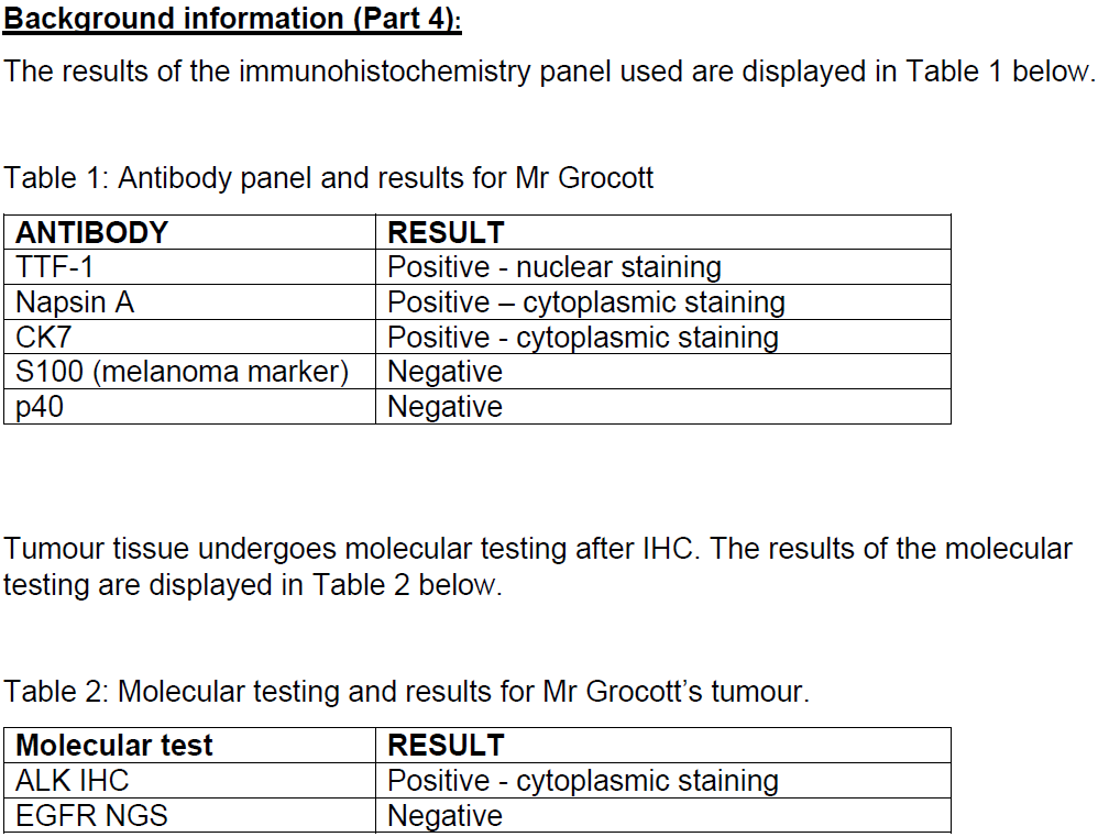
Question 4A: Analyse the immunohistochemistry results presented in Table 1 (see image). Explain how the findings support the most likely tissue origin of the tumour and help to exclude other possible diagnoses. State and justify the most likely diagnosis
Mr Grocott has a liver tumour & a previous history of malignant melanoma, making it important to determine whether the tumour represents metastatic melanoma or an adenocarcinoma originating from another organ
The IHC antibody panel demonstrates pos staining for TTF-1, Napsin A & CK7, alongside neg staining for S100 & p40, providing evidence to establish the tumour’s likely tissue origin
TTF-1 positivity, demonstrated by nuclear staining, strongly supports a pulmonary origin, particularly lung adenocarcinoma
Napsin A positivity, demonstrated by cytoplasmic staining, supports lung adenocarcinoma, as Napsin A is commonly expressed in pulmonary glandular epithelial cells
CK7 positivity, demonstrated by cytoplasmic staining in the case study, supports epithelial differentiation & is consistent with a primary lung adenocarcinoma
The combined expression of TTF-1, Napsin A & CK7 provides stronger evidence of a pulmonary adenocarcinoma than any individual marker alone
S100 negativity argues against metastatic malignant melanoma, an important differential diagnosis given the patient’s previous history of melanoma
p40 negativity argues against squamous cell carcinoma, as p40 is a nuclear marker associated with squamous differentiation
The most likely diagnosis is metastatic lung adenocarcinoma involving the liver, supported by pos TTF-1, Napsin A & CK7 staining, together with neg S100 & p40 staining
These findings are consistent with the previous FNA diagnosis of adenocarcinoma & when interpreted alongside the patient’s clinical history, tumour morphology & imaging, support the conclusion that the liver tumour most likely originated in the lung

Case study 1
Part 4:
Question 4B: Explain the relationship between the ALK immunohistochemistry result in Table 2 (see image) and an ALK gene rearrangement or fusion. In your answer, explain how the IHC result relates to the abnormal protein product, why molecular or in situ hybridisation testing may be used for confirmation, and how the result of the confirmatory test would be interpreted
Mr Grocott’s tumour demonstrates pos ALK immunohistochemistry (IHC), with cytoplasmic staining, indicating the presence of ALK protein within the tumour cells
ALK IHC acts as a screening test, using antibodies to detect the abnormal ALK protein product, which may indicate an underlying ALK gene rearrangement or fusion
An ALK gene rearrangement occurs when the ALK gene undergoes a structural alteration, commonly resulting in a gene fusion with EML4, forming the EML4-ALK fusion gene
The EML4-ALK fusion gene produces an abnormal ALK fusion protein, which possesses abnormal tyrosine kinase signalling activity that can promote uncontrolled tumour cell growth & proliferation
Pos cytoplasmic ALK staining supports the presence of an abnormal ALK protein but does not directly demonstrate the underlying genetic rearrangement. Therefore, confirmatory molecular testing may be performed
Fluorescence in situ hybridisation (FISH) may be used to confirm an ALK gene rearrangement by detecting structural changes in the ALK gene using fluorescently labelled DNA probes
ALK break-apart FISH uses two differently coloured fluorescent probes that bind to DNA sequences on opposite sides of the ALK gene’s rearrangement region
If an ALK gene rearrangement is present, the fluorescent signals become separated or split, indicating disruption of the normal ALK gene arrangement & supporting a pos rearrangement result
If no ALK rearrangement is present, the fluorescent signals remain close together or appear fused, indicating an intact ALK gene arrangement & supporting a neg rearrangement result
A pos ALK IHC result supported by a confirmatory FISH rearrangement result would identify an ALK-rearranged lung adenocarcinoma, providing info relevant to targeted therapy. The separate neg EGFR next-generation sequencing (NGS) result indicates that no EGFR mutation was detected by the assay & does not exclude an ALK rearrangement


Case study 2
Background info (Part 1):
Mrs Bailey is a 67-year-old woman with a long history of cigarette smoking. She presents with a persistent cough & intermittent haemoptysis (coughing up blood). A CT scan reveals a centrally located mass in the right upper lobe. Bronchoscopy is performed & a bronchial brushing specimen is submitted for cytological examination. The final cytology report identifies squamous cell carcinoma.
She was previously treated for colorectal adenocarcinoma, so the origin & histological subtype of the new tumour must be carefully assessed. Representative cytology images are shown in Figures 1A & 1B.
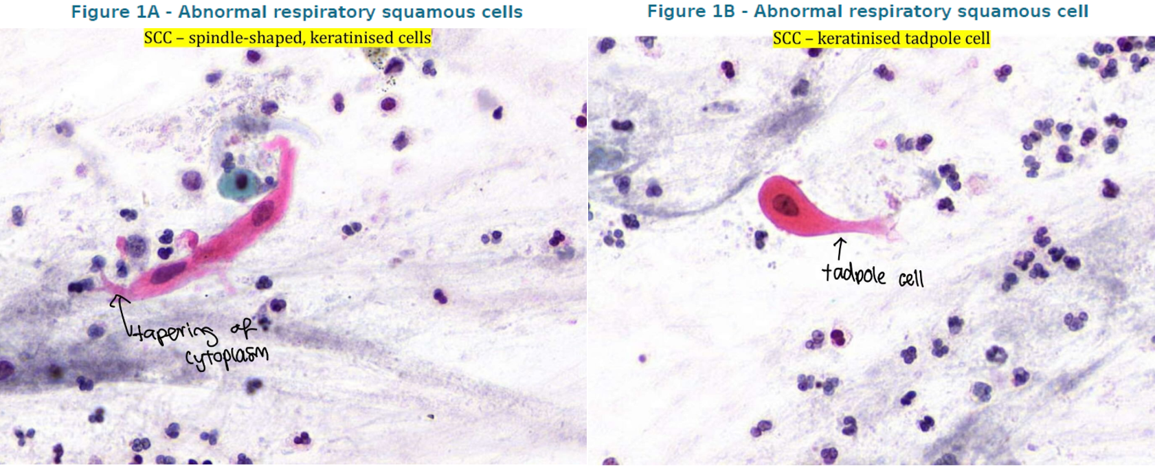
Question 1A: Provide a detailed, systematic cytological description of the cells known in Figures 1A & 1B. Include cell presentation, shape, nuclear features & cytoplasmic features
The specimen contains predominantly isolated abnormal squamous epithelial cells, with scattered inflammatory cells present in the background
The abnormal cells demonstrate marked variation in shape, including elongated, spindle-shaped & tadpole-shaped cells with tapering cytoplasmic projections
The nuclei are round, oval or elongated, with variable nuclear size & shape. Some appear hyperchromatic or pyknotic, particularly within the keratinised cells
The nuclei demonstrate nuclear pleomorphism & atypical chromatin features, with variable N:C ratios. Nucleoli are not conspicuous in the prominently keratinised cells
The cells possess dense, brightly eosinophilic or orangeophilic cytoplasm, indicating prominent keratinisation, with sharply defined cell borders & elongated cytoplasmic extensions
Case study 2
Background info (Part 1):
Mrs Bailey is a 67-year-old woman with a long history of cigarette smoking. She presents with a persistent cough & intermittent haemoptysis (coughing up blood). A CT scan reveals a centrally located mass in the right upper lobe. Bronchoscopy is performed & a bronchial brushing specimen is submitted for cytological examination. The final cytology report identifies squamous cell carcinoma.
She was previously treated for colorectal adenocarcinoma, so the origin & histological subtype of the new tumour must be carefully assessed. Representative cytology images are shown in Figures 1A & 1B.
Question 1B: Analyse how the observed cytological features support the reported diagnosis of squamous cell carcinoma. For each feature, explain its diagnostic significance
The presence of dense, orangeophilic keratinised cytoplasm supports squamous epithelial differentiation, a characteristic feature of keratinising squamous cell carcinoma
The presence of tadpole-shaped & spindle-shaped cells with elongated cytoplasmic processes represents abnormal squamous cell morphology & supports a well-differentiated keratinising squamous cell carcinoma
Nuclear pleomorphism, including variation in nuclear size & shape, supports malignancy by demonstrating abnormal cellular differentiation
Hyperchromatic & irregular nuclei suggest nuclear atypia & disordered cellular activity, supporting the diagnosis of a malignant epithelial tumour
The combination of isolated bizarre squamous cells, prominent keratinisation & nuclear atypia, together with the patient’s smoking history & centrally located lung mass, supports the reported diagnosis of squamous cell carcinoma
Case study 2
Background info (Part 1):
Mrs Bailey is a 67-year-old woman with a long history of cigarette smoking. She presents with a persistent cough & intermittent haemoptysis (coughing up blood). A CT scan reveals a centrally located mass in the right upper lobe. Bronchoscopy is performed & a bronchial brushing specimen is submitted for cytological examination. The final cytology report identifies squamous cell carcinoma.
She was previously treated for colorectal adenocarcinoma, so the origin & histological subtype of the new tumour must be carefully assessed.
Question 1C: Compare bronchial brushing with sputum exfoliative cytology. Discuss collection, slide preparation & fixation, and explain how Papanicolaou & Diff-Quik staining can contribute differently to cytological evaluation
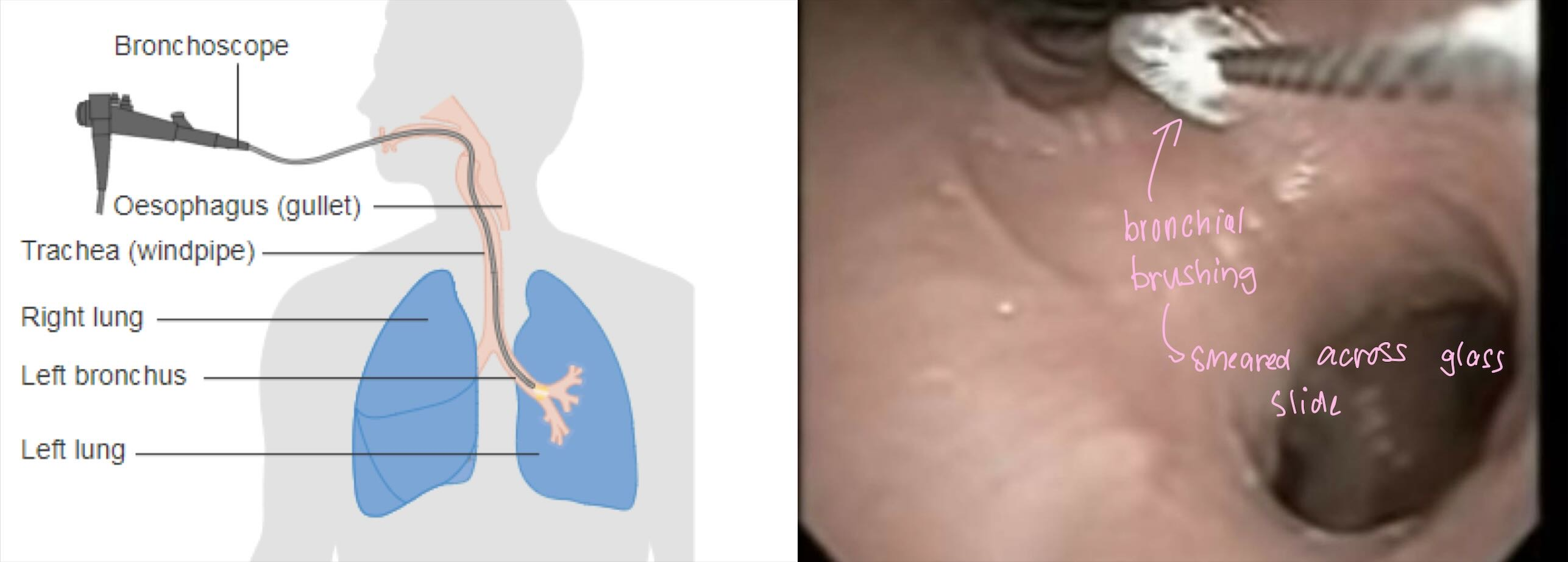
Bronchial brushing is performed during bronchoscopy, where a cytology brush is passed over a suspicious bronchial lesion to directly collect epithelial cells from the abnormal area
Sputum exfoliative cytology examines cells that are naturally shed from the respiratory tract & collected through deep coughing, rather than directly sampling a visible lesion
In bronchial brushing, collected material is transferred onto glass slides, commonly by rolling the brush across the slide, to produce smears for cytological examination
In sputum cytology, suspicious or blood-stained areas of the specimen are selected & smeared directly onto glass slides to produce thin, representative cell preparation
Pap-stained smears undergo immediate wet fixation in 95% ethanol, preserving nuclear morphology & preventing air-drying artefacts
Diff-Quik stained smears are air-dried & subsequently methanol-fixed, allowing rapid Romanowsky-type staining & evaluation
Pap staining provides excellent nuclear detail, allowing assessment of nuclear membranes, chromatin, nucleoli & malignant nuclear abnormalities
Diff-Quik staining enhances cytoplasmic features & the N:C ratio, supporting the identification & preliminary assessment of abnormal cells
Pap staining is generally better for assessing crowded cellular groups & nuclear morphology, whereas Diff-Quik is useful for rapid assessment & cytoplasmic characteristics but is less suitable for interpreting crowded clusters
Therefore, bronchial brushing & sputum cytology differ in specimen collection & preparation, but both rely on appropriate fixation & staining to preserve cellular morphology & support the diagnosis of respiratory malignancies
Case study 2
Background info (Part 2):
Following clinical staging & multidisciplinary review, Mrs Bailey undergoes a right upper lobectomy. The excised lung tissue, containing the bronchial tumour & surrounding non-neoplastic tissue, is transported to the histopathology lab for routine microscopic evaluation.
The surgeon requests an assessment of the tumour size, its relationship to nearby bronchial structures & relevant resection margins.
Question 2: Describe the complete histopathology workflow required to prepare the excised lung specimen for microscopic evaluation. Begin with specimen receipt & verification, then cover fixation, macroscopic examination & sampling, tissue processing, embedding, microtomy & routine H&E staining, through to preparation of suitable finished slides
The excised right upper lung lobe is received in the histopathology lab, where the patient identification, specimen labelling & request form are checked to prevent specimen identification errors
The lab assigns a unique accession number and enters the specimen information into the Lab Info System (LIS). The specimen is assessed for special handling requirements before routine processing
The lung specimen is fixed using 10% neutral buffered formalin (NBF), containing approx. 4% formaldehyde, to preserve cellular morphology & tissue architecture
Formaldehyde produces protein cross-linking, which stabilises tissue components & prevents autolysis, putrefaction & cellular degradation
An adequate volume of formalin, ideally a fixative-to-tissue ratio of approx. 10:1 is used. The specimen may be appropriately opened or sliced to improve fixative penetration, while preserving important anatomical relationships
The lung specimen undergoes macroscopic examination (grossing/cut-up), during which its dimensions, weight, anatomical features & external appearance are noted
The tumour is identified & described, including its size, location, colour, consistency & relationship to the bronchus, surrounding lung tissue, pleura & relevant surgical margins
Representative sections of the tumour, adjacent non-neoplastic lung, tumour-lung interface & relevant surgical margins are selected & placed into appropriately labelled tissue cassettes
Fixed tissue undergoes dehydration using graded concentrations of ethanol, progressing towards absolute alcohol, to remove water from the specimen
Ethanol is replaced by a clearing agent, aka xylene, which is miscible with alcohol & paraffin wax, allowing subsequent wax infiltration
The clearing agent is replaced with molten paraffin wax, which penetrates the tissue & provides structural support for embedding & sectioning
The processing tissue is carefully oriented within an embedding mould, ensuring that the diagnostically important structures, such as the tumour interface or surgical margin, will be appropriately represented during sectioning
The mould is filled with molten paraffin wax & cooled on a cold plate, producing a firm, correctly identified tissue block suitable for microtomy
The paraffin block is cooled & trimmed using a rotary microtome to expose a complete tissue surface & prepare it for sectioning
Thin sections, approx. 3-4 µm thick, are cut up using a sharp microtome blade, producing ribbons of tissue suitable for microscopic examination
Sections are floated on a warm water bath to remove wrinkles, collected onto appropriately labelled glass slides & dried to ensure adhesion before staining
Tissue sections undergo dewaxing with xylene, followed by rehydration through descending alcohol concentrations into water, preparing the tissue for aqueous staining reagents
Haematoxylin stains cell nuclei blue-purple, allowing assessment of nuclear morphology, chromatin & cellular abnormalities. Differentiation & bluing may be performed as required by the staining method
Eosin, an acidic counterstain, stains cytoplasm, muscle & CT in varying shades of pink & red, providing contrast between tissue components
The stained slides are dehydrated through ascending alcohol concentrations, cleared with xylene & coverslipped with DPX. Slide identification & staining quality are checked before microscopic evaluation by the pathologist

Case study 2
Background info (Part 3):
The pathologist confirms a malignant squamous epithelial tumour on H&E sections. In adjacent bronchial luminal debris, the pathologist identifies necrotic material & possible fungal structures. An additional special stain is requested to investigate these structures.
An image has been provided as a reference showing fungal structures demonstrated in a tissue section. It is included as a visual comparison, not as a slide from Mrs Bailey.
Question 3A: Select a suitable special stain to demonstrate the suspected fungal structures. Outline the major staining steps & explain the underlying chemical principles, including the expected staining results
Grocott methenamine silver (GMS) is selected to demonstrate suspected fungal organisms within the necrotic bronchial material, as the technique highlights fungal cell walls & allows their morphology to be assessed
Formalin-fixed, paraffin-embedded tissue sections are dewaxed using xylene & rehydrated through descending alcohol concentrations into water, allowing staining reagents to penetrate the section
Tissue sections are treated with chromic acid, which oxidises carbohydrate components within fungal cell walls to form aldehyde groups capable of participating in the subsequent silver reaction
The sections are treated with sodium metabisulphite, followed by thorough washing, to remove residual oxidising agent & prevent interference with silver impregnation
An alkaline methenamine silver solution, containing a silver complex & prepared with an alkaline reagent such as borax, is applied to demonstrate the oxidised fungal cell wall components
During silver impregnation, the aldehyde-containing fungal wall components promote the reduction of silver ions to visible metallic silver, producing dark staining of fungal structures
Gold chloride is applied as a toning agent to improve staining contrast & produce a more distinct black appearance of the silver-impregnated fungal structures
Sodium thiosulphate removes remaining unreduced silver components, helping prevent non-specific background reactions & improving the stability of the stained preparation
A light green counterstain is applied to provide contrast between the dark fungal structures & the surrounding tissue. Sections are then dehydrated, cleared & coverslipped
The resulting stain demonstrates fungal cell walls in black against a green background, allowing their distribution within the tissue. Organism identification may require additional investigations

Case study 2
Background info (Part 3):
The pathologist confirms a malignant squamous epithelial tumour on H&E sections. In adjacent bronchial luminal debris, the pathologist identifies necrotic material & possible fungal structures. An additional special stain is requested to investigate these structures.
Question 3B: Explain why immunohistochemistry is an appropriate ancillary investigation when establishing tumour subtype & origin in this patient, given her previous malignancy
Mrs Bailey has a history of colorectal adenocarcinoma & has developed a new lung tumour, raising the possibility of either a primary pulmonary malignancy or metastatic disease from her previous colorectal cancer
Although cytological & histological examination supports squamous cell carcinoma, further investigation may be required to establish the tumour’s epithelial differentiation, histological subtype & most likely origin
Immunohistochemistry (IHC) uses specific antibodies to identify antigens expressed by tumour cells, providing evidence about their cellular lineage & differentiation
A targeted IHC panel can distinguish squamous differentiation using markers such as p40 & CK5/6, while markers such as TTF-1, Napsin A & CDX2 help investigate alternative pulmonary or colorectal origins
The combination of IHC findings, tumour morph, clinical history & imaging assists in establishing the most likely diagnosis, distinguishing primary from metastatic disease & guiding subsuquent clincal management

Case study 2
Background info (Part 3):
The pathologist confirms a malignant squamous epithelial tumour on H&E sections. In adjacent bronchial luminal debris, the pathologist identifies necrotic material & possible fungal structures. An additional special stain is requested to investigate these structures.
Question 3C: Outline the major technical stages of IHC & briefly state the purpose of each, from section preparation to visualisation
Thin tissue sections are cut from formalin-fixed, paraffin-embedded blocks, mounted onto glass slides & baked to promote tissue adhesion. They are then dewaxed & rehydrated to prepare them for staining
Antigen retrieval, commonly performed using heat & an appropriate buffer, reduces the masking effects of formalin-induced protein cross-linking, exposing antigenic epitopes for antibody binding
Blocking reagents, such as hydrogen peroxide, inhibit endogenous peroxidase activity, while other blocking steps may minimise non-specific antibody binding & unwanted background staining
A primary antibody binds specifically to the target antigen, followed by an appropriate secondary antibody or polymer detection system, commonly labelled with horseradish peroxidase (HRP), allowing the antibody-antigen complex to be detected
Diaminobenzidine (DAB) is added as a chromogen, reacting through the HRP detection system to produce an insoluble brown precipitate at sites of target antigen expression. The sections are counterstained with haematoxylin, dehydrated, cleared & coverslipped for microscopic evaluation

Case study 2
Background info (Part 4):
The following IHC results were obtained from the excised lung tumour. All results refer to the same specimen
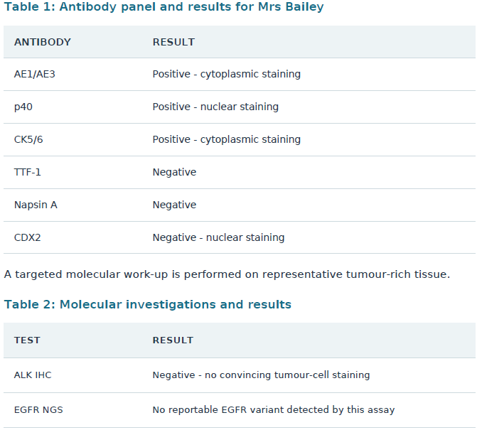
Question 4A: Interpret each of the IHC tables in Table 1 & explain how the combined pos & neg staining patterns support a likely primary tumour subtype & organ of origin, while arguing against important alternatives. State & justify the most likely diagnosis
Mrs Bailey has a centrally located lung tumour & a previous history of colorectal adenocarcinoma, requiring investigation to distinguish a primary pulmonary squamous cell carcinoma from metastatic colorectal cancer or another pulmonary tumour subtype
AE1/AE3 positivity, demonstrated by cytoplasmic staining, supports epithelial differentiation, as this broad-spectrum cytokeratin marker is commonly expressed in carcinomas
p40 positivity, demonstrated by nuclear staining, strongly supports squamous epithelial differentiation, as p40 is an important marker of squamous cell carcinoma
CK5/6 positivity, demonstrated by cytoplasmic staining, supports squamous differentiation, as these high-molecular-weight cytokeratins are commonly expressed in squamous epithelial cells
The combined expression of AE1/AE3, p40 & CK5/6 provides strong evidence of an epithelial malignancy showing squamous differentiation, supporting a diagnosis of squamous cell carcinoma
TTF-1 negativity argues against pulmonary adenocarcinoma, as TTF-1 nuclear expression commonly supports glandular differentiation in primary lung adenocarcinomas
Napsin A negativity further argues against pulmonary adenocarcinoma, as Napsin A is commonly expressed within the cytoplasm of lung adenocarcinoma cells
CDX2 negativity, with no nuclear staining, argues against metastatic colorectal adenocarcinoma, making the patient’s previously diagnosed colorectal malignancy a less likely source of the current tumour
The most likely diagnosis is primary pulmonary squamous cell carcinoma, supported by pos AE1/AE3, p40 & CK5/6 staining & neg TTF-1, Napsin A & CDX2 staining
The IHC findings are consistent with the tumour’s squamous cytological morph, the patient’s long history of smoking & the centrally located lung mass. Together, these findings support a primary lung squamous cell carcinoma rather than metastatic colorectal adenocarcinoma, although the final determination of origin requires clinicopathological correlation
Case study 2
Background info (Part 4):
The following IHC results were obtained from the excised lung tumour. All results refer to the same specimen
Question 4B: Explain the difference between what ALK IHC & EGFR next-gen sequencing (NGS) detect. Describe the importance of choosing tumour-rich FFPE sections & obtaining adequate DNA quality for molecular testing. Explain how ALK break-apart FISH could be used if an ALK protein result were pos or equivocal, how split vs fused signals would be interpreted, and what the two neg results in Table 2 do & do not allow you to conclude
ALK IHC uses antibodies to detect ALK protein expression within tumour cells. Mrs Bailey’s neg ALK IHC result indicates that no convincing ALK protein staining was detected by the assay
EGFR next-gen sequencing (NGS) examines the EGFR gene at the DNA level to identify genetic variations, including clinically relevant mutations. The neg result indicates that no reportable EGFR variant was detected within the assay’s validated scope
The two methods investigate different molecular targets: ALK IHC detects protein expression, whereas EGFR NGS detects DNA sequence alterations. Therefore, a neg result from one assay does not determine the result of the other
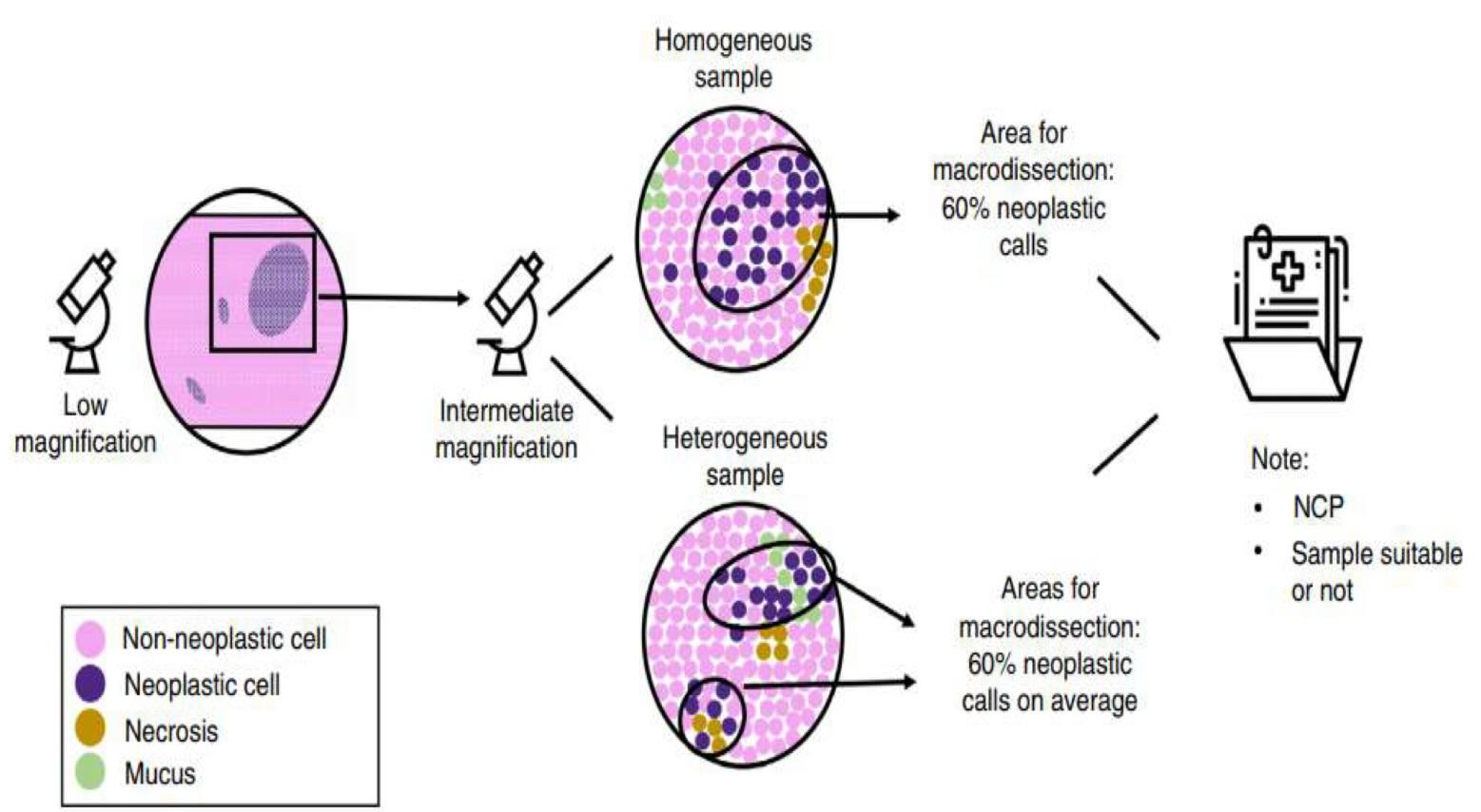
Formalin-fixed, paraffin-embedded (FFPE) tissue sections are selected for molecular testing, with the pathologist reviewing an H&E-stained section to identify areas containing a sufficient proportion of viable tumour cells
Macrodissection may be performed to enrich the tumour-cell population by removing or avoiding areas containing excessive normal tissue, necrosis, inflammation or fibrosis, improving the proportion of tumour DNA available for testing
Adequate DNA concentration, purity & integrity are essential for reliable NGS results. Formalin fixation may cause DNA fragmentation & cross-linking, while insufficient tumour content or degraded DNA may reduce assay sensitivity & increase the risk of false-neg results
If the ALK IHC were pos or equivocal, ALK break-apart fluorescence in situ hybridisation (FISH) could be used when confirmation is indicated. This technique uses two differently coloured DNA probes positioned on opposite sides of the ALK rearrangement region
A pos ALK rearrangement result is suggested when the fluorescent signals are separated or split, indicating that the regions flanking the ALK gene have become physically separated by a structural rearrangement
A neg ALK rearrangement result is supported when the fluorescent signals remain close together or appear fused, indicating that the ALK gene arrangement is intact at the regions assessed. Interpretation needs to follow the validated lab criteria as well
The neg ALK IHC result makes a detectable ALK-associated fusion protein less likely, while the neg EGFR NGS result indicates that no reportable EGFR mutation was detected. However, these results do not exclude every possible ALK alteration, EGFR variant or other oncogenic mutation, and they do not establish that the tumour is free of molecular abnormalities


Case study 3
Background info (Part 1):
Mr Elias Ward is a 64-year-old man with a long history of smoking. He presents with a persistent cough, fatigue & episodes of haemoptysis. Imaging demonstrates a centrally situated left hilar mass with associated mediastinal lymphadenopathy. A sputum sample is collected for cytological evaluation.
Cytology reveals malignant cells, and the final report identifies small-cell carcinoma. Representative images of the relevant cytological morphology are provided (see image).
Question 1A: Provide a detailed, systematic cytological description of the cells shown in Figures 1A and 1B. Include cell presentation, size & shape, nuclear characteristics, N:C ratio, cytoplasmic features & background
The specimen contains small malignant cells arranged in loose clusters & dispersed individually, with some cells appearing in linear arrangements along mucus strands
The cells are relatively small & round to oval, with occasional irregular shapes. They show limited cytoplasmic differentiation, making the nuclei appear particularly prominent
The nuclei are round, oval or irregular, with finely granular or stippled chromatin, often described as a salt-and-pepper pattern. Nucleoli are generally inconspicuous or absent
The cells demonstrate nuclear moulding, where adjacent nuclei conform to one another, and a very high N:C ratio, reflecting the minimal amount of surrounding cytoplasm
The cytoplasm is scant, delicate & occasionally indistinguishable, while the background may contain nuclear smearing artefact & tumour diathesis, including cellular & nuclear debris associated with fragile malignant cells

Case study 3
Background info (Part 1):
Mr Elias Ward is a 64-year-old man with a long history of smoking. He presents with a persistent cough, fatigue & episodes of haemoptysis. Imaging demonstrates a centrally situated left hilar mass with associated mediastinal lymphadenopathy. A sputum sample is collected for cytological evaluation.
Cytology reveals malignant cells, and the final report identifies small-cell carcinoma. Representative images of the relevant cytological morphology are provided (see image).
Question 1B: Analyse how the observed features support the reported diagnosis of small-cell carcinoma. Explain the diagnostic importance of each feature, rather than merely describing its appearance
The presence of small malignant cells with scant cytoplasm & a high N:C ratio is characteristic of small-cell carcinoma, reflecting the poorly differentiated nature of this malignant tumour
Nuclear moulding, where adjacent nuclei become compressed against one another, is a particularly important diagnostic feature supporting small-cell carcinoma
Finely stippled salt-and-pepper chromatin & inconspicuous nucleoli support a neuroendocrine tumour, consistent with the nuclear morph of small-cell carcinoma
Nuclear smearing artefact & tumour diathesis support the presence of fragile malignant cells undergoing cellular breakdown, which commonly occurs in small-cell carcinoma specimens
The combination of small cells, nuclear moulding, high N:C ratio, stippled chromatin & scant cytoplasm, together with the patient’s smoking history & centrally located lung mass, strongly supports the reported diagnosis of small-cell lung carcinoma
Case study 3
Background info (Part 1):
Mr Elias Ward is a 64-year-old man with a long history of smoking. He presents with a persistent cough, fatigue & episodes of haemoptysis. Imaging demonstrates a centrally situated left hilar mass with associated mediastinal lymphadenopathy. A sputum sample is collected for cytological evaluation.
Cytology reveals malignant cells, and the final report identifies small-cell carcinoma.
Question 1C: Compare sputum cytology with pleural-effusion fluid cytology. Discuss specimen collection, selection & concentration of material, smear/cytospin preparation, wet fixation & air drying, and how Papanicolaou & Diff-Quik staining assist cytological evaluation
Sputum cytology is a non-invasive exfoliative technique in which cells naturally shed from the respiratory tract are collected through deep coughing, allowing examination of cells originating from the airways
Pleural-effusion cytology involves collecting fluid from the pleural cavity, usually through thoracentesis (removing excess fluid from the pleural space), to investigate abnormal cells present within an accumulated body fluid
In sputum cytology, the specimen is macroscopically assessed, and blood-stained, discoloured or solid flecks are selected for direct smear preparation using techniques such as pick-and-smear or pull-smear
In pleural-effusion cytology, the fluid is typically centrifuged to concentrate suspended cells, producing a cell pellet from which appropriate cytological preparations can be produced
Cytocentrifugation (cytospin) may be used to concentrate a low-cellularity pleural-fluid specimen onto a defined area of a glass slide, producing a preparation suitable for microscopic evaluation
Pap-stained preparations undergo immediate wet fixation in 95% ethanol, preventing air-drying artefacts & preserving nuclear morph. This is particularly important when preparing sputum smears
Diff-Quik preparations are air-dried & subsequently methanol-fixed, allowing rapid Romanowsky-type staining of cells obtained from appropriate cytological preparations, including cytospins
Pap staining provides excellent nuclear detail, allowing assessment of chromatin patterns, nuclear membranes, nucleoli & cellular abnormalities, while also assisting with the interpretation of crowded cellular groups
Diff-Quik staining enhances cytoplasmic characteristics, cellular detail & the N:C ratio, making it useful for rapid evaluation, although it is generally less suitable than Pap staining for interpreting crowded clusters
Therefore, sputum & pleural-effusion cytology differ in collection method & specimen collection, with sputum relying mainly on selected direct smears & pleural fluid often requiring centrifugation. Both use appropriate fixation, preparation & complementary staining techniques to support cytological diagnosis

Case study 3
Background info (Part 2):
Mr Ward undergoes bronchoscopy, during which several small endobronchial biopsy fragments are obtained from the left hilar mass. The tissue is submitted to this histopathology lab for routine microscopic evaluation & potential ancillary testing.
Because the samples are small & may be needed for immunohistochemistry (IHC) & molecular investigations, correct handling, orientation where relevant, processing, identification & conservation of the remaining tissue are important.
Question 2: Describe the complete histopathology workflow required to produce diagnostic H&E-stained sections from the endobronchial biopsy fragments. Begin with specimen receipt & identification, then explain fixation, macroscopic examination & cassette selection, dehydration, clearing, infiltration, embedding, microtomy, mounting, H&E staining & QC. Discuss the specific precautions required for small biopsy fragments
The endobronchial biopsy fragments are received in the histopathology lab, where patient identification, specimen labelling, anatomical site & request form details are verified to ensure correct specimen identification
A unique accession number is assigned, and specimen details are entered into the Lab Info System (LIS). The tissue is assessed for any additional testing requirements, including IHC & molecular investigations
The biopsy fragments are fixed in 10% neutral buffered formalin (NBF), containing approx. 4% formaldehyde, to preserve cellular morp & tissue architecture
Formaldehyde forms cross-links between proteins, stabilising tissue structures & preventing autolysis, putrefaction & cellular degradation
Because the biopsies are small, adequate but not excessive fixation is important to preserve morph & support subsequent ancillary investigations. The fragments must remain fully immersed in fixative & be handled carefully to prevent tissue loss
The specimen undergoes macroscopic examination (grossing), during which the number, dimensions, colour & appearance of the biopsy fragments are noted
As the tissue fragments are small, they are generally submitted entirely for processing rather than extensively dissected, ensuring that diagnostically important tissue is not discarded
The fragments are placed into appropriately labelled tissue cassettes, using biopsy sponges, mesh or biopsy paper where appropriate to prevent small pieces from escaping during processing
Fixed tissue undergoes dehydration using graded concentrations of ethanol, progressing towards absolute alcohol, to remove water from the specimen
Alcohol is replaced by a clearing agent, aka xylene, which is miscible with ethanol & paraffin wax, allowing subsequent wax infiltration
The clearing agent is replaced with molten paraffin wax, which infiltrates the tissue & provides structural support. A suitable processing schedule for small biopsy fragments is selected to avoid inadequate processing or excessive tissue hardening
The processed biopsy fragments are carefully oriented within an embedding mould, ensuring that representative tissue surfaces can be examined during microtomy
Molten paraffin wax is added & solidified on a cold plate, producing a firm, correctly labelled tissue block. Care is taken to retain every small biopsy fragment during embedding
The paraffin block is cooled & carefully trimmed using a rotary microtome to expose the biopsy fragments while minimising unnecessary loss of tissue
Thin tissue sections, approx. 3-4 µm thick, are cut using a sharp microtome blade to produce sections suitable for microscopic examination
Sections are floated on a warm water bath, collected onto appropriately labelled glass slides & dried to ensure adhesion. Excessive trimming & unnecessary deeper sections are avoided, preserving tissue for IHC & molecular testing
The sections undergo dewaxing using xylene, followed by rehydration through descending alcohol concentrations into water, preparing them for aqueous staining reagents
Haematoxylin stains cell nuclei blue-purple, allowing assessment of nuclear morph, chromatin & cellular abnormalities. Differentiation & bluing may be performed as required
Eosin, an acidic counterstain, stains cytoplasm & extracellular tissue components in varying shades of pink & red, allowing the different cellular & structural components to be distinguished
The sections are dehydrated through ascending alcohol concentrations, cleared using xylene & coverslipped with DPX. Slide identification, section completeness, staining quality & artefacts are checked before microscopic evaluation, while remaining tissue is conserved for additional investigations

Case study 3
Background info (Part 3):
On review of Mr Ward’s sputum, scattered alveolar macrophages contain coarse brown-black intracellular granules. Given the history of haemoptysis, the pathologist wishes to determine whether some of this pigment represents haemosiderin (ferric iron) rather than inhaled carbon. A cell-block preparation is available for special staining.
The image given provides an example of pigment-containing respiratory macrophages. It is a visual reference, not from the patient themselves.
The separate bronchial biopsy demonstrates a poorly differentiated malignant tumour. IHC is requested to distinguish an epithelial carcinoma from potential histological mimics & to assist in determining its subtype.
Question 3A: Select an appropriate special stain to investigate ferric iron-containing pigment in the macrophages. Outline the preparation & major staining steps, explain the chemical principle, describe the role of key reagents & state the expected results. Explain how the stain distinguishes haemosiderin from some other dark pigments
Perls’ Prussian blue stain is selected to identify ferric iron (Fe3+) within pigment-containing alveolar macrophages, allowing the pathologist to investigate the presence of haemosiderin associated with previous bleeding
Sections prepared from the formalin-fixed, paraffin-embedded cell block are dewaxed using xylene & rehydrated through descending alcohol concentrations into water, allowing the staining reagents to penetrate the specimen
Hydrochloric acid (HCl) is applied to release ferric iron ions (Fe3+) from their protein-bound form within haemosiderin deposits
Potassium ferrocyanide reacts with the released ferric iron ions, producing an insoluble ferric ferrocyanide complex, commonly known as Prussian blue
The formation of this blue reaction product at sites containing ferric iron is the underlying histochemical principle of Perl’s staining, allowing iron-containing deposits to be demonstrated microscopically
Following application of the freshly prepared acid-ferrocyanide staining reagent, the sections are thoroughly washed in distilled water to remove excess reagents & reduce unwanted background staining
Nuclear fast red is applied as a counterstain, staining cell nuclei red & providing contrast against the blue haemosiderin deposits
The stained sections are dehydrated through graded alcohols, cleared with xylene & coverslipped with DPX, allowing microscopic evaluation & preservation of the preparation
Haemosiderin containing ferric iron stains blue, while nuclei appear red. In contrast, inhaled carbon pigment does not produce the Prussian blue reaction & may retain its original dark appearance
A pos Perls’ reaction in alveolar macrophages supports the presence of haemosiderin-laden macrophages (which have engulfed & broken down RBCs, leaving behind deposits), which may reflect previous alveolar haemorrhage. A known pos-control tissue should be stained alongside the specimen to confirm that the staining procedure has worked correctly

Case study 3
Background info (Part 3):
On review of Mr Ward’s sputum, scattered alveolar macrophages contain coarse brown-black intracellular granules. Given the history of haemoptysis, the pathologist wishes to determine whether some of this pigment represents haemosiderin (ferric iron) rather than inhaled carbon. A cell-block preparation is available for special staining.
The separate bronchial biopsy demonstrates a poorly differentiated malignant tumour. IHC is requested to distinguish an epithelial carcinoma from potential histological mimics & to assist in determining its subtype.
Question 3B: Explain why IHC is appropriate for this poorly differentiated lung tumour. Discuss its role in evaluating lineage & distinguishing small-cell carcinoma from important differential diagnoses, including lymphoma & poorly differentiated squamous carcinoma
Mr Ward has a poorly differentiated malignant lung tumour, making it important to establish the tumour’s cellular lineage & histological subtype, particularly because poorly differentiated malignancies may share similar morphological appearances
The differential diagnoses include small-cell carcinoma, lymphoma & poorly differentiated squamous cell carcinoma, which many demonstrate overlapping features on routine H&E staining
IHC uses specific antibodies to detect cellular antigens, allowing the pathologist to identify the tumour’s lineage & differentiation pattern
Cytokeratin markers, such as AE1/AE3, support epithelial differentiation, while neuroendocrine markers such as synaptophysin support small-cell carcinoma. In contrast, CD45 helps investigate lymphoma & p40 help identify squamous differentiation
The combined IHC staining pattern, interpreted alongside cytomorph, histological findings & clinical history, helps distinguish small-cell carcinoma from its differential diagnoses & supports an accurate diagnosis

Case study 3
Background info (Part 3):
On review of Mr Ward’s sputum, scattered alveolar macrophages contain coarse brown-black intracellular granules. Given the history of haemoptysis, the pathologist wishes to determine whether some of this pigment represents haemosiderin (ferric iron) rather than inhaled carbon. A cell-block preparation is available for special staining.
The separate bronchial biopsy demonstrates a poorly differentiated malignant tumour. IHC is requested to distinguish an epithelial carcinoma from potential histological mimics & to assist in determining its subtype.
Question 3C: Outline the major steps involved in an indirect enzyme-based IHC method, from tissue-section preparation to chromogen development & counterstaining. Briefly state the purpose of each step
Thin tissue sections are cut from formalin-fixed, paraffin-embedded tissue blocks, mounted onto glass slides & baked to promote tissue adhesion. The sections are subsequently dewaxed & rehydrated to prepare them for immunostaining
Antigen retrieval, commonly performed using heat & an appropriate buffer, reduces the masking effects of formalin-induced protein cross-linking, exposing antigenic epitopes for antibody binding
Blocking reagents, including hydrogen peroxide, are used to inhibit endogenous peroxidase activity, while other blocking steps may reduce non-specific antibody binding & unwanted background staining
A primary antibody binds specifically to the target antigen, followed by an appropriate enzyme-labelled secondary antibody, commonly conjugated to horseradish peroxidase (HRP), which allows the antigen-antibody complex to be detected
Diaminobenzidine (DAB) is added as a chromogen, producing an insoluble brown precipitate at sites of antibody binding through the HRP-catalysed reaction. The tissue is then counterstained with haematoxylin, dehydrated, cleared & coverslipped for microscopic examination

Case study 3
Background info (Part 4):
An IHC panel is performed on the endobronchial biopsy. For this case, synaptophysin is a cytoplasmic marker supporting neuroendocrine differentiation; the remaining markers are used to assess epithelial, pulmonary, squamous, glandular, or haematolymphoid differentiation.
Targeted DNA sequencing is then performed using the tumour biopsy. For this question, TP53 & RB1 are tumour-suppressor genes involved in controlling abnormal cell proliferation. These results provide an interpretation of DNA-level results, rather than a treatment recommendation.
Question 4A: Analyse each IHC result in Table 1 & explain how the pos & neg staining pattern supports the tumour’s likely lineage, subtype & tissue of origin. Explain how the panel helps assess important alternative diagnoses. State & justify the most likely diagnosis, integrating cytomorph & clinical history
Mr Ward has a centrally located left hilar lung mass, a significant smoking history & cytological findings consistent with small-cell carcinoma, requiring IHC confirmation of the tumour’s epithelial & neuroendocrine differentiation
AE1/AE3 postivity, demonstrated by cytoplasmic & partly dot-like staining, supports epithelial differentiation, as AE1/AE3 detects cytokeratins expressed by carcinomas
Synaptophysin positivity, demonstrated by cytoplasmic staining, supports neuroendocrine differentiation, which is characteristic of small-cell carcinoma
TTF-1 positivity, demonstrated by nuclear staining, is consistent with a pulmonary origin in the appropriate clinical context & may be expressed in small-cell lung carcinoma
The combined expression of AE1/AE3, synaptophysin & TTF-1 supports a pulmonary neuroendocrine carcinoma, particularly when interpreted alongside the tumour’s characteristic small-cell morphology
p40 negativity argues against squamous cell carcinoma, as p40 is an important nuclear marker of squamous epithelial differentiation
Napsin A negativity argues against pulmonary adenocarcinoma, as Napsin A is commonly expressed within the cytoplasm of lung adenocarcinoma cells
CD45 negativity argues against lymphoma or another haematolymphoid malignancy, as CD45 is a broad marker of haematolymphoid differentiation
The most likely diagnosis is small-cell lung carcinoma (SCLC), supported by pos AE1/AE3, synaptophysin & TTF-1 staining, together with neg p40, Napsin A & CD45 staining
The IHC findings are consistent with the characteristic cytological features of small cells, nuclear moulding, finely stippled chromatin, scant cytoplasm & high N:C ratios, as well as the patient’s smoking history & centrally located lung tumour. Together, these findings strongly support a diagnosis of primary pulmonary small-cell carcinoma


Case study 3
Background info (Part 4):
An IHC panel is performed on the endobronchial biopsy. For this case, synaptophysin is a cytoplasmic marker supporting neuroendocrine differentiation; the remaining markers are used to assess epithelial, pulmonary, squamous, glandular, or haematolymphoid differentiation.
Targeted DNA sequencing is then performed using the tumour biopsy. For this question, TP53 & RB1 are tumour-suppressor genes involved in controlling abnormal cell proliferation. These results provide an interpretation of DNA-level results, rather than a treatment recommendation.
Question 4B: Explain what next-gen sequencing (NGS) detects & how that differs from IHC protein detection. Describe the importance of selecting tumour-rich FFPE tissue, estimating the neoplastic-cell %, performing macrodissection where appropriate & assessing DNA quality. Interpret the TP53 & RB1 results in Table 2, discuss what these findings do & do not establish about the tumour, and explain why tumour fraction, assay scope & analytical limitations matter for confidence in molecular results
Next-gen sequencing (NGS) is a molecular technique that allows multiple DNA sequences or genes to be analysed simultaneously, identifying genetic alterations such as single-nucleotide variants & small insertions or deletions, depending on the assay
NGS detects genetic alterations at the DNA level, whereas IHC detects protein expression using antibodies. Therefore, the two methods provide different but complementary info about tumour biology
Formalin-fixed, paraffin-embedded (FFPE) tissue may be used for molecular testing. An H&E-stained section is reviewed to identify representative areas containing viable tumour cells & avoid excessive non-neoplastic tissue
The neoplastic-cell % (NCP) is estimated by comparing the number of tumour cells with the total number of nucleated cells. Mr Ward’s specimen contains approx. 20% tumour cells, which may limit the sensitivity of molecular testing depending on the assay
Macrodissection may be performed to enrich the tumour-cell population by selectively collecting areas containing higher proportions of tumour cells while avoiding normal tissue, inflammation, necrosis & fibrosis
DNA extracted from FFPE tissue must undergo assessment of DNA quantity, purity & integrity, as formalin fixation may produce DNA fragmentation & chemical alterations that interfere with sequencing
The specimen’s sequencing QCs have passed, indicating that the assay met its specified analytical requirements. However, the approx. 20% tumour fraction & the assay’s detection limits must still be considered when interpreting results
The detection of a pathogenic TP53 variant indicates an alteration affecting a tumour-suppressor gene involved in controlling abnormal cell proliferation. TP53 alterations are commonly associated with small-cell lung carcinoma but are not exclusive to this tumour type
The likely pathogenic RB1 loss-of-function variant suggests disruption of another important tumour-suppressor pathway involved in cell cycle regulation. The combined presence of TP53 & RB1 alterations supports the molecular characteristics commonly associated with small-cell carcinoma, although the findings are not independently diagnostic
The combined TP53 & RB1 findings support the diagnosis of small-cell lung carcinoma when interpreted alongside the cytomorph, IHC profile & clinical history. However, these results alone do not establish the tumour’s tissue of origin, prove that both copies of either gene are inactivated, or exclude additional genetic abnormalities. Tumour fraction, sequencing quality, assay scope & detection limits must be considered when interpreting the molecular findings