Cardiac ultrasound review
 Call Kai
 Learn
 Practice Test
 Spaced Repetition
 Match
 Flashcards
 Knowt Play
1/340

There's no tags or description
Looks like no tags are added yet.
341 Terms
POM for:
PLAX
PSAX
A4CH
A2CH
A3CH
SC
SSN
PLAX - 10 oclock
PSAX - 1 oclock
A4CH - 3 oclock
A2CH - 1 oclock (45 degrees from A4CH)
A3CH - 12 oclock
SC - 3 oclock
SSN - 1 oclock
The LV has __ papillary muscles called…
The RV has __ papillary muscles called…
What is their role?
LV has 2 papillary muscles: Posteromedial, Anterolateral
“prime minister at lunch”
RV has 3 papillary muscles: Anterior, Posterior, Septal
They connect to the chordae tendiniae, which connect to the AV valves (mitral + tricuspid). During ventricular systole, they contract to ensure the valve stays closed + prevent backflow (MR/TR)
Name the mitral and tricuspid valve leaflets
Mitral: AMVL, PMVL
Tricuspid: Anterior, Posterior, Septal
Name the pulmonary valve leaflets, and aortic valve cusps
Pulmonary: Right, Left, Anterior
AoV: RCC, LCC, NCC
Layers of the heart (outside to inside)
Fibrous pericardium (adheres to diaphragm)
Pericardium (parietal - pericardial fluid - visceral/epicardium)
Myocardium
Endocardium
How does the heart sit in the adult thorax?
The VENTRICLES and RIGHT side sit slightly more anteriorly in the chest
The society standardizing rules and measurements for echo scanning in US/Canada is _____.
What is the equivalent society in Europe?
In Switzerland?
ASE: The American Society of Echocardiography
ESC: The European Society of Cardiology
SSC: The Swiss Society of Cardiology
Normal measurement IVS and PW
<11mm (male)
<10mm (female)
*measured at ED, outer-outer
10-12mm (mildly abnormal), 13-15mm (moderately abnormal), and >15mm (severely abnormal)
*at 11-12mm they start calling it LVH
IVS and PW should measure similarly, and PW should never be larger than IVS. If IVS is significally larger than PW (Ratio of 1.3 - 1.5 for IVS diam/PW diam, there may be ASH - a sign of HCM, a genetic disease)
If both IVS and PW are thicker than normal, that means….
If only the IVS is thick, and PW is normal, that means…
Will PW ever be thicker than the IVS?
Both walls thick: ?LVH/concentric hypertrophy (is there evidence of increased afterload, like AS or HTN? Otherwise, it may be caused by a genetic disease - concentric HCM)
Only IVS thick (with a ratio ≥1.3 for IVS/PW): ?ASH - a variant of the genetic HCM
No, PW should never be thicker than IVS, unless caused by PW hypertrophy of a mass
Normal measurement LVIDd
<59 (male)
<53 (female)
*measured at ED, inner-inner + perpindicular to LV LAX just beyond tips of MV leaflets and in line with IVS and PW measurements
If LV linear measurements are larger than this then ask yourself whether the LV is dilated
Normal diameter, BSA, and height indexed AoSV (sinus of valsalva) vale
Diameter:
<38 (male) <34 (female)
*measured at ED, leading to leading, at the widest part of the sinuses
BSA-indexed:
<2.0 cm/m2
Height-indexed (more accurate for obese patients, to avoid missing a dilation):
<2.2 cm/m2
Normal measurement LVIDs
<40 (male)
<35 (female)
*measured at ES, inner-inner, at the same place where you measured LVIDd
NOTE: not routinely measured anymore??
Normal measurement LA
<41 (male)
<39 (female)
*measure at ES, inner-inner, perpindicular to LA LAX and in line with AoV cusps
Normal measurement LVOT
18-22 mm
*measured at MS, inner-inner, 3-10mm proximal to AoV annulus
Normal diameter and BSA-indexed measurement of AoV annulus
not routine - except for WHEN
Diameter:
<30 (male) <26 (female)
*measured at MS, inner-inner, AT the AoV annulus
BSA-indexed:
<1.5 cm/m2
Especially relevant for pre-TAVI procedure! Determine size of the annulus to choose the correct valve size
Normal diameter of the aortic STJ (sino-tubular junction)
not routine
<33 (male) <30 (female)
*measured at ED, leading - leading, just distal to the SoV
Normal diameter and BSA-indexed measurement for ascending aorta
Diameter:
<35 (male) <32 (female)
measure at ED, leading to leading
*An ascending aorta ≥40mm is universally classified as dilated
*ASE defines an ascending aortic aneurysm as ≥48mm (male) ≥52mm (female)
*often don’t operate until 55mm (normal pt), 50mm (bicuspid valve), 45mm (marfan) due to their different risks of dissection/rupture. OR if the aorta grows >5mm per year.
BSA-Indexed:
<1.8 cm/m2 (male) <2.0 cm/m2 (female)
What is RWT, and what is a normal value?
A unit-less value calculated using 2D ED measurements, which tells us about the relative thickness between the LV walls and the size of the internal cavity
RWT = 2PW/LVIDd
Normal RWT ≤ 0.4
A RWT >0.4 means there is either concentric remodeling (if LV mass index is normal), or concentric hypertrophy (if LV mass index is elevated)
What is LV mass index, and what is the normal vale?
It considers the patient’s size when assessing the LV mass (calculated using your 2D linear measurements)
Normal ≤ 115 g/m2 (males) ≤ 95 g/m2 (females)
How is the RVOT measured - if clinically relevant, or if you suspect right heart dilation?
not routine
What are the normal values?
Ideally, prox and dist RVOT are measured in PSAX. Regardless, RVOT is always measured at ED, inner-inner
In PSAX….
Normal prox RVOT: <36mm
Normal dist RVOT: <28mm (measured JUST proximal to PV annulus)
In PLAX (not ideal)…
Normal RVOT: <31mm
How is the PA measured? What are the normal values?
not routine
In PSAX, measure mid-way between PV and MPA bifurcation, perpindicular to the PA. At ED, inner-inner
Normal < 25mm
How is the right heart measured, from the RV-focused A4CH view?
Here, you will measure basal RV diameter (basal 1/3 of chamber, near leaflets, at ED inner-inner). You will also measure the RA major (length), RA minor (width, at mid-atrium), and RA volume/volume index
Normal basal RV diameter measurement?
<41mm
Measure inner-inner, at ED, using the RV-focused A4CH view
Normal RA diameter measurements?
RA minor: <42 mm - inner-inner at ES, using RV-focused A4CH view. Place calipers at mid-atrial level, perpindicular to LAX of RA
RA major: <54mm - inner-inner at ES, calipers be parallel to the IAS and end at the center of the TV
RA volume index: <30mL/m2
_______ and ______ are used to assess RV systolic function. What are the normal values?
TAPSE ≥ 17mm
(13mm or below is moderately decreased, and 10mm or below is severely decreased)
TDI ≥ 9.5 cm/s
Normal LA volume index?
≤34 mL/m2
Trace volume of LA in A4CH and A2CH at ES, excluding the PVs and LAA from your trace!
*42 or more is moderately dilated, and 48 or more is severely dilated
Normal EF ranges for women and men?
Men: 52-72%
Women: 54-74%
40-50% = mildly down
30-40% = moderately down
<30% = severely down
When tracing the LV to use Simpson’s Biplane EF, the LENGTHS of the LV at ED in A4CH and A2CH must be within ____ of each other!
10% or 5mm
What is SV (stroke volume)?
What are the 3 factors determining stroke volume?
The mL of blood ejected per beat
Normal ~ 70mL
Preload (EDV) - high HR or significant regurg will decrease preload → decreased SV
Afterload - HTN or AS increase afterload → decrease SV
Contractility - heart disease (myocardial damage/scarring), ischemia, and drugs → decreased SV
Increased afterload (due to HTN or AS) increases wall stress on the LV. The compensate for this, the LV does what?
Forms thicker walls over time
Increasing the wall radius decreases the wall stress on the LV
What is CO (cardiac output)?
CO = SV x HR
The L of blood ejected per minute
Normal = 4-8mL/min
A low CO (BAD) can be caused by decreased SV or decreased HR
Concentric remodeling
A structural change occuring in response to chronic stress on the LV (HTN, MI, longstanding pressure/volume overload)
Defined as normal LV mass index, but abnormally high RWT - so the walls are thick, but the actual LV cavity is normal or small. The ventricle may look more rounded than bullet-shaped. Often systolic and diastolic dysfunction is seen.
LV mass index ≤115 g/m2 (males) ≤95 g/m2 (females)
RWT > 0.42 - abnormal!!
Concentric LVH
Thickening of the LV walls in response to a chronic (systemic) pressure overload, causing an increased afterload - HTN, AS. The myocardium must thicken in order to normalize the wall stress. Often has diastolic dysfunction and abnormal strain.
Defined as an abnormally elevated LV mass index, and an abnormally elevated RWT
LV mass index > 115 g/m2 (males) > 95 g/m2 (females) - abnormal!!
RWT > 0.42 - abnormal!!
Concentric LVH and Concentric HCM may mimic eachother, although LVH is caused by HTN or AS and HCM is genetic. What are some ways to differentiate?
Concentric HCM is rare (<30% of cases), it is usually ASH
LVH will be thick, but usually <15mm. HCM is often >15mm.
Does the patient have known, long-standing HTN or do you see AS?
Is there any RV thickening? LVH is usually restricted to the LV, HCM is frequently associated with RV hypertrophy as well!
Eccentric LVH
The LV walls or normal thickness, but the LV chamber has dilated in response to a chronic volume overload - usually due to AI, or high CO (eg. endurance training). Cardiac function often normal, or mildly decreased systolic/diastolic.
Defined as an abnormally high LV mass index, and a normal RWT:
LV mass index > 115 g/m2 (males) > 95 g/m2 (females) - abnormal!!
RWT ≤ 0.42
Normal IVC diameter, and how is it used to calculate RAP?
Normal IVC < 2.1cm
If IVC diameter is normal, and it collapses >50% with a sniff/inspiration - RAP = 3mmHg
If one of the two is abnormal - RAP = 8mmHg
If both are abnormal (IVC is dilated, and does NOT collapse >50% with a sniff) - RAP = 15mmHg
How are the aortic arch and descending aorta measured, and what are the normal values?
Normal AA measures <37mm
*at ED, leading-leading, just proximal of BCA in SSN
Normal desc Ao measures <31mm
*at ED, leading-leading, just distal of LSA in SSN
What conditions increase risk of aortic aneurysm?
Age (just due to wear over time)
HTN
AS (mild)
Bicuspid AoV (the sructure of the aorta is inherently different!)
Connective tissue disorders (Marfan’s, Ehlers Danlos, Loeys-Dietz)
AAA threshhold measurement
You suspect a AAA in the abdominal aorta if it measures ≥3.0 cm AP, outer-outer
Sinus of Valsalva Aneurysm (SOVA)
Which sinus of valsalva is most commonly affected?
Rare, usually congenital - but may be acquired due to IE, connective tissue disorders, etc..
Patient is born with weakened or absent media layer in the sinus of valsalva, leading dilation/aneusysm of one of the aortic sinuses. Gives a “windsock” appearance, best appreciated in PSAX.
Often associated with VSD + Bicuspid AoV
Most commonly RSOVA > > NCSOVA > LSOVA
Right sinus of valsalva = most common by far!

What are the consequences of an unruptured SOVA (often found incidentally on TTE)?
Risk of rupture → Often closely monitored to make sure they are stable.
May compress nearby chambers/structures…
RSOVA → may compress RVOT, may compress conduction system → arrhythmia/heart block. Can also compress RCA → acute MI or result in RCA dissection!
NCSOVA → may compress AV node → arrhythmia/heart block
LSOVA → may compress LA

What are the consequences of an RUPTURED SOVA (often found incidentally on TTE)?
Basically, it creates a shunt/fistula between the aorta and one of the heart chambers → sudden HF, murmur, chest pain, and (if ruptures into the pericardium) tamponade.
RSOVA ruptures into the RA or RV, or may rupture into pericardium → tamponade
NCSOVA ruptures into LA, LV, or RA
LSOVA ruptures into the LA or RA
Basically, figure out which SOV is aneurysmal, and add CDI to confirm whether a shunt is present, and if so - which chamber it’s going into!

An aortic dissection involves an intimal tear → blood enters a “false lumen” with either an entry point, or an entry and exit point so that there is now a “true lumen” and a “false lumen”
If you suspect a dissection, alert the cardiologist immediately
How do you classify a dissection based on the Stanford classification?
Type A - Root / Ascending aorta
Type B - Descending thoracic + abdominal aorta
Blunt force trauma (eg. MVA) risks hematoma or dissection especially in the _______ of the aorta. It is also most likely to damage which chamber?
ISTHMUS
Most likely to damage the right ventricle which sits most anteriorly in the chest.

Which leaflets are seen in PLAX?
AoV: RCC, NCC
MV: AMVL, PMVL
What are the landmarks for basal / mid / apical LV wall segments?
Basal = MV annulus → tips of papillar muscles
Mid = tips → base of papillary muscles
Apex = base of papillary muscles → apex
Which LV wall segments are seen in PLAX?
Inferolateral wall (basal + mid)
Anteroseptal wall (basal + mid)
Which LV wall segments are seen in A4CH?
Anterolateral wall (basal + mid), lateral apical (POM)
Inferoseptal wall (basal + mid), septal apical
Which LV wall segments are seen in A2CH?
Anterior wall (basal + mid), anterior apical (POM)
Inferior wall (basal + mid), inferior apical
Which LV wall semgents are seen in A3CH?
Anteroseptal wall (basal + mid), anterior apex (POM)
Inferolateral wall (basal + mid), lateral apex
The most common type of CAD affects the _____ artery. After this, _____ and _____ are the second and third most common.
LAD ***
RCA
Cx
Which LV wall segments are typically “down” if a patient has an “LAD territory blockage”?
Entire apex, Anterior wall, Anterior septum + mid inferior septum
For a heart with RWMAs which follow the LAD territory pattern, you will notice the abnormalities as follows:
In PLAX, the entire septum (anteroseptal wall) will be down
In PSAX, the anterior + septal portions are down, as well as the entire apex
In A4CH, the entire apex, and mid-apical inferoseptal wall is down
In A2CH, the anterior wall and entire apex is down
In A3CH (same as PLAX), the anteroseptal wall and apex will be down
**BE VERY CAREFUL to check for apical thrombus with this type of RWMAs!
Which LV wall segments are typically “down” if a patient has a “RCA territory blockage”?
Inferior wall, basal inferior septum (± mid), RV free wall
also supplies PM papillary muscle and SA + AV nodes → arrhythmias
*apex will be pretty normal!
For a heart with RWMAs which follow the RCA territory pattern, you will notice the abnormalities as follows:
In PLAX, the inferolateral will be down
In A2CH, basal + mid inferior wall will be down (most obvious point)
Check to see if basal ± mid inferoseptal wall is down in A4CH
In A3CH (like plax, the inferolateral wall will be down, but the apex is normal
Check to see if RV free wall is moving normally
Which LV wall segments are typically “down” if a patient has a “Circumflex territory blockage”
This can be more difficult to detect, due to collateral supply at all portions supplied by the LCx, but generally the circumflex artery supplies the Lateral wall and LA
The most obvious abnormality will be:
In A4CH, the anterolateral wall will be down - this is a classic sign of circumflex disease
Other than this, check the inferolateral walls (PLAX and A3CH)
The PM papillary muscle is supplied by the ______. The AL papillary muscle is supplied by _______.
PM → RCA
AL → LAD + Cx
Features. of a good quality PLAX image?
IVS horizontal on the screen (correct a diving septum by moving to a higher rib space)
STJ visible
Apex excluded
Ensure AoV + MV leaflets close centrally

From PLAX, you angle towards the patient’s ____ to get RVIT and towards the patient’s ____ to get RVOT
hip - RVIT
shoulder - RVOT
For assessing the IAS with colour, you should drop your colour baseline to ____cm/s. In which other views should you drop it?
30-40cm/s
pulmonary veins
hepatic veins
Typical ASD velocity? Typical VSD velocity?
ASD: 0.5 - 1.5 cm/s
VSD: 5 cm/s
Much higher, as the PG between the ventricles is much higher than the PG between the atria!
Crista Terminalis
A normal variant in the RA. A muscular ridge along the posterior wall from SVC to IVC. Seen in A4CH.
*If you add colour it should flow right over it, not “around it” like a clot/mass - however a clot in this area would be very abnormal anyways…

Eustachian Ridge
A normal variant seen in the RA. A muscular ridge running “horizontally” (on A4CH) across the RA, between the IVC and coronary sinus

Eustachian Valve
A normal variant seen in the RA / fetal remnant. Between the IVC and IAS. Best seen in the RVIT view as an extension of the wall, between the IVC (below) and the CS (above)

Chiari Netwoek
A normal variant seen in RA. A highly mobile “ball of yarn”, best seen in RVIT view.

Coumadin Ridge
A partition between the LAA and LUPV, seen when you angle slightly off PLAX
Lipomatous Hypertrophy of IAS
A normal variant. Fatty infiltration of IAS with sparing at the FO. Resulting in a dumbell-shaped IAS.
False Tendons
Fibrous tendons traversing the LV cavity. NCS.

Valve Excrescences / Lambl’s Excrescences
Thin filaments always UPSTREAM of valves. Called “Lambl’s” for semilunar valves.

Atrial Septal Aneurysm
A normal variant (not worrisome), but high risk of PFO.
Excursion of the IAS >10mm in one direction, or >15mm in both directions.
EPSS
E-point Septal Separation
On an M-mode image through the MV in PLAX, it’s the measurement between the “E” and the IVS. An abnormal (large) EPSS is a sign of abnormal/low EF..
Normal EPSS ≤ 6mm
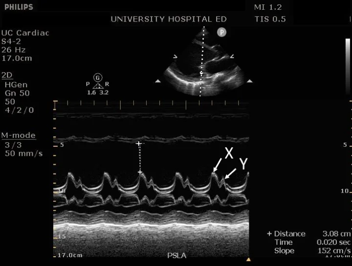
Aortic Root Excursion
On an M-mode image through the AoV in PLAX, the aortic root in a heart with normal systolic function will move anteriorly during systole (excursion). If the aortic root stays in a flat line, it’s a sign of abnormal/low EF.

TAPSE and TDI (S’) are used to assess RV systolic function. What exactly are they measuring?
TAPSE uses m-mode to measure HOW FAR (mm) the tricuspid annulus moves towards the apex during systole. This tells us about longitudinal fiber shortening of the RV.
S’ uses TDI to measure the VELOCITY (cm/s) of the RV annulus during systole, as it contracts.
Ridges in the LV and RV apical compartments are called __________
Trabeculations / Trabeculae Carnae

What are the 3 phases of systole?
IVCT (Isovolumic Contraction - all valves are closed, but immediately after ED the ventricles contract against closed semilunar valves, until the LV pressure exceeds the Ao pressure and the valve opens!)
Rapid ejection (large PG)
Reduced ejection (small PG)
What are the 4 phases of diastole?
IVRT (Isovolumic Relaxation - all valves are closed, so no change in ventricular volume, but immediately after ES the ventricles relax until the LA pressure excees the LV pressure and the valve opens)
Early Rapid Filling (large PG = E wave → 80% of blood enters LV)
Diastasis (LV and LA pressures start to equalize → small PG = time between the E and A peaks on the MV inflow PW)
Atrial kick (as the atria contracts, the PR increases so more blood flows into the LV = A wave → 20% of blood enters LV)
What is “Coronary Artery Dominance”? What is the most common form?
Is the PDA supplied by the RCA or Cx?
70% of people are “right dominant”, meaning that the PDA is supplied solely by the RCA. RWMAs are as expected for an “RCA pattern” blockage… inferior + infero-septal LV walls will be down.
20% of people are “co-dominant”, meaning that the PDA is supplied by both the RCA and Cx. Often milder/patchier RWMAs due to the collateral supply.
10% of people are “left dominant”, meaning that the PDA is supplied by solely the Cx. RWMAs will not fit the expected pattern.. Cx blockages could result in inferior RWMAs, and RCA blockages will not.
Grading RWMAs
Grade 1 = Normal (LV walls thicken >40% during systole)
Grade 2 = Hypokinetic (LV walls thicken during systole, but <40%)
Grade 3 = Akinetic (LV walls do not thicken during systole. Most commonly this is due to a past MI in the coronary artery supplying this wall segment → wall will appear thin + echogenic/scarred here. If the wall thickness is preserved, it may be due to “myocardial hypernation” from a severe blockage, infiltrative diseases, takotsubo cardiomyopathy, or DCM)
Grade 4 = Dyskinetic (LV walls bulge outward during systole. As above, most commonly this is at a site of an old MI → thin/echogenic/scarred LV wall. However other causes with normal wall thickness are possible)
Grade 5 = Aneurysmal (LV walls have remodeled into a pouch / aneurysm shape. Outwards bulging is present at all parts of the cardiac cycle, unlike dyskinesis. Again, walls most commonly - but not always - are dead + thin.
What is the main reason a “stress echo” is done?
To un-mask RWMAs that may not be present at rest, but are present during excercise due to the increased oxygen demand of the heart
Even patients with severe CAD may have a normal echo at rest.
NOTE: patient can either be riding a bike, or dobutamine can be used. Target HR is 220-age.

A subset of stress echos is called a “Viability Study” - what is this?
For patients with RWMAs at rest, to test whether walls with are “down” are viable myocardium are not. Is revascularization surgery worth it for this patient?
Dobutamine given at a low dose → increases contractility
If myocardium is truly infarcted (dead), still no wall thickening
If myocardium is viable (stunned/hibernating), wall thickening seen!
A viability study is the best predictor of the recovery of LV function following revascularization
Stunned myocardium vs. Hibernating myocardium
Both of these appear as RWMAs (hypo-akinetic-dyskinetic) at rest, but will improve contractility with low dose dobutamine (viability study).
Stunned = the muscle cells remained “stunned” / motionless for days-weeks after a short but severe event of ischemia. This may be a temporary blockage that cleared on it’s own, or a heart attack that was quickly treated. The myocardium is not dead, but “stunned”.
Hibernating = when the muscle cells go into “hibernation” due a severe narrowing of the coronary artery which supplies them. They stop contracting to preserve energy, but are not dead. If adequate O2 supply resumes, they become totally normal again.
(Traditional) Coronary Angiogram
The gold standard for assessing the coronary arteries for % blockages. They are done ± treatment (PCI = Percutaneous Coronary Intervention = angioplasty + stenting → inflate a ballon to open up the walls, then place a mesh tube to hold it open) during the same procedure!
Done in a cath lab, taking about 30-60 minutes. The patient is (usually) sedated, and a catheter is fed up towards the heart through either the femoral (groin) or brachial (arm) artery. Therefore, it’s an invasive test
A contrast dye is injected, which fills the arteries - less so or not at all in areas of partial or complete blockages. The dye is detected by an XRay. % blockages of each artery are reported.

CT Coronary Angiogram
A purely diagnostic test (unlike a traditional angiogram), where contrast dye is injected intravenously and a CT scan is taken to detect blockages.
Unlike a traditional angiogram, it is non-invasive (+) but does not have the option to treat… they will have to book a separate PCI test after!

MIBI Scan
A method to assess for CAD using nuclear medicine. A nuclear isotope is injected intravenously. The patient is scanned with CT. In a normal heart, the isotope will distribute evenly. In a patient with blockages, certain sections supplies by this artery will be without the isotope.
Often done at rest AND with stress, to unmask RWMAs absent at rest

Cardiac MRI
Very expensive, but VERY detailed (GOLD STANDARD) + non-invasive. Also, avoids the radiation used in Xray or CT. Used for patients with medium + high probability of CAD.
Looks at everything, not just blood supply. Can see scar tissue. Can confirm that a section of tissue is getting lots of blood flow despite a mild blockage (avoiding unnecessary surgery)
Takotsubo Cardiomyopathy
aka: “stress cardiomyopathy” / “broken heart syndrome”
A reversible aneurysmal appearance (no thickening) to the LV apex triggered by a severe emotional stress (eg. death of a loved one)
Symptoms mimic a heart attack - sudden severe chest pain
Resolves weeks - months. At risk (10%) of LV apical thrombus formation.

Kawasaki Disease
Inflammation + aneurysm of the arteries (including coronary arteries) of the body, usually affecting children < 5 years old. Increases risk of heart attack as adult.
When a patient has a heart attack (acute MI), why might some patients be sent to the cath lab for a PCI (angioplasty with stent), while others are sent to the OR for open heart surgery (CABG = coronary artery bypass graft)?
For a simple blockage (1-2 vessels), PCI is preferred. For a complex (≥3 vessels) or major (eg. LCA) blockage, CABG may be preferred - get it all done at once, with better patient outcomes.
For older/frail patients, PCI is preferred (less invasive).
If the MI resulted in other complications (eg. ruptured papilllary muscle, ruptured wall, etc), open heart surgery will be needed anyways to repair these.
A common vessel harvested for a CABG is ______________
GSV (Greater Saphenous Vein)

Define Ischemia
Imbalance between oxygen supply and demand to the myocardium. Either the supply goes down (atherosclerosis) or demand goes up (LVH, tachycardia)
#1 cause = atherosclerosis
Symptom = angina, fatigue, dyspnea
________ is chest pain and tightness that occurs predictable, when O2 demand increases (eg, exercise) and is relieved by rest or meds.
__________ is chest pain that occurs “randomly” (at rest), most commonly due to the rupture of an atherosclerotic plaque + sudden blockage. It is also known as ________.
Stable Angina
Unstable Angina = Acute Coronary Syndrome (ACS)
An MI is usually caused by CAD (atherosclerosis → narrowing of vessel walls), but what are 2 other (rarer) possible causes?
Ruptured atherosclerotic plaque → Unstable angina / ACS
Coronary artery spasm
The 2 biomarkers which elevate in the blood after a heart attack are ________ and _________.
Troponin
Creatine Kinase (CK)
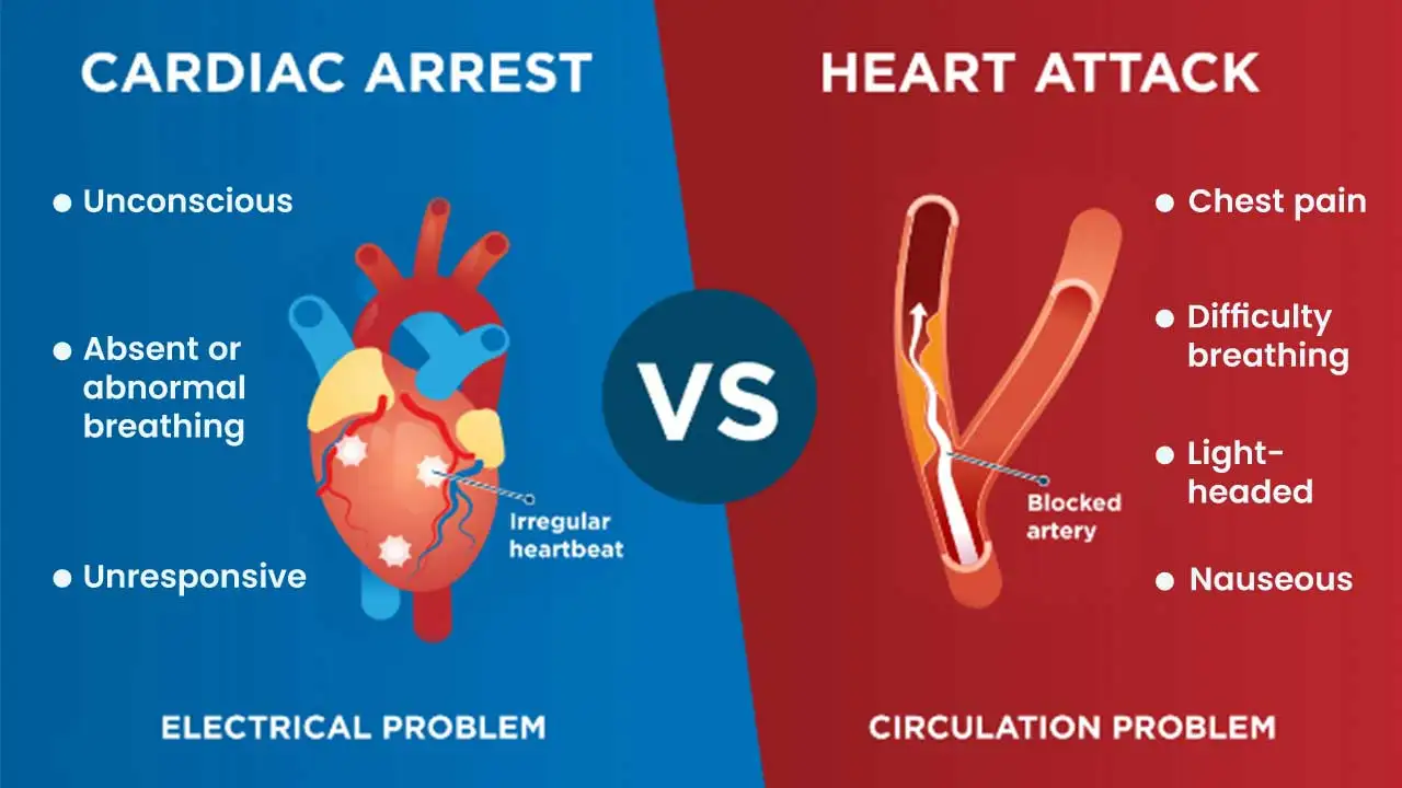
What is the difference between a heart attack (MI) and cardiac arrest?
In an MI, a partial (N-STEMI) or complete (STEMI) blockage of a coronary artery causes the heart to pump very poorly. Patient will have chest pain, SOB, arrhythmias → Call 911 or take patient to ER. Treated with PCI or CABG.
Until they go into cardiac arrest, they are conscious and have a pulse
Cardiac arrest is when the heart stops completely due to a fatal arrhythmia. The patient is unconscious with no pulse. You must perform CPR, ideally + AED.
Fatal arrhythmia may be a sequellae of an MI, or occur on its own (PEA arrest, shock, HCM)
Certain fatal arrhythmias are considered cardiac arrest (pulseless VTachy, VFib, PEA)
When do you perform CPR + AED
ONLY perform CPR ± AED if the patient satisfies ALL of the following criteria:
Unconscious/unresponsive
Not breathing
No pulse
These patients are considered to be in cardiac arrest , even if the ECG machine shows some signals (fatal arrhythmias such as VFib or PEA, or pulseless VTachy)…. as the heart is either barely or not pumping (ensure no pulse) in response to these!
What is “Cardiogenic Shock”
Low CO → organ hypoperfusion → organ failure
Most common cause = MI
Rarer causes = acute severe MR, ruptured wall
Paradoxical Septal Motion
The movement of the IVS away from the LV free wall during systole
Caused by:
Post open heart surgery
LBBB
Septal RWMA
RV pressure or volume overload → septal flattening
Pericardial diseases
Tamponade, Constrictive pericarditis → “swinging septum”

What are some complications (other than RWMAs) that can arise post-MI
LV aneurysm or pseudoaneurysm
LV myocardial rupture (free wall or IVS)
Papillary muscle rupture
Thrombus (if akinetic apex)
New MR
New arrhythmias
Pericarditis or Dressler’s
Aneurysm vs. Pseudoaneurysm post-MI
Aneurysm is common post-MI, pseudoaneurysms are rare
Aneurysms is damage + stretching of all 3 layers of heart wall, whereas psueodoaneurysm is epicardium only
Aneurysms are in the apex, pseudoaneurysms can be anywhere, but usually LV free wall
Only pseudoaneurysm has a “neck”
Pseudoaneurysms at ++ risk of spontaneous rupture

What are the 2 common locations for cardiac thrombus to form, and under what circumstances?
Forms due to the stasis of blood in a location
LV apex → if apex akinetic, or aneurysmal
LAA (or less commonly, LA) → if patient has AFib

How long after an MI does a myocardial rupture (free wall, IVS, papillary muscle) occur? However, _________ can occur a bit later.
Is it rare?
3-5 days post-MI
Free wall rupture may occur a bit later (2 weeks post-MI)
RARE - <0.5% of patients post-MI will have a myocardial rupture
LV free wall myocardial rupture post-MI
Blood leaks into pericardial space → pericardial effusion → tamponnade → medical emergency
