Clinical Presentation & Diagnostic Considerations of the Small Animal Cardiac Patient
 Call Kai
 Learn
 Practice Test
 Spaced Repetition
 Match
 Flashcards
 Knowt Play
1/28

There's no tags or description
Looks like no tags are added yet.
29 Terms
Describe the normal cardiac auscultation.
Sounds
ECG
• S1: Mitral and tricuspid valves closure (AV valves)
Beginning of systole
Is longer, louder, lower pitched
• S2: Aortic and pulmonic valves closure.
→ Lub(S1) - dup (S2)
Is shorter, not as loud
Beginning of diastole
S4 & S3 - usually not heard in small animal patients
Atrial depolarization = S4
Then Isovolumetric contraction - pressure in left and right ventricles rises
Until aortic valve opens (Systole)
Pressure falls, aortic and pulmonic valve close or blood would reenter the ventricle
Isovolumetric relaxation

What are the main qualities of a heart murmur?
Timing
Intensity
Location
Quality
Pitch
**First three to important to recognize
Describe the features of the timing in regard to a heart murmur.
• Determined by the phase of the cardiac cycle, when did it occur?
• Systolic (If produced from S1 to S2)
S1 - beginning of systole
• Diastolic (S2 to S1)
S2 - beginning of diastole
• Continuous (machinery)
Hear it interrupted, throughout
• Early, Mid, Holo (through whole phase)
More nuanced, can be difficult to hear with high HR
Describe the features of the intensity in regard to a heart murmur.
• How loud is the murmur?
• Classification of grade of intensity from I to VI
Degree of disease correlated to intensity of the murmur
Describe the heart murmur scale - grades 1-6 and the definition for each.
Definition 1 - Heart Sounds
Definition 2 - Localization (How far are you from the source)

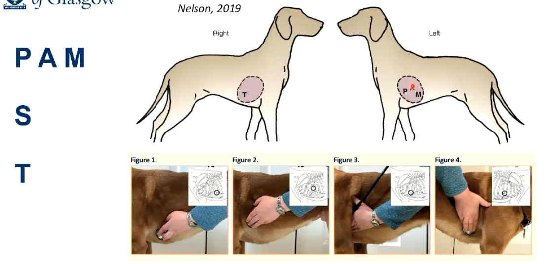
Describe the features of the location in regard to a heart murmur.
Where is the loudest murmur present?
Left: Apical (Mitral), Basal (Aortic and Pulmonic)-
Right: Apical (Tricuspid)
PAM → Side Change → T
Describe the features of the pitch and quality in regard to a heart murmur.
Describe how it behaves
• Pitch describes the auscultator's perception of the frequency of the murmur: high / medium / low
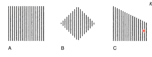
• Quality describes the "shape" of the murmur:
• Regurgitant (also called plateau shaped or rectangular, A)
• Ejection (crescendo-decrescendo or diamond shaped, B)
• Blowing (decrescendo, C)
How can you diagnose an arrhythmia?
What is a normal rhythm?
• Auscultation provides the first clue to the presence of an arrhythmia and in some cases the rhythm diagnosis.
• When an arrhythmia is auscultated or suspected, however, an ECG (Electrocardiogram) should always be obtained to confirm the diagnosis.
• Normal finding: Sinus rhythm
Regular rhythm, characterized by a monotonous S1-S2 cadence.
Approximately 60 - 160 bpm in dogs, 140 - 220 in cats.
What are normal arrhythmias?
• Normal finding: Sinus arrhythmia
• Regularly irregular pattern, associated with breathing.
• Normal or slow heart rate.
• Vagal Tone (VTo) induced:
• Inspiratory → Vto decreases, HR faster.
In healthy, relaxed dogs, means heart is saving energy
• Cats = May be a sign of disease with increased vagal tone
Since are usually stressed at the vet
What are some abnormal arrhythmias?
• Atrial fibrillation:
• Generally rapid, irregularly irregular rhythm.
• Variable loudness of heart sounds.
ECG - random appearance of peaks
What are the features of normal respiratory sounds?
• Normal lung sounds are created by the turbulent movement of air in the tracheobronchial tree in large (> 2 mm diameter) airways → Transmission to the chest.
• «Bronchovesicular» and «vesicular breath sounds» implies «made in the alveoli» but are transmitted from the large airways.
• Inspiratory: Lobar and large subsegmental bronchi.
• Exhalation: Trachea and the first one to two divisions of bronchi.
Why do abnormal lung sounds occur?
• Disease state → Altered sound transmission →
Efficient high-frequency sound transmission.
• Pleural disease/Pneumothorax → increased reflection of sound
Reduced intensity of lung sounds.
Not allowing sound waves to reverberate as they should.
What produces adventitious lung sounds?
• Abnormal lung sounds that are superimposed on normal (or increased/decreased) lung sounds.
• Three categories:
Crackles
Fine
Coarse
Wheezes
Ronchi

What do “fine crackles” sound like upon auscultation?
Common causes?
• High pitch (>800 Hz)
• Quickly muffled, unmusical lung sounds.
• Usually at the beginning/end of breathing.
Findings, not specific for disease, high overlap:
Bronchitis
Lung oedema
Pneumonia
Lung fibrosis
Fine Crackles
Brief, popping and high pitched, similar to wood burning in a fireplace
Heard in both phases, sometimes heard more in one phase than another
What do “course crackles” sound like upon auscultation?
• Usually lower pitch (≥700-800 Hz) and longer duration (<50 msec) than fine crackles.
• You can hear them over the trachea/mouth with/without stethoscopes.
These indicate the presence of fluid in the large airways!
Bubbling sound quality, popping, low-pitched, longer and louder, heard in both phases
What do “wheezes” sound like upon auscultation?
• Longer duration (>80-100 msec), relatively high frequency (oft >1000 Hz) compared to crackles.
• Sinusoidal (musical), narrowing of airway, caused by vibration of small bronchial walls.
What do “ronchus/ronchi” sound like upon auscultation?
• Longer duration(>100 ms), but in contrast to Wheezes they have deeper frequency (<2-300 Hz).
• Vibration of large airways or air- fluid interface disruption.
• Snoring sound.
Describe the sounds characteristic of stridor.
• Harsh high-pitched sound produced by obstruction of upper airways:
Nasal stridor
Pharyngeal stridor
Laryngeal stridor
Tracheal stridor
Describe the sounds characteristic of stertor?
• Low pitch nasopharyngeal obstruction.
• Brachycephalic syndrome!
Describe how the pulse is evaluated.
Pulse Pressure: Difference between systolic and diastolic pressure
Wide difference: Strong, hyperkinetic.
Small difference: Weak, hypokinetic (can be high or low)
• Anacrotic Pulse (parvus et tardus): weak and delayed.
• Pulsus alternans: Strong/Weak alternation.
• Pulsus paradoxus: Weak arterial pulse during inspiration, strong during expiration.
Sometimes with pericardial effusion or tamponade
• Pulsus bigeminus: Regular alteration of ventricular contractionised by premature
Additional beat or pulse wave

What does the hepatojugular reflex tell you?
Hepatojugular Reflex: cranial part of abdomen squeezed, enters right side, jugular vein becomes enlarged
• Right heart failure: Increase RA → Hydrostatic Pressure Caval Veins
→ Backwards Failure
• Pressure on the liver → rise in venous pressure.
May also see ascites - intravascular hydrostatic pressure increases, fluid leaks out
What does CRT evaluation allow you to assess?
• Capillary Refill Time (CRT) parameter to evaluate peripheral perfusion. Normal is below two seconds.
• Cyanosis: bluish discoloration of the mucous membranes
Central: systemic deoxygenation of arterial blood, not from lungs
Peripheral: localized reduction in oxygenated haemoglobin.
Differential: deoxygenation of caudal part of body, with normal oxygenation of head and forelimbs, certain region of body
What is a cough and how relevant is it in cardiac disease?
• Sudden expiratory effort in which a loud expulsion of air occurs.
• Defensive Mechanism (Stimulus: Irritation).
• Larynx, Trachea, Bronchi, Mediastinum.
• Cardiac disease? NOT in cats!
Can produce a cough in dogs, receptors in upper areas or larynx and, trachea and large bronchi
Pulomary oedema in alveoli should not trigger coughing reflex, unless it reaches bronchi
• Haemoptysis: Bloody Cough.

What are some potential differentials for coughs?

Describe the main features of respiratory distress?
• Eupnoea: normal, unlaboured breathing.
• Hyperpnea (more volume) / Tachypnoea (higher frequency)
• Dyspnoea: respiratory difficult
• Paradoxical breathing: Breathing pattern in which the chest moves in on inhalation and out on exhalation.
Lung depend on _____ and _____.
Ventilation, Circulation
V & Q
What are differential diagnosis for dyspnoea?

What is syncope?
• Defined as transient loss of consciousness (TLOC) due to cerebral hypoperfusion, characterized by a rapid onset, short duration, and spontaneous complete recovery.
• Presyncope (near-syncope): used to indicate symptoms and signs that occur before unconsciousness in syncope.
Prodrome of syncope
Not followed by loss of consciousness
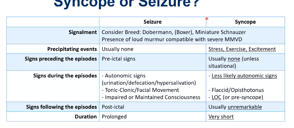
How can you distinguish a seizure form a syncope event?