OMS1- Anatomy practical 3
 Call Kai
 Learn
 Practice Test
 Spaced Repetition
 Match
 Flashcards
 Knowt Play
1/76

There's no tags or description
Looks like no tags are added yet.
77 Terms
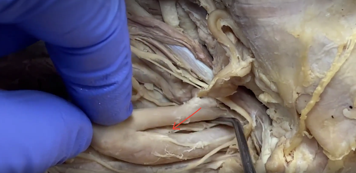
sternoclavomastoid muslce
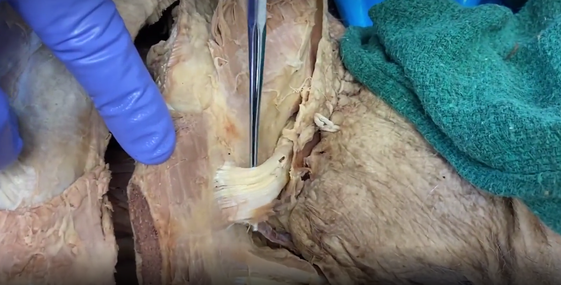
transversis thoracis muscle
on inner side of ribcage
fibers move from costal cartilage down 1 level below towards sternum
most superficial when looking at the back of ribcage
depressed ribs in forced expiration
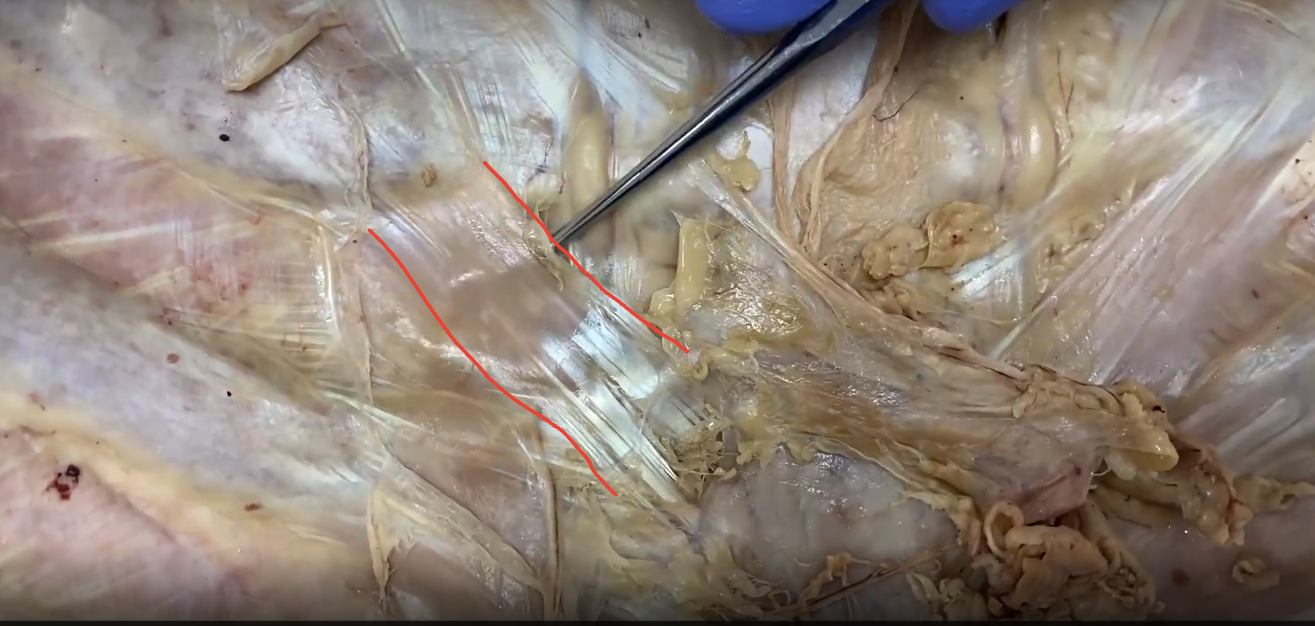
internal thoracic artery
can be seen through the thin muscles of ribcage
runs on either side of the sternum
veins are located to the side of the artery
as it gets to the xyphoid process, it will branch into superior epigastric artery and musculophrenic artery

intercostal nerve
runs on inner side of a rib
anterior intercostal arteries and veins run with them

superior epigastric artery and musculophrenic artery
superior epigastric will go straight down
musculophrenic hugs intercostal margin, will move laterally

fiberous pericardium
visualized with lungs removed
covers heart and ascending aorta+pulmonary trunk, sits directly on diaphram
fiberous is most superficial layer
on inner side, is shiny-er and called serous parietal pericardium

visceral pericardium
sits directly on heart under the fiberous/serious parietal pericardium. aka epicardium.

pulmonary trunk
exits right ventrical, is the the anterior aspect of heart
within pulmary trunk is the semilunar palmonic valve, had 3 cusps
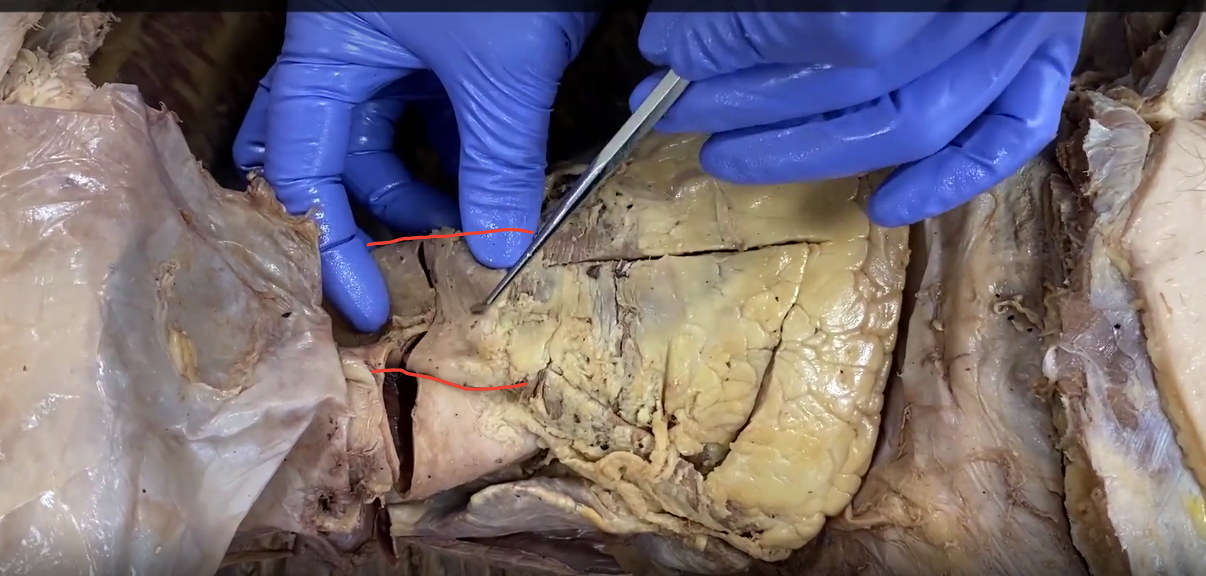
right atrium of heart

ascending aorta
within aorta is the semilunar aortic valve

superior vena cava
enters right atrium
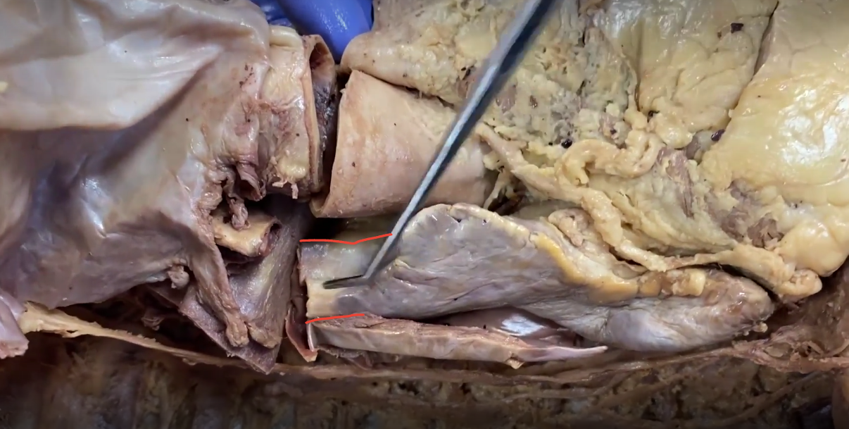
left ventrical
forms left boarder of the heart
forms apex of heart
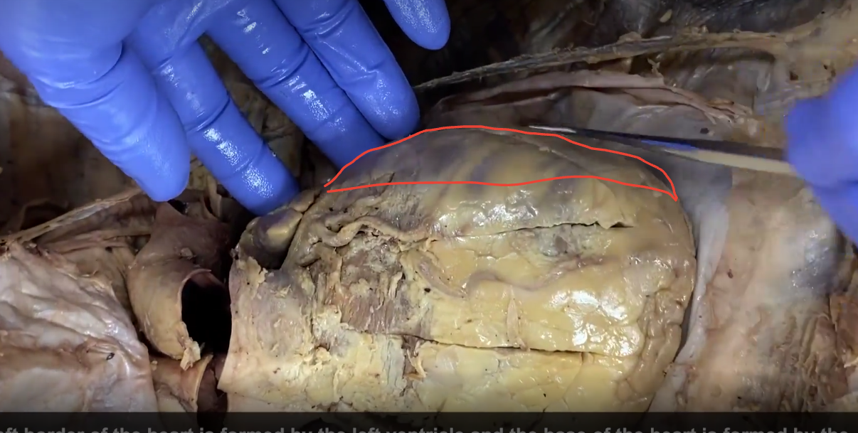
left atrium
superior to left ventrical

transverse sinus
under ascending aorta and pulmonary trunk

inferior vena cava
can be seen under apex of heart

oblique pericardial sinus
where hand is
under apex of heart to just under pulmanary veins (where pericardial sac ends)

anterior interventricular artery
runs in anterior interventricular sulcus (between right and left ventricals on anterior aspect)
aka left anterior descending artery
the great cardiac vein runs with it before continuing with circumflex artery to coronary sinus.

left coronary artery
first branch off aorta on left side
has three branches: circumflex (right most); diagonal artery (middle); anterior interventricular artery (left most)
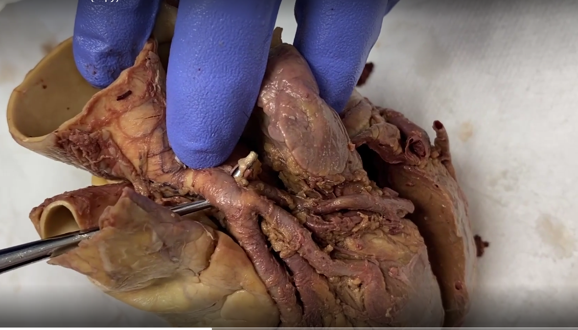
coronary sinus
on posterior aspect of heart
in artioventricular sulcus under left atrium
drains into right atrium

right coronary artery
first branch off aorta on right side

SA nodal artery
branch off right coronary artery

right marginal coronary artery
branch off coronary artery
small cardian vein runs with it, will drain into coronary sinus

posterior descending artery
can branch off right or left circumflex coronary artery. Or both. This picture is a right coronary artery branch
right is called a right dominant heart and vise versa
AV node artery will be a continuation of right coronary artery, diving between atriums

middle cardiac vein
drains into coronary sinus, where it drains forms the crux of the heart (where AV nodal artery dives too)

external intercostal muscles
directly below intercostal membrane, can be fused
slope down to midline
elevate ribs for breathing
innervated by intercostal nerves, supplied by anterior/posterior intercostal arteries

internal and innermost intercostals
deep to external intercostals, fused. fibers run upwards towards midline
depress ribs
forced expiration
between them sits the intercostal neurovasculatiue bundle
diaphragm
seperated thoracic and abdominal cavity
primary respirator
pericardophrenic vessels with phrenic nerve
one both sides of heart

hilum of right lung
main bronchus
pulmanary artery
pulmanary veins

hilum of left lung
left lung will have an impression for the aortic arch

pectinate muscles of right atrium

Crista terminalis right atrium
runs from the junction of SVC
Area of SA node

fossa ovalis in right atrium
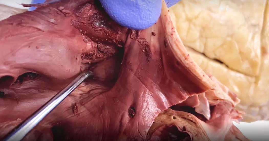
tricuspid valve into right ventrical

Chordae tendinae
run between AV valves and papillary muscles

moderator band
in right ventrical
connects anterior papillary muscles to interventricular wall.
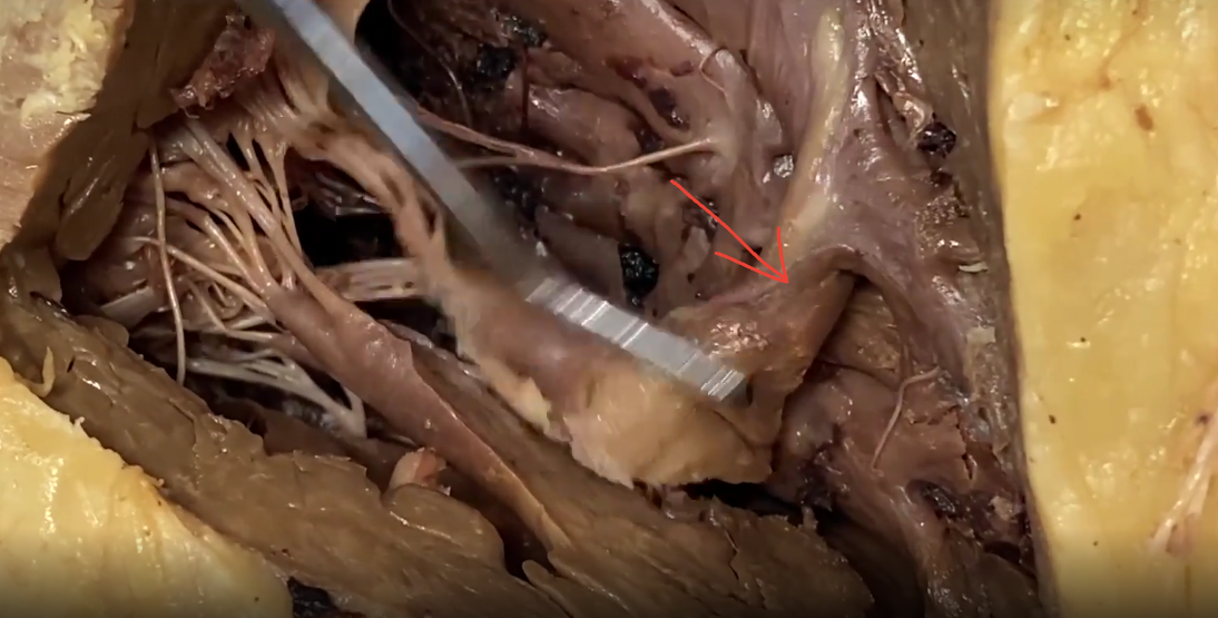
septal papillary msucle
right ventrical

left ventricals anterolateral papillary msucle
directly on lateral wall
one other papillary muscle in left ventricals- posterior medial papillary muscle

left auricle of left atrium
only part of atrium that is not smooth msucle
has pectinae muscles

pericardiophrenic neurovasculatiue bundle
contains phrenic nerve and pericardiophenic vessels

ascending aorta
left to superior vena cava
anterior to pulmonary trunk
3 brances of right to left: brachiocephalic trunk, left common carotid artery, left subclavian artery

pulmonary trunk
most posterior of great vessels

ligamentum arteriosum
connects aorta to pulmonary trunk
on left side of pt
near vagus nerve
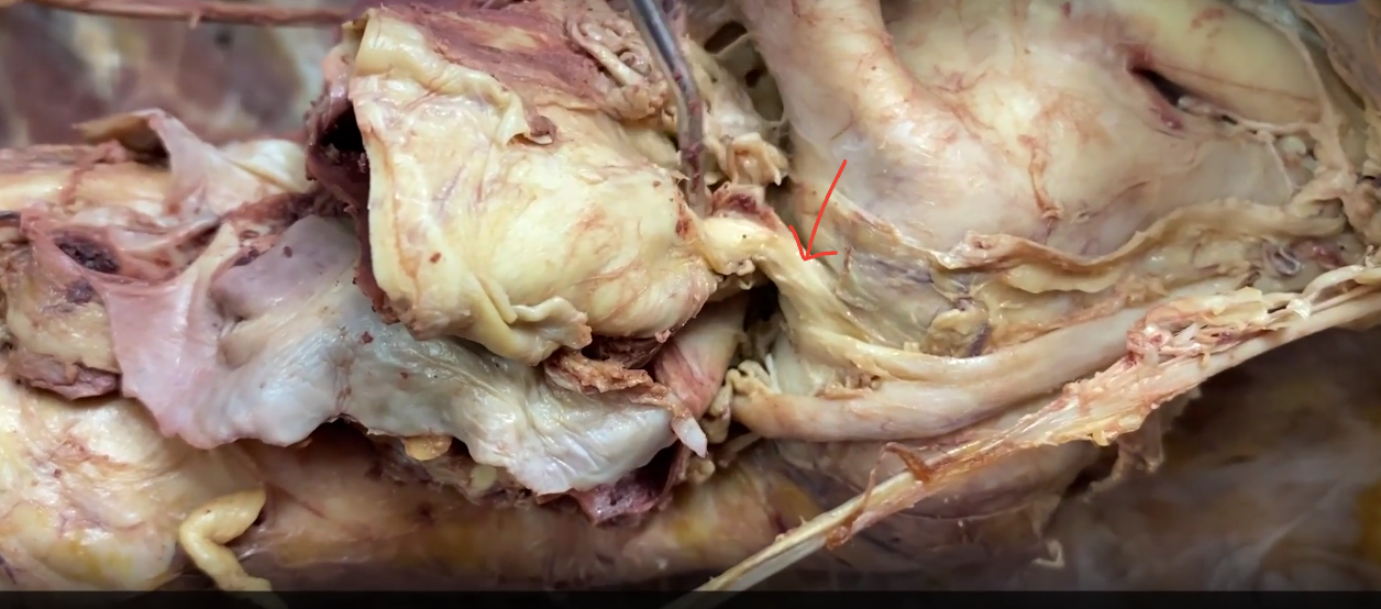
L recurrent laryngeal, left vegus nerve
Vagus nerve travels to esophagus behind lung root
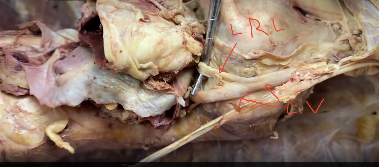
esophageal plexus
composed of right and left vagus nerve

descending thoracic aorta

left azygous system
hemiazygous vein- on left side, startes inferior and will cross right to drain into azygous vein
accessory hemizygous vein- more superior. Will cross to right side to drain into azygous vein

sympathetic chain
ganglia are buldges

right azygous system
azygous vein- other veins drain into it

brachiocephalic veins
join to form superior vena cava

arch of azygous vein
drains into superior vena cava
wraps above right mrain bronchous (cut, posterior) and right pulmonary artery (cut, anterior)

right vagus nerve
runs with trachea
near the superior thoracic aperture, will give off right recurrent laryngeal nerve

platysma muscle
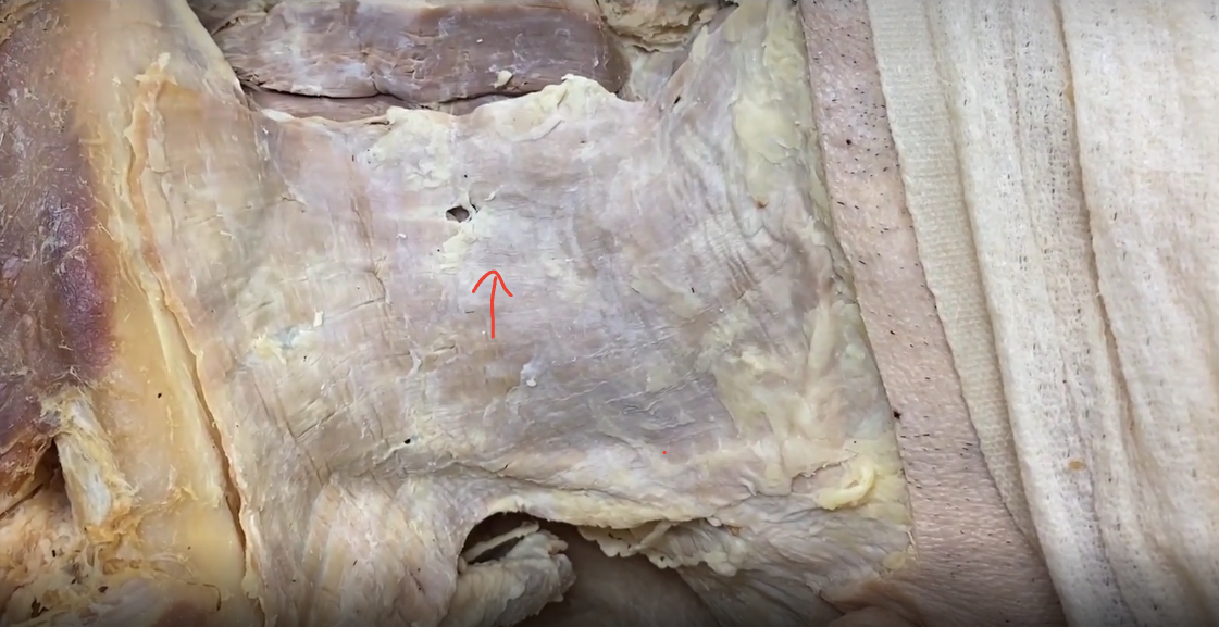
external jugular vein

posterior triangle
Levator scapulae
posterior scalene
middle scalene
inferirior omohyoid

spinal accessory nerve
runs between SCM and Trapezius

supraclavicular nn

transverse cervical nerve

great auricular nerve

superior belly omohyoid

anterior jugular vein

muscular triangle
sternohyoid,
sternothyroid
thyrohyoid

cricothyroid muscle
deep in muscular triangle

thyroid gland
inferior to cricothyroid muscle

submandibular gland
facial artery wraps around this

digastric and stylohyoid msucle
intermediate digastric tendon prices stylohyoid muscle

mylohyoid muscle
probe is touching hypoglossal nerve
inferior to nerve is hyoglosis muscle

carotid aa
hypoglassal nerve passes over both branches
travels with internal jugular vein and vegus nerve in carotid sheath

ansa cervicalis
originates where the the hypoglossal nerve does at the neck
innervates omohyoid, sternohyoid, sternothyroid
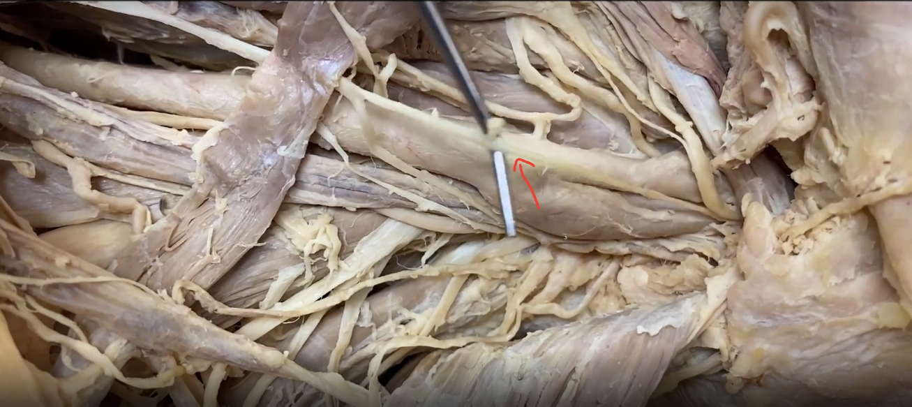
carotid sheath (Carotid a, internal jugular v, vagus n)
Carotid a
internal jugular v
vagus n

carotid body and sinus
sinus is on the internal carotid artery
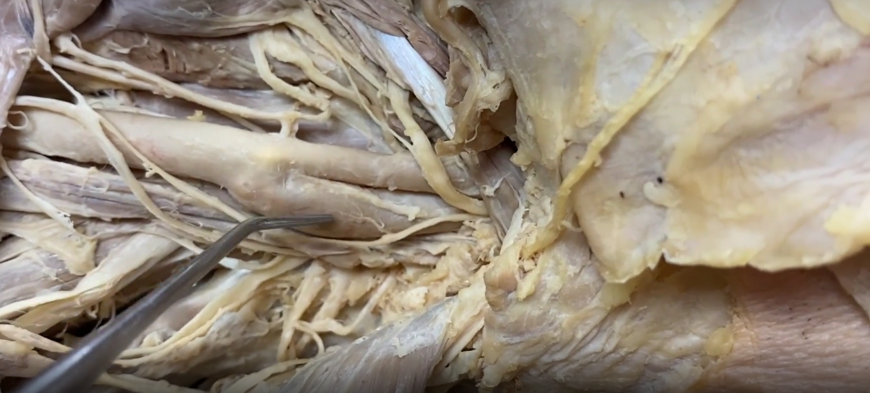
glossalpharyngeal nerve
on internal carotid artery

superior thyroid artery
first branch of extneral carotid artery
superior larngeal branches off this and goes to larynx more superiorly
thyroid artery will go inferiorly to supply thyroid

internal laryngeal nerve
a branch of superior laryngeal nerve
runs with superior larngeal artery

external larengeal nerve
inferior to internal laryngeal nerve
innervate cricothyroid muscle

lingual artery
second branch off external carotid
third branch, facial artery, is directly superior

ascending pharyngeal artery
between the external and internal branches of carotid artery