Pathological Conditions of the Biliary System
 Call Kai
 Learn
Learn Practice Test
 Spaced Repetition
 Match
Match Flashcards
 Knowt Play
1/123

There's no tags or description
Looks like no tags are added yet.
Name | Mastery | Learn | Test | Matching | Spaced | Call with Kai | Chat |
|---|
No analytics yet
Send a link to your students to track their progress
124 Terms
what are 6 other causes for obstructive jaundiced and biliary duct dilation
pancreatic head carcinoma
cholangiocarcinoma
sclerosing cholangitis
hepatoma
GB cancer
biliary atresia
what are 5 symptoms of obstructive jaundiced and biliary duct dilitation
yellow skin and sclera
weight loss
elevated bilirubin and alk phos
RUQ pain
pruritis
cancer in the bile ducts is called ____
cholangiocarcinoma
sclerosing cholangitis
inflammation of bile ducts that causes a narrowing
pruritis
skin itchiness from jaundice
what are 3 levels where obstruction can occur in the biliary system
CBD
Ampulla of Vater
GB alone
obstruction at the level of the ampulla of vater will cause dilatation of both ____ and ____ duct
biliary and pancreatic
ampulla of vater obstructions most commonly are due to _____
pancreatic head carcinoma or acute pancreatitis
what are some causes for intrinsic biliary obstruction
stones
biliary atresia/choledochal cyst
carcinoma of ampula of vater
cholangiocarcinoma
pancreatitis which causes duct stricture
post liver TX bile duct stenosis
what are some causes for extrinsic biliary obstruction
carcinoma of head of the pancreas
pancreatitis
lymphadenopathy at porta hepatis
masses at porta hepatis
intrahepatic tumors
what are 2 diffuse hepatic conditions that cause biliary obstruction
sclerosing cholangitis
caroli’s disease
a normal CHD has internal diameter of ___, ____ is borderline, and ____ requires further investigation
<4, 5, 6mm
can a pt have a normal size hepatic duct and still have a distal obstruction
yes
a normal CBD has internal diameter slightly greater than the hepatic duct, ____ is borderline, and ____ is dilated
>6, >10 mm
bile ducts can _____ after cholecystectomy
enlarge
Obstruction of biliary ductal system is diagnosed by ultrasound when the sonographer finds the presence of _____
ductal dilation
what finding is seen when intrahepatic ducts are dilated
“too many tubes“ or “shotgun“ sign
what effect can fibrosed or infiltrative disease of the liver have on biliary obstruction
can prevent intrahepatic dilation (due too lack of complaince of the hepatic parenchyma)
biliary obstruction may present as ____
jaundice
when is painful jaundice seen
with acute obstruction or infection that may invade biliary tree
what are the 3 primary areas for biliary obstruction
porta hepatic
suprapancreatic
intrapancreatic (most distal)
porta hepatic biliary obstruction is usually the result of a ______
neoplasm
what does the intrahepatic ducts and the CBD look like with porta hepatic biliary obstruction
dilated intrahepatic ducts, normal CBD
_____ of the gallbladder may be present with porta heptatic biliary obstruction
hydrops
suprapancreatic biliary obstruction originated between the ________
pancreas and the porta hepatis
what do the intrapancreatic duct, pancreatic duct, and the head of the pancreas look like with the presence of suprapancreatic biliary obstruction
normal
the most common cause for suprapancreatic biliary obstruction is _____
malignancy or adenopathy

what is being shown in this image
suprapancreatic biliary obstruction (bile duct is dilated due to mass)
what 3 conditions cause the majority of biliary obstruction at the level of the distal duct and cause the extrahepatic duct to entirely dilate
pancreatic carcinoma/cancer of ampulla of vater
choledocholithiasis
chronic pancreatitis with stricture formation
uncommon cause for extrahepatic biliary obstruction as a result of an impacted stone in the cystic duct, which creates extrinsic mechanical compression of the common hepatic duct
mirizzi syndrome
what is the anatomy behind mirizzi syndrome
cystic duct inserts unusually low into CHD so they run parallel to each other
what is the sonographic appearance for mirizzi syndrome
immoble calculus in cystic duct or neck and dilation of intrahepatic duct
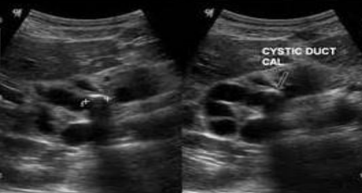
what is this image showing
mirizzi syndrome

what is this image showing
intrahepatic ductil dilatation (shot gun sign)

what is this image showing
intrahepatic ductil dilatation
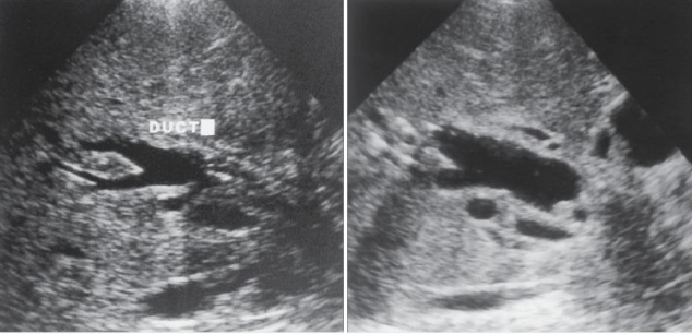
what is this image showing
dilated intrahepatic ducts secondary to mass in porta hepatis (irregular walled, dilated bile ducts)
_____ is stones in the bile duct
choledocholithiasis
what is one of the most common causes of biliary obstruction
choledocholithiasis
___% of pts with GB stones will have choledocholithiasis
15%
choledochalithiasis is associated with _____
calculous cholecystitis (GB inflammation due to gallstones in cystic duct)
the cystic duct is most common area for choledocholithiasis but can commonly be found in the distal portion of the _____ near the pancreatic head
CBD
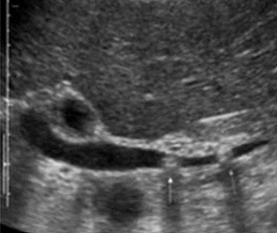
what is this image showing
choledocholithiasis (in CBD)

what is this image showing
choledocholithiasis
what is cholangitis
inflammation of the bile ducts (caused by baterial infection, acquirred or congenital)
cholangitis put pt at risk for ______
cholangiocarcinoma
what is charcots triad
fever
jaundice
RUQ pain
what are the clinical symptoms for cholangitis
biliary colic, charots triad, elevated serum leukocytosis,bilirubin,and alkaline phosphate, leukocytosis
the cholangitis sonography is ____ bile ducts and ____ formation
thickening, pus

what is this showing
cholangitis (note bile duct walls are thickened)

what is this showing
cholangitis (note bile duct walls are thickened)
A chronic hepatobiliary disease in which the walls of the bile ducts become inflamed which causes narrowing
primary sclerosing cholangitis
what causes primary sclerosing cholangitis
often idiopathic (may be associated with inflammatory bowel disease)
what are the clinical symptoms for primary sclerosing cholangitis
jaundice, pruritis, fatigue, gallstones can be present
what is the sonography for primary sclerosing cholangitis
thick bile ducts
hyperechoic bile ducts throughout liver, ductal strictures or dilations

what is this image showing
primary sclerosing cholangitis

what is this image showing
primary sclerosing cholangitis
______ is a disease caused by the parasitic roundworm ascaris lumbricoides
ascariasis
ascariasis is contracted via ______
fecal-oral route (fecal matter)
ascariasis worms are usually _____cm long
20-30cm
ascariasis grows in the _____ before entering biliary tree through ampulla of vater
small bowel
ascariasis causes _____
acute biliary obstruction
what are the clinical symptoms for ascariasis
asymptomatic or biliary colic

what is this image showing
ascariasis

what are these images showing
ascariasis (dilated duct showes movement)
intrahepatic calculi occurs in patients with ________
recurrent pyogenic cholangitits (aka oriental cholangiohepatitis/intrahepatic pigment stone disease)
intrahepatic calculi is very common in what continent
asia
what are choledochal cysts
congenital, focal or diffuse cystic dilation of the biliary tree
_____ may result from pancreatic juices refluzing into the bile duct
choledochal cysts
what are the symptoms for choledochal cysts
abdominal mass
pain
fever
jaundice
what are the classifications for choledochal cysts
Type I, Type II, Type III, Type IVa, Type IVb, Type V (caroli’s disease)
type I choledochal cysts are ____ dilation of the common bile duct
fusiform
type II choledochal cysts are true ____ of the bile ducts
diverticuli
type III choledochal cysts (_________) are confined to the intraduodenal portion of the CBD
choledochoceles
type IVa choledochal cysts are condinded to the ____ biliary tree
extrahepatic
type V choledochal cysts are classified as _____ disease
caroli’s
what is caroli’s disease
congenital disease where bile ducts dilate and can become infected and seperate from the bile duct to form choledochal cysts
caroli’s disease has been associated with _____
cystic kidney disease
caroli’s disease is usually found in _____
childhood or early adulthood
what is the sonography for caroli’s disease
multiple biliary cysts continuous with the biliary tree, stones and echoes may appear in tree

what is shown in this image
caroli’s disease (cystic structures continuous with biliary tree)

what is shown in this image
caroli’s disease (cystic structures continuous with biliary tree)

what is shown in this image
choledochal cysts, diverticuli projections off biliary tree

what is shown in this image
choledochal cyst
what is clonorchiasis
parasitic worm from raw freshwater fish
what happens to the bile ducts with clonorchiasis
dilatation of bile ducts with wall thickening and echogenic focus within
intrahepatic biliary tumores are RARE and primarily limited to ______ and _____
cystadenoma and cystadenocarcinoma
what is the sonographic appearance of intrahepatic biliary neoplasms
cystic mass with multiple septa and papillary excrescences
what are the differential diagnoses for intrahepatic biliary neoplasms
hemorrhaic cyst or infection, echinococcal cyst, abscess, or cystic metastasis
cholangiocarcinoma is also known as _______
biliary adenocarcinoma
cholangiocarcinoma can mimic ___
HCC (hepatocellular carcinoma)
cholangiocarcinoma arises from the ______
wall of the bile duct
cholangiocarcinoma can occur anywhere in the biliary system, however it is most commonly found _______
at the union of the common hepatic duct (inbetween the rt and lt hepatic ducts)
what is the cholangiocarcinoma called when found at the union of the common hepatic duct
klatskin tumor
what are the clinical symptoms for cholangiocarcinoma
RUQ pain, malaise, anorexia, jaundice, biliary colic
what is the sonography for cholangiocarcinoma
large hepatic mass (hypo to hyperechoic), masses within the ducts may be outlined, duct wall thickening, focal duct stricture or duct termination
what is the sonography of klatskin tumors
very difficult to detect, mass at the hilum

what is shown in this image
intrahepatic cholangiocarcinoma (dilated ducts with abrupt termination)
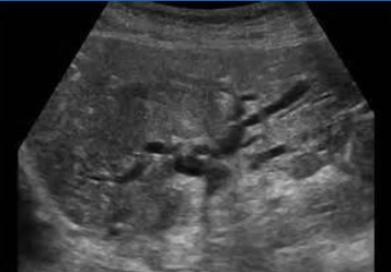
what is shown in this image
intrahepatic cholangiocarcinoma (dilated ducts with ill defined mass
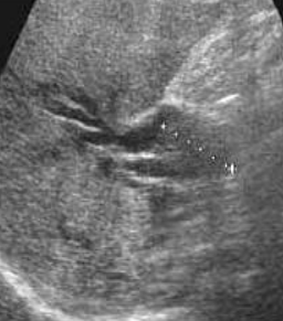
what is shown in this image
klatskin tumor (mass with dilated ducts that terminate)

what is shown in this image
klatskin tumor (mass with dilated ducts that terminate)