W7L1: Higher Visual Cortices (II) - Visual Disorders
 Call Kai
 Learn
 Practice Test
 Spaced Repetition
 Match
 Flashcards
 Knowt Play
1/34

There's no tags or description
Looks like no tags are added yet.
35 Terms
Describe the receptive fields of simple cells, in terms of:
What they are made of
Their shape
What they are tuned for
The relationship b/w ON/OFF zones
What they detect
Made of concentric receptive fields
Receptive fields are elongated
Tuned for orientation
Separate ON/OFF zones
Detect bars/edges of correct orientation & polarity

Describe the receptive fields of simple cells, in terms of:
What they are made of
Their shape
What they are tuned for
The relationship b/w ON/OFF zones
Made of concentric simple cell receptive fields
Receptive fields are elongated
Tuned for orientation
Overlapping ON/OFF zones
Detect bars/edges of correct orientation & any polarity

What are the implications of the visual pathway being very large?
Means there are many things that can go wrong/many places for things to go wrong
Define & describe amblyopia
Amblyopia: reduced vision in an eye in the absence of any ophthalmologically detectable retinal anomaly or any disorder of the afferent visual pathways that might cause defect
Absence of adequate symmetrical stimuli to the two eyes → binocular reflexes cannot be developed
Something goes wrong at an early age → two eyes stop working together
When are children usually most susceptible to amblyopia?
Before the age of 6, but is particularly pronounced within the first few years of development (e.g. 1-2 y/o)
Provide some examples of what can contribute to amblyopia, & types of intervention to resolve the issue
Causes:
Amblyogenic anisometropia
Constant unilateral or bilateral esotropia or exotropia
Amblyogenic bilateral isometropia
Amylogenic unilateral or bilateral astigmatism
Image degradation
I.e. anything that leads to the imbalance/mis-integration of the input to 2 eyes
Intervention:
Caught early: put eye patch over good eye
Example: even having an eye swollen for the week as an infant can cause amblyopia
Describe the effects of monocular deprivation on ocular dominance columns in V1
Suture one eye shut soon after birth → block form vision from that eye
Non-deprived eye: wider ocular dominance columns
Deprived eye: thinner ocular dominance columns
Decreased V1 input (less colour input)
Describe why the timing of monocular deprivastion is critical
Very pronounced effects at age 2-4 weeks
Effects less pronounced at older ages (e.g. 10 weeks -14 months)
Older age + shorter deprivation period → slight effect but not as pronounced

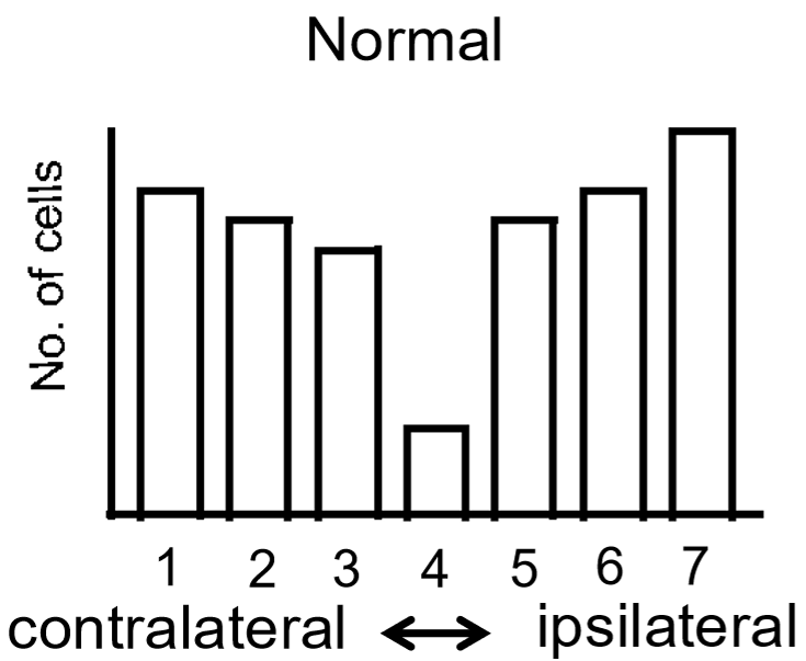
What is the Hubel & Wiesel Ocular Dominance Scale?
Quantifies the electrophysiological responses of V1 cells to stimuli applies to the L/R eye
X-axis showing the ocular dominance class
Y-axis showing the number of cells responding to the stimulus
V1 cells are generally very strongly biased towards one eye or the other
1 = input only from contralateral eye
4 = equal responses to both eyes
7 = input only from ipsilateral eye
Describe the Hubel & Wiesel Ocular Dominance Scale following monocular deprivation
Vast majority of cells are driven exclusively by the non-deprived eye

Describe the changes that occur during the development of V1 in infants
Initially:
V1 initially has no retinotopic map
Long, straggly branches → spread over very wide areas of the cortex
No segregation of signals from the L & R eyes; all cells sending random branches everywhere
Development:
Active connection of cells
Spontaneous, coordinated waves of neural activity in infants → groups of cells become more active → signal propagates in the retina → groups of cells in one eye become active together → strengthen the circuit of that group of cells
Segregation of separate zones b/w the L & r eyes is forced through differential strengthening of gaps of cells in L & R eyes → help build retinotopic map
