Cardio Physiology 4 - Cardiac Cycle
 Call Kai
 Learn
Learn Practice Test
 Spaced Repetition
 Match
Match Flashcards
 Knowt Play
1/70

There's no tags or description
Looks like no tags are added yet.
Name | Mastery | Learn | Test | Matching | Spaced | Call with Kai | Chat |
|---|
No analytics yet
Send a link to your students to track their progress
71 Terms
How is the cardiac cycle divided
Systole and diastole
How long the cardiac cycle length
Period of time from beginning of one heartbeat to the beginning of the next
Why is diastole longer than systole
Allows heart to fill with blood when ventricles are contracted
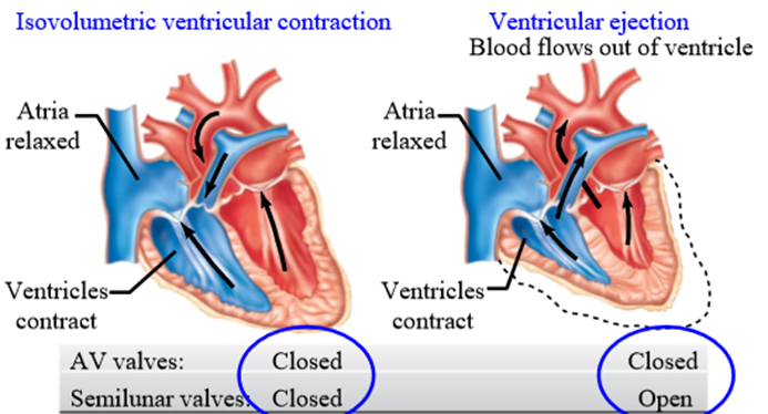
Two phases of systole
Isovolumetric contraction, ventricular ejection
How is blood flow created
Ventricles squeeze blood in their chamber, generating pressure
Isovolumetric contraction
Ventricles contract with all heart valves closed. Blood volume is constant, but the pressure rises. Ventricular muscle fibers cannot shorten
Ventricular ejection
Pressure generated by ventricles during contraction exceeds artery pressure. Opens semi lunar valves and pumps blood into the artery
What valve is closed during ventricular ejection
AV valve is closed via chordae tendineae and papillary muscles
Stroke volume
Volume of blood ejected from each ventricle during systole (contraction)
Which ventricle contracts more blood
Both ventricles contract the same amount of blood, but the left ventricle does this with more force (pressure)
Is the entire volume of blood in ventricle ejected?
No - allows heart to change the amount of blood pumped based on body's needs
2 phases of diastole
Isovolumic relaxation phase, Ventricular filling phase
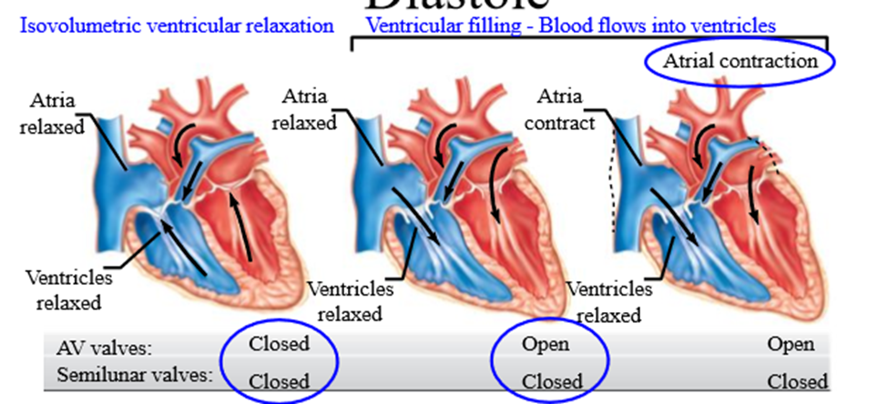
Isovolumetric relaxation
All heart valves are closed. Blood volume is constant, but pressure drops due to myocardium relaxation
Ventricular filling
AV valves open, blood flows into relaxed ventricles from atria (passive)
How do the AV valves open in ventricular filling
Atria in diastole (relaxed) - fills with blood. Pressure in atria is greater than ventricles, opening up the AV valve (forward pressure gradient)
2 phases of ventricular filling
Passive ventricular filling and atrial contraction
Passive ventricular filling
Ventricles receive 70% of blood volume from atria - both atria and ventricles are relaxed. Blood moves passively
Atrial contraction
Completes ventricular filling by contracting last amount of blodd
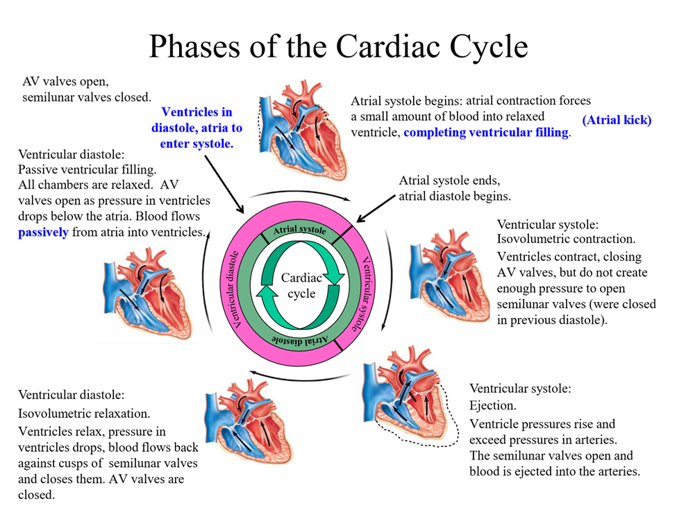
Cardiac cycle
Rhythmical contraction and relaxation of the hearts chambers coordinated by heart electrical activity. Represents events in a single heartbeat
Steps of cardiac cycle:
· Ventricles in diastole, atria about to enter systole. Ventricles received 70% of blood through passive filling (AV valves open, semilunar closed)
· Atria contract (kick), completing ventricular filling. Atria complete systole, enter diastole
· Ventricles enter systole - ventricles contract, pushing against AV valve, closing it.
· Mycardium contracts while both valves are closed = isovolumetric contraction - not enough pressure to open semilunar valve
· Ventricular ejection - continued ventricular contracting opens Semilinar valve - blood flows into the arteries (right - pulmonary truck, left - aorta). AV valves are closed due to chordae tendineae and papillary muscles
· Once blood pushed into arteries = diastole. Pressure drops, closing semilunar valves (blood flows back towards ventricle from artery, shutting semilunar valves). At this point, both AV and SL valves closed
· Ventricles enter isovolumetric diastole - ventricles have lower pressure than atria, AV valve opens, and blood flows from relaxed atria to relaxed ventricle (passive ventricular filling). Forward pressure gradient allows blood to flow from atria to ventricle. Semilunar valve is closed
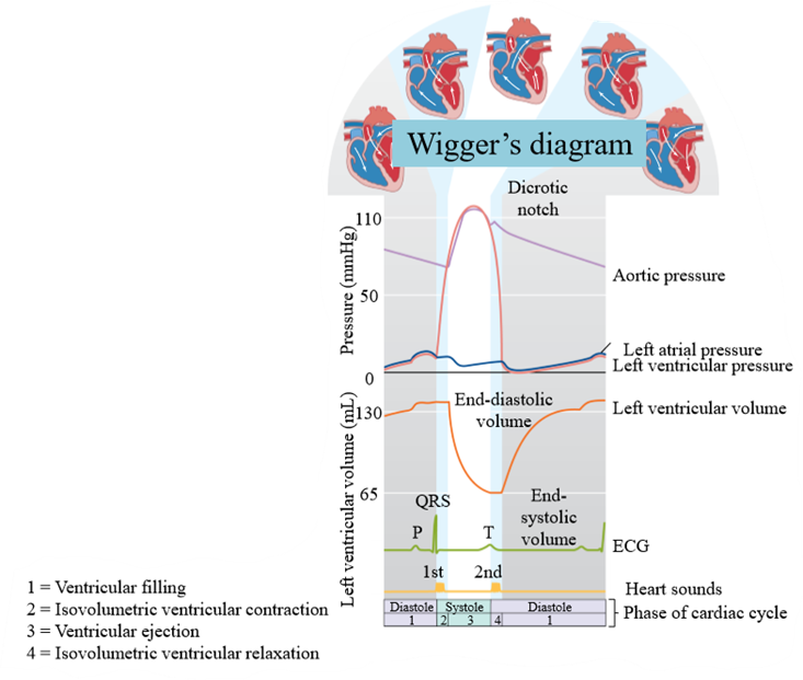
Wiggers diagram
relates electrical events of heart cycle to the mechanical, pressure, and sound changes that occur during a cardiac cycle
How are valves opened in the heart
Forward pressure
How are valves closed in the heart
Backward pressure
End-diastolic volume
Volume of blood in each ventricle at the end of ventricular diastole
End-systolic volume
Volume of blood in each ventricle at the end of ventricular systole
Stroke volume (SV)
Volume of blood pumped out of each ventricle during systole
How to calculate stroke volume
SV = EDV - ESV
Typical stroke volume
70 mL
Reading of Wiggers diagram in the cardiac cycle
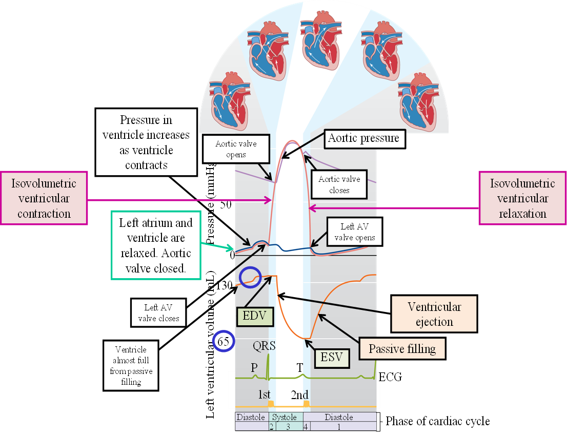
1. AV valve open, atrium and ventricle relaxed. Blood flows from atrium to ventricle passively, filling up to 70%.
2. P wave - atrium contraction, fills up the rest of the ventricle
3. Ventricle volume is full = EDV. Atrium in diastole for rest of cycle
4. QRS compelx - ventricular depolarization and contraction. Ventricular pressure increases rapidly, forces AV valve closed (prevent backflow). Ventricle continues to contract, increasing pressure. This causes aortic valve to open.
5. Ventricular ejection begins - fill aorta with blood. Rapid ejection (loss of volume), and then slowing. ESV = blood volume at end of cotntraction (60-65 mL). Ventricle does not eject entire volume of blood
6. Blood flows into aorta, increasing aortic pressure. Volume and pressure rate decrease as less blood comes out of ventricles and more blood goes into tissues
7. T wave = ventricle into diastole. Reduced pressure causes aortic valve to close. AV valve still closed due to higher pressure in ventricle than aorrta
8. Isovolumetric ventricular relaxation - muscle contracts, blood volume remains constant. Pressure falls below atrium pressure, which allows AV valve to open. Blood in atria received from veins in diastole
9. AV valve opened, blood passively flows into ventricles (down pressure gradient)
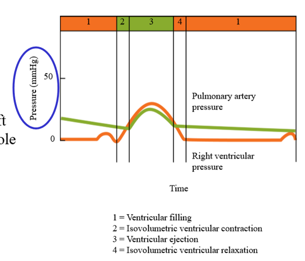
Difference in Wigger's diagram for left and right side of the heart
Same ECG, same cardiac cycle phases, but the pressure created by the right side of the heart (50 mmHg) is less than the left (130 mmHg)
Heart sounds
Lub dub
Lub sound
AV vales closing - beginning of isovolumetric ventricular contraction - signifiies onset of ventricular systole
Dub sound
Semilunar valves closing - signifies onset of ventricular diastole
What do the heart sounds represent
Turbulence when valves snap shut as pressures change
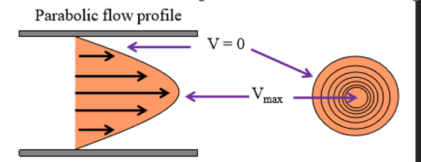
Laminar flow sound
No sound - blood moves in concentric layers, minimizing viscous interactions with vessel wall
Where is the highest velocity of blood flow
The center of the vessel (no interaction with vessel wall)
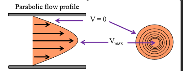
Where is the slowest velocity of blood flow
Nearest to the vessel wall (blood interacts with vessel wall)
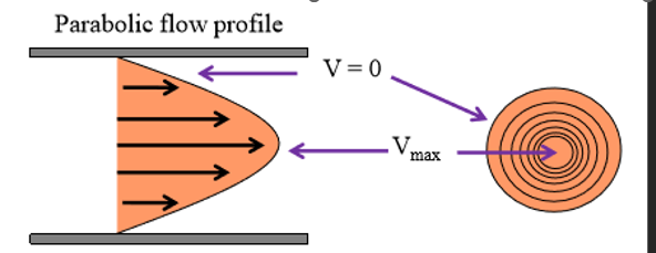
Shape of laminar flow
parabolic
How do we know if blood flow is turbulent
Makes a sound = heart murmer
2 causes of heart murmers
Stenotic valve, insufficient valve
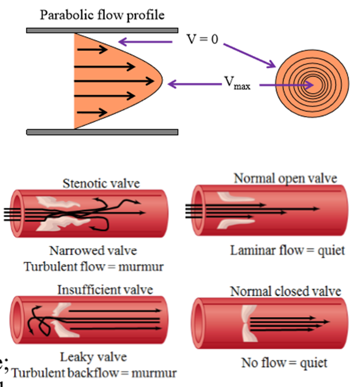
Stenotic valve
Valve leaflets do not open freely, causing turbulent blood flow
What causes stenotic valves
Stiff leaflets due to calcium deposits or scarring
Insufficient valve
Valve does not close completely, causing blood to flow backwards. Causes turbulent flow
What causes insufficient valve
Widening of aorta, scarring of valve
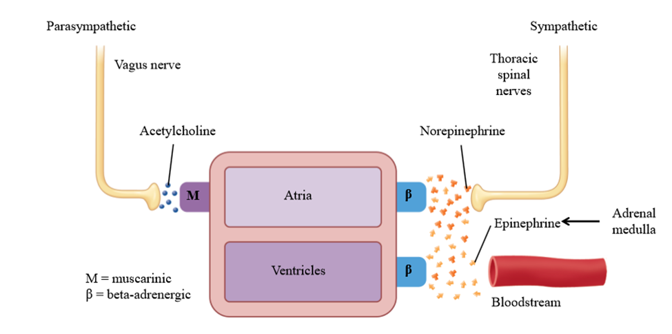
What nerve sympathetically innervates the heart
Thoracic spinal nerve
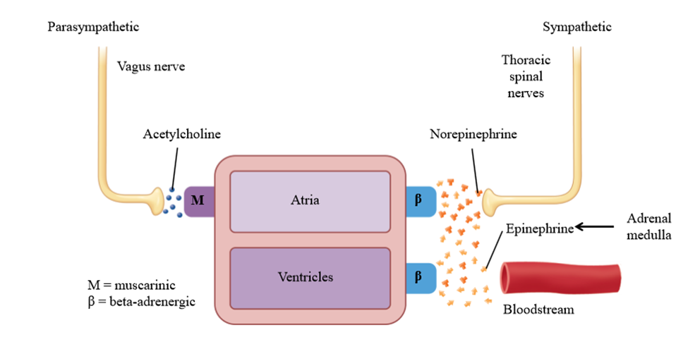
What nerve parasympathetically innervates the heart
Vagus nerve
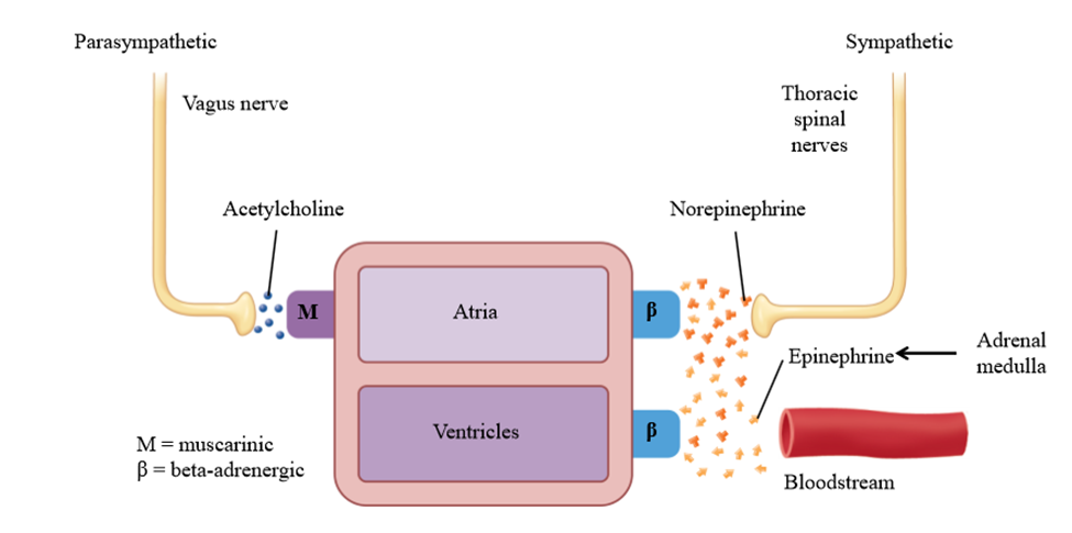
Neurotransmitter released by the thoracic spinal nerve to the heart
Norepinephrine
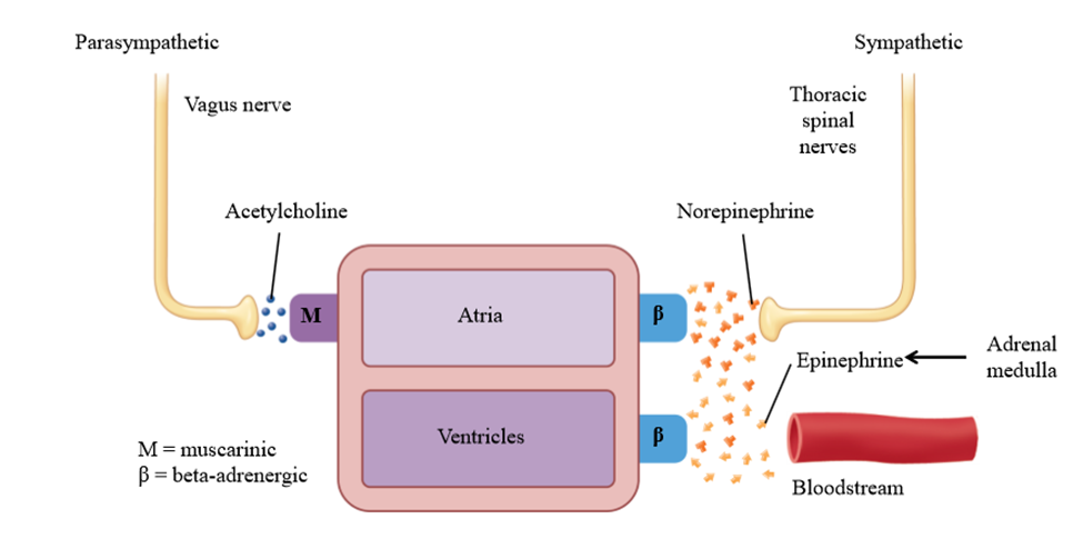
Neurotransmitter released by vagus nerve to the heart
Acetylcholine
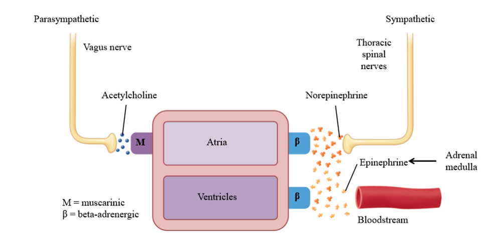
What parts of the heart are sympathetically stimulated
Atria and ventricles
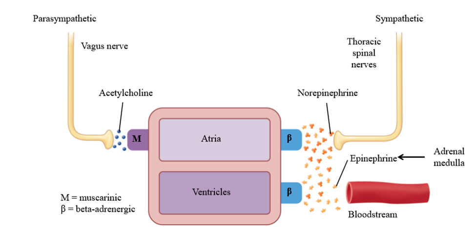
What parts of the heart are parasympathetically stimulated
Atria only - ventricular myocardium is not effected by parasympathetic activity
Parasympathetic stimulation of the heart
- Decrease rate of SA node depolarization = lower HR
- Decrease AV node conduction, increase AV nodal delay
- Decrease atrial muscle contractility
- No effect of ventricle muscle contractility
Sympathetic stimulation of the heart
- Increase rate of SA node depolarization = higher HR
- Increase AV node conduction, decrease AV node delay
- Increase atrial muscle contractility
- Increase ventricle muscle contractility
Cardiac output
Amount of blood pumped by each ventricle in one mintute
How to calculate cardiac output
CO = HR x SV
Average cardiac output
72 BPM x 70mL blood per beat = 5L blood per min
How to alter cardiac output
Alter Heart rate (SA node), alter stroke volume (ventricular myocardium)
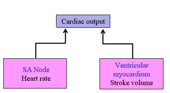
How to alter heart rate for cardiac output
Alter activity of the sinoatrial node
How to alter stroke volume for cardiac output
Vary the strength of the contraction of the ventricular myocardium. Increased contraction = increased SV (and vice versa)
Change in autonomic nervous system to increase heart rate
Increase in sympathetic stimulation with a decrease in parasympathetic stimulation
Sympathetic/Parasympathetic tone
Steady Background "level" the autonomic nervous system runs at. When one system is activated, the rate of firing raises above a tonic level, and the other system falls below
How does the sympathetic nervous system increase heart rate
Increased thoracic nerve activity and increase epinephrine from the adrenal medulla increase activity of the SA node, increasing HR and CO
How does the parasympathetic nervous system affect heart rate?
Increased vagus nerve activity and increased acetylcholine decrease activity of the SA node, decreasing HR and CO
How is heart rate initiated
Conducting myocytes in the SA node generate action potentials (heart's pacemaker)
Sympathetic stimulation of the SA node
Increase slope of the pacemaker potential, causing faster depolarization to threshold.
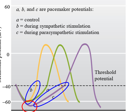
What causes the slope of the pacemaker potential to increase (sympathetic stimulation)
Increase permeability to F-type (sodium enters myocyte) and T-type (calcium enters myocyte) channels.
Parasympathetic stimulation of the SA node
Decrease slope of the pacemaker potential, causing slower depolarization to threshold
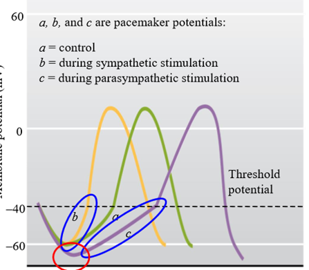
What causes the slope of the pacemaker potential to decrease (parasympathetic stimulation)
Decrease F-type (sodium channel) permeability, reducing sodium entering the cell
Increase K+ channel permeability, causing K+ to leave the cell
Effect of nervous system on pacemaker potential
Increases/Decreases plateau length of pacemaker potential, increasing/decreasing heart rate
How to increase HR
increased plasma epinephrine, increased release of norepinephrine from sympathetic nerves and decreased release of acetylcholine from parasympathetic nerves acts on the SAN to increase HR
How to decrease HR
decreased plasma epinephrine, decreased release of norepinephrine from sympathetic nerves and increased release of acetylcholine from parasympathetic nerves acts on the SAN to decrease HR
Does heart still beat without the ANs?
Yes - SA node initiates action potentials without the need for hormones