CHE 4341 Exam 2
 Call Kai
 Learn
Learn Practice Test
 Spaced Repetition
 Match
Match Flashcards
 Knowt Play
1/151

There's no tags or description
Looks like no tags are added yet.
Name | Mastery | Learn | Test | Matching | Spaced | Call with Kai | Chat |
|---|
No analytics yet
Send a link to your students to track their progress
152 Terms
Primary Structure of Proteins
amino acid sequence & location of S-S bonds
Main bonding interaction for Primary Structure
Covalent bonds
Secondary Structure of Proteins
Interaction of amino acids close to each other to form specific patterns (eg alpha-helix, beta-sheet)
Tertiary Structure of Proteins
3D structure of polypeptide
Main bonding interaction for Tertiary Structure
Hydrophobic effect → other bonding interactions still help
Quartenary Structure of Proteins
arrangement of multiple folded protein molecules (subunits) in a multi-subunit protein
Main bonding interaction for Quatenary Structure
Hydrophobic effect → other bonding interactions still help BUT no covalent bonds or disulfide bonds between subunits
Peptide bond has _______
40% DB character (so behaves like double bond)
Nearly all peptide bonds are
TRANS; 10% that follow proline may be cis
Peptide bonds are
PLANAR → no rotation → cis/trans isomerism
Why are peptide bonds mostly trans?
Trans bonds are more stable than cis
Since no rotation can occur around a peptide bond,
rotation can only occur around ALPHA carbon
Pauling and Corey figured out the
structure of the peptide bond
Phi (Φ) Angle
For Ca-N = +180º
Psi (ψ) bonds
For Ca-C = -180º
Certain conformations (rotations around ψ and Φ are forbidden
b/c of steric hindrance
Ramachandran Plot
Calculated Plot of all sterically allowed ψ & Φ angles for a polypeptide chain

Beta sheets are on located on
top left side of the Ramachandran Plot
Green terrain
for GLYCINE; has more conformations than rest of AAs
Blue terrain
for rest of AAs (excluding glycine)
What is Protein 2º Structure?
general three-dimensional form of local segments of biopolymers such as proteins and nucleic acids (DNA/RNA) to give regular, recurring local conformations
Types of bonding in Protein 2º Structure?
H-bonding between backbone amide NH of one AA & backbone carbonyl C=O of another

Linus Pauling with Robert Corey proposed
Alpha helix (1951)
In alpha helixes, _____ form between adjacent AA’s to give __________
H-bonds; curled polypeptide helix
Hydrogen bonds in Alpha helices occur between
n and n+4 residues
In Alpha helices, R groups point
outward ~ perpendicular to axis!
Pitch of Alpha Helices
5.4Å (0.54nm)
How many resides does it take to make one turn in a alpha helix?
3.6 residues/turn1.
What is rise?
how far the backbone moves forward along the helix's axis with each new amino acid
Rise per AA in alpha helix
1.5Å per AA
H-bond in alpha helices are
parallel to helix axis
Most alpha helices are/have
RIGHT-handed; have a dipole (polarity)
Alpha Helix Dipole
All C=O groups point toward the C-terminus give entire helix is a dipole with (+) N, (-) C-termini
Stable α-helices typically end (C-term) with a
+ charged AA to neutralize the dipole moment (C=Od-)
Some proteins can be PREDOMINANTLY α-helices
eg: Myoglobin

Destabilizers of α-helix
Proline (Pro) not likely found in α-helix
Steric/charge repulsion is caused by adjacent bulky amino acids; also by like charged amino acids
R-groups too large (tryptophan, tyrosine)
or too small (glycine)
Amphipathic α-helices
Hydrophobic residues on one side and hydrophilic residues on opposite side
α-Keratin
coiled coil* motif of 2 amphipathic alpha helices
Where is α-Keratin found?
key structural material in hair, skin, & nails; horns that cover core of live bone
α-Keratin makes up hair via
Keratin Dimer
Keratin
disulfide crosslinks → α-Keratin in Cys → form -S-S- bonds
How do perms work (same method for relaxing curly hair)?
Reduce -S-S- bonds → 2RSH; Next curl; then re-oxidize to –S-S-
α-Keratin can be used to
detect Heavy Metal Poisoning
β-pleated strand
polypeptide chains almost fully extended
β-pleated strand are found in
β sheets
β sheets are
multiple β strands arranged side-by-side; can come from same or different polypeptide
β strands are in ______ and H-bonds occur between AAs on ________ perpendicular to strand direction
X-plane; adjacent strands
Side chains in β sheets
alternate in pointing above & below plane perpendicular to H-bonds AND strands → Z axis
β sheets can easily
accommodate adjacent bulky / like-charged residues
Antiparallel β sheets
strands run in opposite N to C- terminal direction
Parallel β sheets
strands run in the same N- to C- direction
β sheet structure is ______ conformation with ______ residues involved per strand
Extended zig-zag (ruffled); 6 to 12 residues
In β sheets, there is a repeating unit of
2 AA residues w/ distance = 7 Å (3.5 Å per AA)
β sheets may be amphipathic because
one surface of β sheet may consist of hydrophobic side chains (R gps) while other can have hydrophilic ones
Some proteins are predominantly β sheets
E.g. Concanavalin A

Silk Fibroin has
Extended layers of antiparallel b sheets of (-Gly-Ser-Gly-Ala-Gly-Ala-) → roughly alternate with Ala and Gly
The Ala in slik fibroin
Ala from 1 sheet interdigitates w/ Ala from another sheet:
Spider silk is
biocompatible → because it is protein
What is collagen?
main structural protein in the extracellular matrix (ECM) in connective tissues in animals
What is the most abundant protein in mammals which makes up 1/3 of total protein mass in large animals?
Collagen
Collagen is present in
Bone matrix, tendons, cartilage, blood vessels, skin
The Collagen Triple Helix (“Proline Helix”)
3 strands in a triple helical coil (Strong); made up of general repeating unit in each strand = G-P-P/Hyp
Why GLY in Collagen Triple Helix?
Gly residues invariant; located along central axis of triple helix and very small
Main bonding interaction in Collagen Triple Helix
H-bonding only; INTERCHAIN bonding involves HydroxyPro (Hyp) also helps stabilize helix
Collagen Primary Structure
General Repeating unit in each strand = …G-P-P/Hyp…

Osteogenesis imperfecta (Brittle bone disease)
a mutation in a gene for collagen leads to the substitution of Gly for another amino acid → prevents the normal production of mature collage, causing brittle bones
Formation of hydroxyPro (by prolylhydroxylase) and hydroxyLys (by lyslylhydroxylase)
requires O2 and ascorbic acid (vitamin C)
What does Vitamin C deficiency (scurvy) impact?
lack of hydroxyPro
not enough interchain H-bonding
weak collagen (skin lesions, fragile blood vessels, bleeding gums)
Collagen Fibrils are also strengthened by
intrachain lysine-lysine and interchain hydroxypyridinium covalent crosslinks
Collage cross links accumulate over time, which causes
↑ cross-links = less elastic collagen = more brittle bones /tendons
Non-regular secondary structure turns and loops allow _____ and are found on _______
chain reversal; surface of globular protein (hydrophillic)
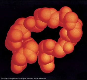
The β turn allows
polypeptide in antiparallel b-sheet to reverse direction
β turns are made up of
carbonyl O’s are H-bonded to amide H’s between residues 1 and 4
What is the similarity between Type I and Type II beta turns?
Pro often 2nd residue in both types
Tertiary Structure is a result of
folding of a polypeptide chain into a closely-packed three-dimensional (3D) structure mainly b/c of hydrophobic interactions/effect
What MAY occur due to formation of 3º structure?
Amino acids far apart in the primary structure may be brought together
3º Structure Globular (Cytosolic) Proteins are
Hydrophilic outside; hydrophobic inside

What is X-ray crystallography?
Reveals 3D structure in atomic detail → Need crystals of protein
Method of Calculating Protein Structure
diffraction pattern → Fourier transform → Computer fit map to protein sequence → Fixed (frozen) picture of protein
Resolution is measured in
Å (= 10-10m) → Smaller the better
Negatives of X-ray crystallography
For X-ray crystallography → need protein crystals → difficult to get for many proteins
Limitations of 2D NOESY NMR spectroscopy
need high concentrations of protein, time consuming, only for small (low molecular weight) proteins
NMR Structure of
64 residue polypeptide of Src protein SH3 domain; proteins are dynamic (‘breathe’ → can be displaced up to 2Å)
What is Cryo-Electron Microscopy (Cryo-EM)?
form of transmission electron microscopy (TEM) where the sample is studied at cryogenic temperatures (generally liquid N2 temp. = -196 oC!)
How are proteins categorized?
Globular, Fibrous and Membrane
Globular Proteins are
Aqueous (water soluble)
Fibrous (Rod/fiber-like) Proteins are
Normally insoluble
Protein denaturation occurs via
heating and chaotropic agents which disrupt interactions that stabilize 3º structure → Some (but not all) proteins can be refolded or renatured
Misfolded proteins get refolded or proteolyzed immediately (by proteasome) via
Polyubiquitination (Ubiquitin conjugation) and Protein degradation (proteolysis)
Who made discovered Denaturation and Renaturation of Ribonuclease A (RNase A)?
Christian Anfinsen: Showed that 1º structure determines 3ª structure →
What happened when urea and most β-ME were dialyzed out (leaving only a trace of β-ME)?
The protein spontaneously refolded into its native, catalytically active conformation. The trace β-ME prevented incorrect disulfide pairs from locking in, allowing the correct –S–S– bonds to form.
What reagents were used to denature and reduce RNase A, and what did each do?
8 M urea → disrupts non-covalent interactions (H-bonds, hydrophobic effects) → unfolds the protein
β-mercaptoethanol (β-ME) → reduces disulfide bonds → converts –S–S– to free –SH groups
Its ______ to fold in vitro
EASY → where folding by dilution in buffer
Its ______ to fold in vivo
DIFFICULT → folding occurs via co-translational folding
Why difficult to fold in vivo?
cellular environment is crowded → high potential for misfolding and precipitation (aggregation)
Chaperone
proteins that help other proteins fold in vivo (although most proteins can fold without them) → also called Heat Shock Proteins (HSPs)
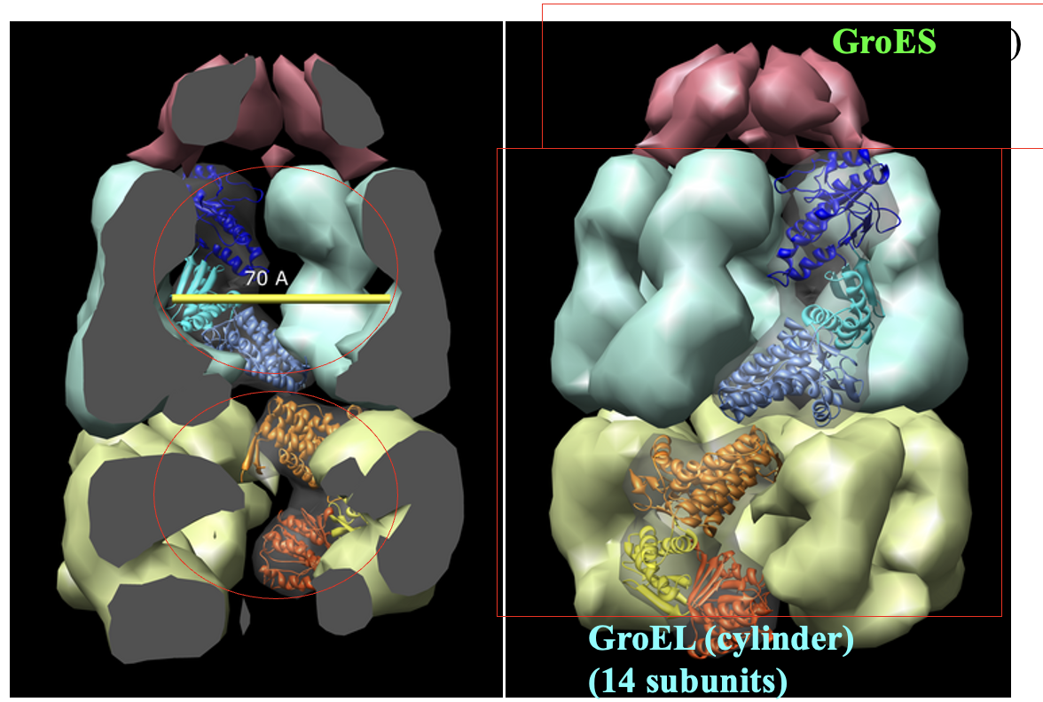
Large Chaperones called Chaperonins like Hsp60 (GroEL/GroES complex in E. coli) actively fold
proteins in prokaryotes, cytosol of eukaryotes & mitochondria.
Precipitation (aggregation) is prevented/folding assisted by
DnaJ&DnaK coating the unfolded protein and preventing denaturation/precipitation
GroEL/GroES Folding Mechanism
requires energy from ATP hydrolysis
Protein disulfide isomerase facilitates
formation of correct disulfide bridges