Simple stain
 Call Kai
 Learn
Learn Practice Test
 Spaced Repetition
 Match
Match Flashcards
 Knowt Play
1/26

There's no tags or description
Looks like no tags are added yet.
Name | Mastery | Learn | Test | Matching | Spaced | Call with Kai | Chat |
|---|
No analytics yet
Send a link to your students to track their progress
27 Terms
Which of the following reasons is why slides should be labeled with a wax pencil before staining?
To identify which bacterium/ bacteria are placed on the slide,
to ensure the slide is in the correction orientation when placed on the stage,
to give a focusing reference before attemting to find the bacterial smear
Which staining reagent is used to stain the Staphylococcus epidermidis bacteria?
methylene blue
Which side of the slide should face the opening in the incinerator when heat-fixing the smear?
Bottom (side without bacteria)
Which of the following can not be determined using the simple stain?
If the bacteria are Gram positive of Gram negative, If the bacteria have flagella
How long should methylene blue be left on the bacterial smear before rinsing the slide with water?
60 seconds
Which type of stains have a positively charged chromophores that interact with the negatively charge components found the in the cell wall of bacteria?
basic stains
Why do we stain?
The morphology of individual cells may be examined by looking at individual microorganisms in living, unstained material or by observing dead cells that have been colored with dyes. Many live microorganisms do not contrast greatly with their clear background, making them difficult to observe. Staining with colored dyes will increase their contrast, making them more readily visible. Additionally, certain specific stains can make various structures visible
Basic stains
those in which the colored ion is the cation (positively charged ion),
acidic stain
contain a colored anion (negatively charged ion)
Methylene blue is an example of a direct stain—
the stain directly interacts with the cells.
Before bacteria can be stained, a _____ of the cells must be made and fixed to a slide
smear
How is the smear prepared
by spreading the cells on a clean slide and allowing them to air dry.
The cells are then fixed to the slide by rapidly passing it through a Bunsen burner flame two to three times to heat-fix the bacteria to the glass slide. Heat-fixing cells ensures that the cells are killed and are stuck to the slide.
Alternatively, the dry smear can be fixed chemically by covering the smear with an alcohol solution. Fixing enhances the adherence of the cells to the slide for the staining procedure.
Which of the following are the two most important functions of heat fixing a bacterial smear before staining?
ensures that the cells are killed and are stuck to the slide
enhances the adherence of the cells to the slide for the staining procedure, so they are not washed off during the staining and rinsing steps of the staining procedure.
Why do we have to let the smear completly air dry before fixing?
it will cause the cells to rupture and clump together.
Simple stain instructions
1. If staining bacteria grown on solid media, place a loopful of water onto a clean glass slide. (A drop of water will take much longer to dry than a loopful of water). Flame your loop again to sterilize it.
NOTE: Once a smear dries it can be difficult to see where it is. Tilt the slide in the light and your smear will appear as a haze on the slide.
2. Using aseptic technique, obtain a small portion of the sample with your metal loop—either a loopful of bacteria from a liquid broth culture or a small amount from a single, well-isolated colony on a plate.
NOTE: A 1 mm x 1 mm patch of cells (actual size is close to the size of this box = □) can fit more than 1 million bacteria, so DO NOT use the loop to pull up all of the available cells!!!
3. Smear the cells around in the loopful of water on the slide to make a thin, milky film of diluted cells suspended in the water.
4. Allow the film to air dry. (DO NOT speed this up by heating it in your flame, as this tends to cause the cells to rupture and clump together.)
5. Pass the slide quickly (with the smear side pointing up) over the flame of a Bunsen burner two to three times or hold in front of the opening of an incinerator (smear side pointing outward) for 10 seconds to heat fix the cells. The slide will become quite hot, so be careful not to touch the glass. Always use a slide holder, never bare fingers.
NOTE: If the slides you are using have a frosted edge, make sure you write on the textured side when you label them, as this will help you avoid staining and viewing the wrong side of the slide.
6. Place the slide (smear side up) on a rack over a staining tray or just hold it above the staining tray with a slide holder
7. Flood each smear with a few drops of the stain solution.
For methylene blue or safranin, stain for 60 seconds.
For crystal violet, stain for 30 seconds.
Note: If you leave the stain on for too long (especially crystal violet), the background will stain, making it difficult to observe the bacteria.
8. Hold the slide at an angle and use water to gently rinse off the excess stain from the front and back of the slide.
9. Gently blot water from the slide with pieces of bibulous paper, being careful not to rub off the stained smear. Alternatively, tap the slide onto a paper towel to “shake” the water drops off, and let it air dry.
10. Center and focus on the smear with the scanning 4X objective. Then switch to the 10X objective and move the slide around to find an area where the bacteria cells are uniformly spread around and not clumped together. Selecting such an area will allow easier observation at higher powers. Once focused, switch to the high dry 40X objective and focus the specimen (remember on the high dry and oil objectives to never use the coarse adjustment knob; only use the fine adjustment knob).
11. Once your specimen is in focus under the 40X objective, rotate the objectives such that your 40X and 100X objectives straddle the center of your slide (your specimen is between the two objectives). Add a drop of oil to your specimen and then slowly slide the 100X oil objective into place. Do not lower the stage to add the drop of oil; your specimen will then be out of focus and you will have to start over. Remember, NEVER use the 40X objective while oil is present on the slide.
NOTE: You must always observe bacteria under the 100X oil objective because they are so small. You will be able to see them under the 40X objective, but will not be able to discern any real details.
12. Record your observations of the stained cells viewed only under the oil immersion objective in the spaces provided. Make a drawing of a representative microscopic field. Describe the cells according to their morphology, arrangements, and size.
13. When you have finished viewing your slides and recording your results, you may dispose of the slides you prepared according to the instructions given by your instructor. You must then also thoroughly blot the oil off of your 100X objective using lens paper and clean off any oil that has contaminated other parts of your microscope, such as the 40X objective, the condenser lens, or the stage. Use lens cleaning solution if necessary.
What does the simple stain tell you?
morphology, arrangements, and size of cells

What are common shapes and arrangements? cocci
cocci (spherical) singular = coccus
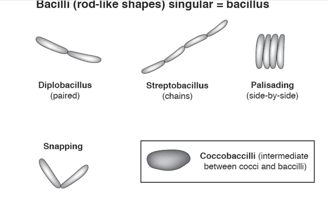
What are common shapes and arrangements? bacilli
bacilli (rod-like shapes) singular = bacillus

What are common shapes and arrangements? spilla
spirilla (spiral-like shapes)
Staphylococcus epidermidis:
cell shape:
cell arrangement:
cell shape: coccus, cocci
cell arrangement: staphyloccocus, staphylococci
Escherichia coli:
cell shape:
cell arrangement:
cell shape:
bacillus
bacilli
coccobacillus
coccobacilli
cell arrangement:
individual
scattered
individual, scattered
individual and scattered
individual/scattered
individual/ scattered
individual /scattered
invidual,scattered
Which part of bacterial cell is the simple stain actually staining?
cell wall
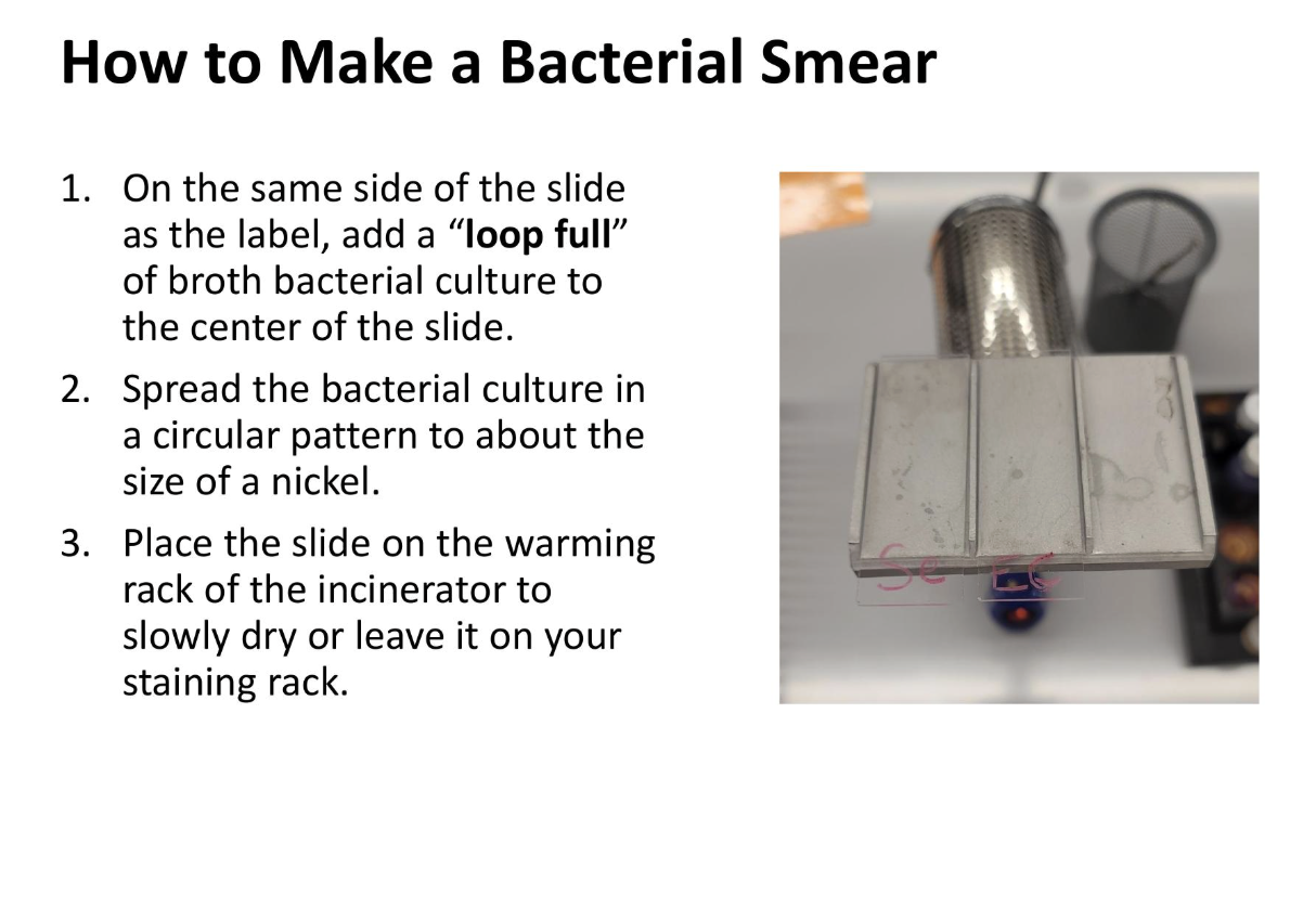
how to make a bacterial smear
how to make a bacterial smear
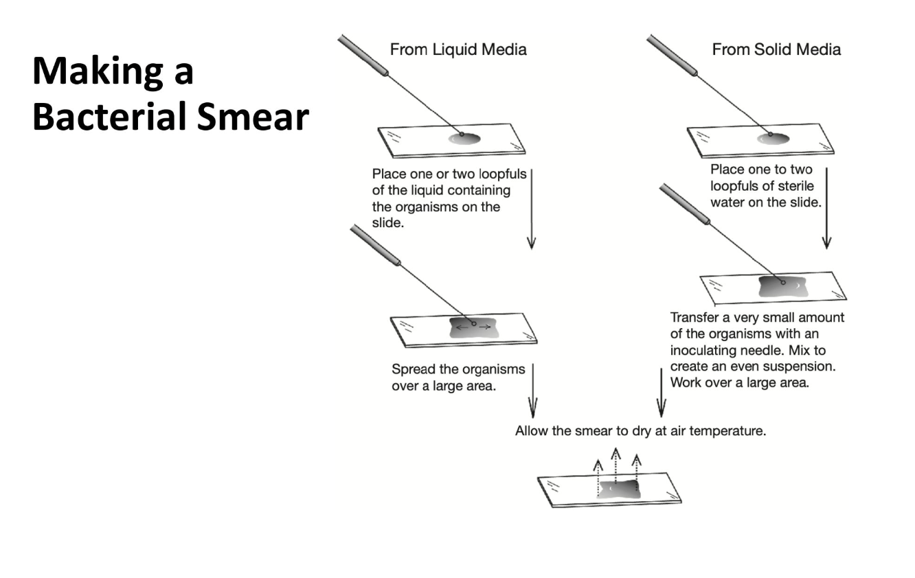
making a bacterial smear
making a bacterial smear
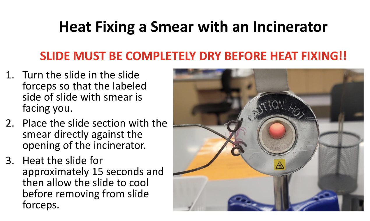
heat fixing a smear with an incinerator
heat fixing a smear with an incinerator

how to perform a simple stain
how to perform a simple stain
no arrangement=
indivdual/scattered