theory for unit 4 aos 1 bio
 Call Kai
 Learn
Learn Practice Test
 Spaced Repetition
 Match
Match Flashcards
 Knowt Play
1/69

There's no tags or description
Looks like no tags are added yet.
Name | Mastery | Learn | Test | Matching | Spaced | Call with Kai | Chat |
|---|
No analytics yet
Send a link to your students to track their progress
70 Terms
summary of cells
Cellular Pathogens: Bacteria:
Bacteria: Small prokaryotic cell, lacks membrane bound organelles, Has singular circular DNA, Replicate via binary fission. Prokaryotic (single-celled) organism
How do bacteria cause disease?
Enter the host and begin to reproduce.
Bacteria can produce toxins which cause harm to the body. (toxins can inhibit protein synthesis, damage cell membranes, interfere with nerve function, cause fever, cause diarrhoea).
Bacterial enzymes can destroy cells and tissues.
Irritation from bacterial waste products.
Exaggerated immune responses to bacterial cells.
Cellular, eukaryotic, Unicellular/multicellular:
Protist: eukaryotic organism, unicellular, that cannot be classified as an animal, plant, or fungus
Protozoans (Can reproduce in their host’s cell or outside of a host, can evade the host’s immune system).
Cellular, eukaryotic, unicellular/ Multicellular:
Can be ectoparasites (external) or endoparasites (internal).
Arthropods: invertebrate animal, ticks, fleas, lice, mites, Plants: psyllids, flies, wasps and thrips can produce galls on leaves and stems
Worms: tape worm, can infect plants and animals, large eukaryotic cell, very large.
Fungi: varied group of small eukaryotic organisms (yeasts and moulds) that get their food from living or dead organic matter, large eukaryotic cell, have cell walls.
Oomycetes: Cause downy mildew on plants and life-threatening infections in animals.
Non-Cellular Pathogens: Viruses:
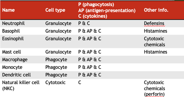
Very small, contain nucleic acid (RNA / DNA) surrounded by a protein coat (capsid).
Cannot function outside of a host cell, can’t reproduce on their own and hijack the host’s machinery to replicate.
Genetic changes / mutations can change the antigens on their surface. (common cold, herpes, mumps, rubella, varicella-zoster (causes chickenpox), influenza, HIV (causes AIDS), COVID-19).
Prions (Non-Cellular Pathogens)
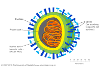
Misfolded version of protein, Do not contain any nucleic acid.
Causes healthy proteins to refold and become infectious. Slow process, thus onset of prion related diseases are very delayed.
When an abnormal prion enters the host, it touches the normal protein and transforms it. Cause neurodegenerative diseases.
Examples of transmissible spongiform encephalopathies (TSE): mad-cow disease (now called Bovine Spongiform Encephalopathy), Creutzfeldt-Jakob Disease, kuru. No treatment is available – prions are extremely resistant to heat and chemicals, and the immune response is not effective.
Viroid: (Non-Cellular Pathogens)
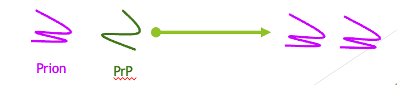
Plant pathogen, Composed of short-circular strand of RNA without a protein coat. Much smaller than a virus and can only reproduce inside a host cell.
A type of self-cleaving RNA enzyme (ribozyme). They form bundles which interfere with the internal structures of plants (like a tumour). Have a very high mutation rate which enables them to avoid the host’s immune system.
Microbiota barrier
In animals: natural gut flora: non-pathogenic bacteria on the skin, in the mouth, nose, throat, gut and vagina. Inhibit the growth of pathogenic bacteria, successful competitors and use resources available as well as produce chemicals that lower the pH of the microenvironment. (beneficial bacteria, fungi)
In plants: Microorganisms on the surface of plant tissue (bacteria and fungi) that live on or inside plant tissues to protect the host against pathogens
- Rhizosphere Microbiome: Microbes clustering around the roots in the soil.
- Endophytes: Beneficial bacteria and fungi living completely inside plant tissues without causing harm
Distinguishing Self from Non-Self: Self-antigens:
Belong to u. Are found on your own cells that tells the body that they are your own. Looking for, and communicate and alert others.
MHC (major histocompatibility complex): a type of self-marker
Type of receptor
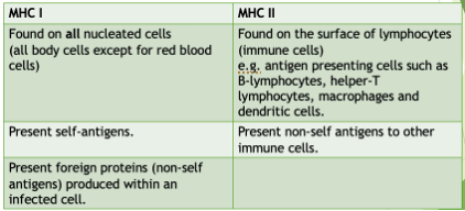
MHC I: found on all nucleated self-cells (all except RBC). Display self-antigens allowing cells to be identified as self/ can display foreign antigens to allow for the identification of infected and tumour cells
MHC II: only on dendritic cells, macrophages and B lymphocytes. display fragments of pathogenic/foreign antigens and present them to other cells of the immune system/ initiating the adaptive immune response/ activating helper T-cells
Blood group antigens (only on RBC), types, antigen, antibody, blood donors
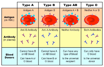
Distinguishing Self from Non-Self: Non-self-antigens
(don’t belong): are found on pathogens/allergens that body recognises as foreign. The body looks for traits on pathogens that wouldn’t be found on normal self cells (e.g. the protein coat on viruses, the cilia of bacteria). When the body recognises a foreign antigen, it initiates an immune response.
What happens if there is a pathogen inside a cell?
Endogenous pathogens:
1. Pathogen invades cell
2. Cell breaks down pathogen into small antigen peptides
3. Peptides are transported to MHC I molecule
4. MHC I molecules present small antigen peptides on the surface of the cell
To alert other immune cells that this cell is infected and that it needs to be destroyed to prevent further spread/replication of the pathogen.
What happens if there is a pathogen outside a cell, invading the bloodstream?
Exogenous pathogens:
1. APCs engulf a pathogen
2. They process it into small antigen fragments
3. Antigens are shown on the cell surface using MHC II molecules
Alerts other immune cells that there is a pathogen infecting the body. Thus, if MHC molecules are empty, body recognises the cell as a self cell. If MHC molecule is carrying an antigen, it knows that the cell/body has been infected.
Inflammatory Response:
Non-specific process (same everytime).
When tissue is damaged or infected due to the production, activation and release of complement proteins and cytokines.
Capillaries dilate and become more permeable, increasing blood flow (increases vasodilation) and allowing fluid, plasma proteins and leukocytes to enter the affected tissue. This causes redness, heat, swelling and pain, while enabling phagocytes to reach the area quickly.
Eliminates the effects of an injury, defends against potential pathogens, clears out cells that may have been damaged or destroyed and initiate’s repair.
Can occur as part of the innate or adaptive immune response. Histamines are released by phagocytes at the area to attract more phagocytes.
Involves three main aspects – initiation, vasodilation and migration.
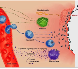
Inflammation process, initiation, vasodilation and migration.
● Initiation - Pathogen enters the body (e.g. from a cut or injury)/tissue is damaged. Injured cells release cytokines that attract neutrophils and causes mast cells to release histamine (degranulate)
● Vasodilation - Histamine increases blood vessel dilation and permeability; leads to increased blood flow to injury site (redness, swelling, warmth) and ‘leaky’ blood vessels
● Migration - Neutrophils (cytokines attract) and macrophages (are activated, release cytokines) leave blood vessels and migrate to injury site; engulf and destroy pathogens. A clot (due to influx of immune cells) forms to seal the area and stop pathogens from spreading further into the body. Once removed, healing begins and inflammation slowly reduces.
Defensive System: Cytokines:
Chemical messengers between immune cells, made by many immune cells. They trigger a variety of responses:
u Promote proliferation of lymphocytes.
u Induce inflammation and fever.
u Promote antibody responses.
u Activate macrophages.
Interferons and chemokines are two different types of cytokines.
Fever:
Increase in body temperatures to about 39ºC. Temperature set by hypothalamus (in the brain) in response to inflammatory cytokines (called interleukins) released by macrophages. A defense mechanism against many pathogens (restricts their functioning and slows the replication, temp not optimum). Also increases the activity and proliferation of leukocytes, allowing the immune system to catch up in the fight against the infection.
summary of immune system
The Lymphatic System:
(highway and service stations of your immune system):
Defending the body against infection. The transport network. Open system that collects tissue fluid and returns it to the blood. Its major role in immune response is:
· One-way system of vessels that collect fluid (lymph) from tissues and carry it back into the blood
· Along the way, it transports immune cells (especially lymphocytes like T and B cells). Providing a place for lymphocytes to mature.
· Also carries pathogens, antigens, and dendritic cells from tissues to lymph nodes
Returning fluid that seeps out of the blood vessels into tissues back to the circulatory system.
Absorbing and transporting fatty acids and fats from the digestive system.
Providing a place for lymphocytes to mature.
Transporting B and T lymphocytes and antigen-presenting cells throughout the body and to the lymph nodes.
Immune response takes place inside the lymph vessels and nodes (pathogens are transported to the lymph nodes).
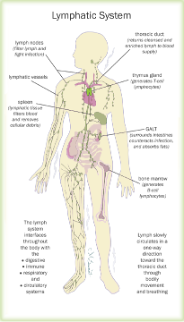
Structure of the Lymphatic System:
- Fine lymphatic capillaries join to form increasingly larger vessels.
- Most vessels cannot contract and have valves to ensure one-way flow of lymph.
- Cells and fluid can pass between the lymphatic and circulatory capillaries through extravasation.
· Lymph= the fluid that carries around immune cells (like T and B cells) and any pathogens or foreign materials they need to respond to. carries debris, pathogens and immune cells. Circulates in one direction throughout the body
- If a person is inactive for long periods of time, lymph collects in areas due to gravity = swelling.
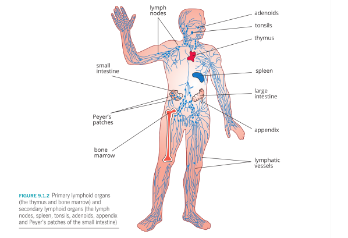
Organs of the Lymphatic System: Primary organs produce new lymphocytes:
- Bone marrow: contains stem cells from which B and T lymphocytes originate. B lymphocytes mainly develop and mature in the bone marrow and can complete their maturation in secondary lymphoid tissues. T lymphocytes are produced in the bone marrow.
- Thymus: T lymphocytes travel to the thymus to mature. Gradually shrinks after puberty and contributes to the higher risk of infection and cancer that comes with age.
Organs of the Lymphatic System: Secondary Organs and Tissues:
adaptive immune responses begin here when lymphocytes are activated. Tonsils, Adenoids, Appendix, Peyer’s patches of the small intestine and:
- The lymph vessels (the roads that transport this lymph fluid): Travel in one direction (to the heart), contain liquid called lymph which drains from nearby tissues. Consists of plasma, white blood cells, memory T and B cells and antigens. Connect to lymphatic tissues and organs. transport antigens and immune cells from tissues to lymph nodes. The lymph vessels: Travel in one direction (to the heart), contain liquid called lymph which drains from nearby tissues. Consists of plasma, white blood cells, memory T and B cells and antigens. Connect to lymphatic tissues and organs.
- Lymph nodes (the service stations along those roads where immune cells gather, talk, and plan attacks against invaders):
. Monitor/Filter lymph fluid
· Trap/remove antigens/pathogens
· Site of antigen presentation and the activation of the adaptive immune response
· Site of clonal selection and expansion
· Store lymphocytes and other immune cells
- Spleen: Controls the number of red blood cells in the body by destroying old and defective red blood cells. Stores up to a quarter of the body’s lymphocytes. One of the sites of B cell maturation.
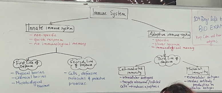
The Third Line of Defence: The Adaptive Immune Response:
Specificity (Cells can recognize and respond exclusively to specific antigens) and Immunological memory (Cells ‘remember’ antigens after primary exposure and mount a larger, more rapid response when exposed to the same antigen in future). 2 main fighters B lymphocytes, T lymphocytes.
Involve: lymphocytes, and clonal selection.
Mechanisms of Adaptive Immune Responses:
Cell-mediated immunity (When the cells become infected….)
(“Like elite soldiers sneaking into enemy-occupied buildings to take them out.”): Involves the action of T lymphocytes and antigen-presenting cells. For intracellular threats (like virus-infected cells or cancer), mainly run by cytotoxic T cells with the help of helper T cells. Involve: Helper T cells (CD4+): like commanders, they activate B cells and cytotoxic T cells. Cytotoxic T cells (CD8+): like assassins, they kill infected cells directly. Memory T cells: stick around to respond faster next time.
T Cells and Cell-Mediated Immunity:
Many different T cells are matured in the thymus – these also recognise many different antigens. After binding with an antigen (= activation), T cells will reproduce and form memory T cells, cytotoxic T cells and helper T cells. T cells do not make antibodies. This form of immunity is called cell-mediated immunity; it involves cells (T cells and phagocytes) and not antibodies.
T Lymphocytes Involved in Cell-Mediated Immunity:
u Helper T lymphocytes (TH cells)
u Cytotoxic T lymphocytes.
u Memory T lymphocytes
Antigen Recognition (b and T cells)by T Cells:
Patrol the lymph nodes, waiting to find and respond to their matching antigen.
T cells check the antigens they come across and differentiate between self and non-self.
APCs present antigens to T cells by engulfing a pathogen and breaking down into small peptides.
These fragments bind to molecules inside the cell and this complex moves to the cell surface to be presented to helper T cells.
Signal transduction takes place in the helper T cell and it is now activated.
The helper T cell proliferates and releases cytokines.T and B cells are constantly circulating through lymph nodes, looking for their specific antigen. If an antigen is recognized:
o T cells
Helper T cells (Th/CD4+): recognise antigen presenting cells and activate B and Tc cells by releasing cytokines.
Cytotoxic T cells (Tc/CD8+): kill infected cells via apoptosis
B cells activate and produce antibodies.
The Steps of Cell-Mediated Immunity:
Antigen presentation and recognition:
- When a foreign antigen (a bacterium) enters the body, a phagocyte will engulf and destroy it.
- The phagocyte then displays the antigen on its cell surface (on MHC-II markers).
- The phagocyte presents the antigen to a T helper cell (called antigen presentation).
- The T helper cell is now activated.
Clonal expansion: Once the T cells are activated, they proliferate (divide rapidly).
), Cytotoxic T cells (TC, target infected or dying cells, body’s assassins), Memory T cells (TM).
Antigen elimination (TH cells):Helper T cells are involved in antigen elimination in the following ways:
- Secrete cytokines to stimulate other T cells.
- Release protein messenger molecules which attract phagocytes.
- Activate B cells.
5. Antigen elimination (TC cells): Release toxins. Trigger the infected cell to self-destruct (apoptosis).
- Destroy foreign eukaryotic cells.
- Kill self-body cells that have been infected with a virus (same job as natural killer (NK) cells).
- They secrete protein granules that punch holes in the membrane of the infected cell and the contents ooze out = cell dies.
- Cytotoxic T cells cannot destroy isolated viruses.
- Some destroy cancer cells.
- They are also involved in organ transplant rejection.
6. Memory: Memory T cells are also produced during differentiation. These remain in circulation for many years, ready to respond if you encounter the same pathogen again.
Mechanisms of Adaptive Immune Responses: Humoral immunity
(“Antibodies are like archers, shooting arrows at invaders in the open”): Involves B lymphocytes. Macromolecules such as complement proteins and antibodies produced by B lymphocytes are secreted into the extracellular fluid (blood and lymph, previously known as the humor). For extracellular threats. Mainly run by B cells and antibodies with the help of helper T cells. B cells detect antigens and activate. They clone into plasma cells (which make antibodies) and memory B cells. Antibodies stick to pathogens, marking them for destruction.
Humoral Immunity: In the humor (body fluids/blood): B Lymphocytes Involved in Humoral Immunity:
Clone cells:
Plasma cells:
B-memory cells:
B Cells and Antibodies:
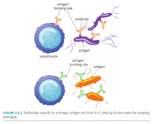
B cells have immunoglobulins on their surfaces – membrane-bound antibodies that are known as B cell receptors (BCRs). BCRs on any individual B lymphocyte are the same, but different B lymphocytes have different BCRs that detect different antigens. Immunoglobulins have a specific structure and recognize only one kind of antigen.
When BCRs bind to antigens, B lymphocytes engulf and process the antigens and function as antigen-presenting cells. The binding of BCRs to antigens also results in the activation and proliferation of B cells with the same specific BCR variants.
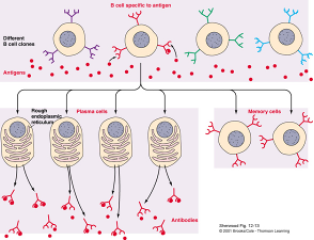
Cytokines released by helper T cells help activate B lymphocytes. Activated B lymphocytes divide and differentiate into either plasma cells or memory B cells.
There are millions of antigens to which the body must respond, but only a few of each kind of B cell are made – so there are millions of different B cells made with different immunoglobulins on their surfaces.
The Steps of Humoral Immunity:
Antigen recognition: B cells patrol the body looking for extracellular antigens. When an antigen enters the body, one B cells will have a specific receptor to match. When they detect their specific antigen, they recognize the antigen and bind to it they:
o Activate (with help from helper T cells)
o Differentiate into plasma cells → these produce antibodies
Clonal expansion: Activated B-lymphocyte proliferation into clone cells
Differentiation: B- clone cells differentiate into Plasma and B-memory cells.
3. Antigen elimination: Plasma cells produce antibodies specific to antigen. These antibodies bind with antigens, Neutralising antigen, Tagging it for destruction (by phagocytes) and Clumping pathogens together for easier removal.
Memory: Memory B cells live for many years, remaining in circulation, faster and better response next time. Now said to be immune to disease.
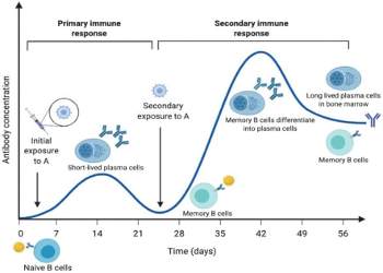
First Exposure
· The body takes time to recognise the pathogen.
· B and T cells are activated.
· B cells produce antibodies, and memory cells are created.
· Response is slow and symptoms usually occur.
IgM antibodies are the predominant antibodies produced in a primary response.
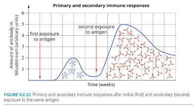
Second Exposure
· Memory B and T cells recognise the pathogen immediately.
· Response is faster, stronger, and more targeted.
· Often, no symptoms occur — the body clears the pathogen quickly.
· IgG antibodies are the predominant antibodies produced in the secondary response.
This is the principle behind vaccination:
· A vaccine mimics the first infection → builds memory cells.
· On real exposure, the immune system responds like it’s a second exposure.
Antibodies (immunoglobulins (Ig))
Structure of an Antibody:
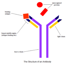
proteins that have a quaternary structure, containing four polypeptide chains:
- two long heavy (polypeptide) chain and two shorter light (polypeptide) chain.
- Free ends are antigen-binding sites: these differ in different antibodies, specific to a particular antigen. Known as the variable regions which are identical in the same antibody.
- Hinge area to form a better link with an antigen.
What do antibodies do?
They find and stick to harmful germs like bacteria and viruses, blocking them from doing harm, tagging them for destruction, or clearing them out of your system.
Cause agglutination: When antibodies bind to antigens on two or more pathogens (forming antigen-antibody complexes), causing the pathogens to clump together. This Rrestricts the movement of the pathogen (neutralization). They are now more readily attacked by phagocytes,. Makes the pathogen more recognizable to phagocytes. Activates complement proteins. Neutralization of bacterial toxins (another type of antigen). Neutralization of pathogens as when antibodies bind to antigens on the surface of the pathogen, the pathogen can be prevented from invading host cells.
Comparing the Innate and Adaptive Immune Response:
Speed of response: Adaptive is a slower response than innate.
Use of antibodies: Adaptive includes the use of antibodies, innate does not.
Forming a barrier: Innate includes the formation of barriers to prevent pathogens from entering the body, adaptive cannot.
Formation of specific memory cells: Adaptive forms memory cells, and therefore has a rapid secondary response, innate does not.
Humoral vs cell mediated
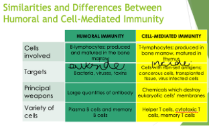
● Humoral involves B cells with antibodies whereas cell-mediated involves cytotoxic T cells to remove the non-self-antigen/pathogen
● Humoral targets (extracellular pathogens/antigens) such as bacteria, bacterial toxins and viruses whereas cell-mediated targets eukaryotic (body) cells presenting non-self-antigens (e.g. virus-infected self-cells - pathogen is intracellular or cancer cells)
Immunoglobulin E (IgE):
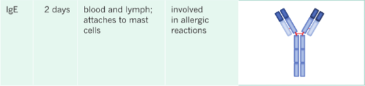
Antibodies produced by plasma cells (more produced in individuals who suffer from allergic reactions). Travel in the bloodstream. Meet mast cells and binds to receptors on mast cells, ready to trigger the allergic response upon second exposure.
Allergic Reactions:
Type 1 (immediate) hypersensitivity = allergy. Due to a rapid and vigorous overreaction of the immune system to antigens (allergens), otherwise be harmless.
(allergic reactions to pollen (called rhinitis / hay fever) most caused by grass and tree pollen).
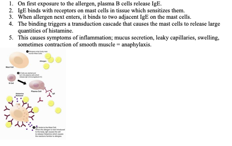
On first exposure to the allergen, plasma B cells release IgE.
IgE binds with receptors on mast cells in tissue which sensitizes them.
When allergen next enters, it binds to two adjacent IgE on the mast cells.
The binding triggers a transduction cascade that causes the mast cells to release large quantities of histamine.
This causes symptoms of inflammation; mucus secretion, leaky capillaries, swelling, sometimes contraction of smooth muscle = anaphylaxis.
Types of Immunity:
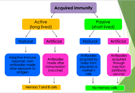
Active immunity: Protection provided by own adaptive immune response. Takes time to develop. Memory B and T lymphocytes produced provide immunological memory for many years, even a lifetime.
Passive immunity: Transfer of antibodies produced by another organism. Immediate. Only protected for a limited time as antibodies degrade over time and are removed. No B and T cells so no immunological memory.
Feature | Active Immunity | Passive Immunity |
What happens? | Your body makes it own antibodies | You’re given antibodies directly |
Time to develop? | Takes longer (days-weeks) | Acts immediately |
Lasts how long? | Long-term (often years or lifelong) | Short-term (weeks to months) |
Memory cells? | Yes - memory cells are made | No - memory cells are not made |
The 4 Types of Immunity:
How you contract the disease | Active - If you produce your own antibodies | Passive - If you’re given the antibodies |
Natural | Natural Active · You catch a disease and recover · E.g. getting chickenpox and becoming immune | Natural Passive · Antibodies passed from mother to baby · E.g. breast milk or through placenta |
Artificial | Artificial Active · You get a vaccine (weakened/inactive pathogen) · E.g. COVID-19 vaccine | Artificial Passive · You’re given antibodies directly · E.g. antivenom for a snake bite |
Suppressing Active Immunity when it is Harmful: Rhesus incompatibility and Haemolytic Disease:
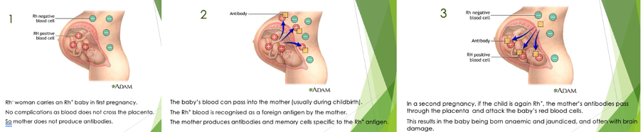
Most people have red blood cells with antigens called the rhesus D antigen (Rh+). If no antigen is present, the person is Rh-. When Rh+ and Rh- come into contact, antibodies will be produced against the other antigen. This can occur when a mother’s natural active immunity causes her immune system to attack the red blood cells of her unborn foetus.
Treatment/Prevention: These days if this situation arises, Rh- mothers are injected with Rh+ antibodies soon after giving birth. These antibodies destroy any foetal Rh antigens that she may have obtained. Therefore, her body does not have time to produce an adaptive immune response, and she does not make her own Rh+ antibodies or memory cells. The next pregnancy should then be problem-free!
Vaccines:
Need to be highly specific to initiate an adaptive immune response resulting in memory. Still successful when given within the incubation period (before the onset of symptoms). Way to train your immune system using weakened, dead or parts of pathogens (antigens). Helps your body produce memory cells so it can respond faster if the real pathogen enters later. (rabies and smallpox; education and vaccination are the main reasons we have these diseases under control). After vaccination, B and T memory cells are created. These cells remember the pathogen and trigger a fast and strong immune response if infected. You often don’t get sick at all or get much milder symptoms if you are infected by the disease.
Vaccination Programs:
Organised programs = high vaccine coverage across the population. Examples: School immunisation programs, national campaigns. They decrease the incidence of many diseases, hospitalisations, and sometimes even eradicate diseases (e.g. smallpox). In Australia, smallpox, polio and measles have either been eliminated or a very rare, due to vaccination.
Types of Vaccines:
Vaccine: Injection that contains altered/weakened/killed pathogens/antigens OR inactivated forms of toxins or proteins (that induce an immune response).
Live attenuated vaccines: weakened form of a living pathogen ( cannot usually cause disease) . Stimulate a strong adaptive immune response, produce antibodies, long-lasting immunity. Risky for People with weakened immune systems, Developing foetuses (as some may cross the placenta). More for viruses than bacteria. (Measles, mumps, rubella and polio vaccines).
Inactivated vaccines: Contain killed pathogens that cannot reproduce / cause disease. Stimulating production of a range of antibodies. Safer for people with weakened immune systems. Stimulate a weaker immune response than live attenuated vaccines, booster doses are often required to maintain immunity. Many vaccines against bacteria are inactivated. (Rabies and hepatitis A vaccines.)
Subunit vaccines: components (antigens) of a pathogen. Do not contain live pathogens, (safer, stable than live attenuated vaccines). Usually require multiple doses/boosters to produce strong immune response. Antigens can be obtained by growing pathogens and extracting components or produced using recombinant DNA technology. Toxoid vaccines are a type of subunit vaccine that use inactivated toxins. (tetanus and diphtheria vaccines).
Nucleic acid vaccines: Contain genetic instructions (DNA or RNA) that code for a pathogen antigen. Host cells take up the nucleic acid and use it to produce the antigen, which triggers an adaptive immune response.
- RNA vaccines: Contain mRNA enclosed in a lipid membrane. mRNA enters cells and is translated by ribosomes to produce pathogen antigens.
- DNA vaccines: Contain DNA instructions for producing antigens. Less commonly used.
- Viral vector vaccines: Use a harmless recombinant virus to deliver genetic instructions for producing pathogen antigens. The modified virus can enter host cells but does not cause the target disease. The antigen produced stimulate an immune response.
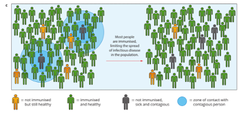
Herd Immunity:
Herd immunity threshold = the % of people needing immunity to stop disease spread (varies by disease).
For an immunization program to be successful, over 90% of people need to be vaccinated = herd immunity. More people vaccinated means fewer potential carriers and so those people who cannot be vaccinated (elderly or newborn babies) or have suppressed immune systems can be safe. Booster vaccinations must also be kept up to date to maintain herd immunity. E.g. whooping cough.
Emerging Disease and Infection Control Strategies:
Emerging Infectious Diseases:
The study and surveillance of newly emerging and re-emerging diseases aim to find ways to predict, prevent and respond to outbreaks of disease.
Emerging infectious diseases may be defined as:
u New or previously unrecognized pathogens and diseases.
u Diseases that have increased in incidence, prevalence or geographic range over the past 20 years.
u Disease that may increase soon.
New diseases can emerge as Mutation (e.g. new virus variants like COVID-19), Zoonotic transfer (animal → human, e.g. Ebola, SARS), Changing environments (climate change, deforestation). These often cause epidemics or pandemics because no one has immunity.
Old diseases can come back due to: Declining vaccination rates (e.g. measles), Antibiotic resistance (e.g. TB), Travel and migration. Even if a disease was once rare or eliminated, it can return.
Uncontrolled or Inappropriate Use of Antimicrobials:
Antimicrobial resistance: has been made worse by overuse of antimicrobial drugs which has resulted in some drug-resistant pathogens. Resistant pathogens survive antimicrobial treatment, reproduce, and pass on resistance genes, making infections harder to control.
Factors that influence the emergence and spread of diseases, including:
- Human migration and demographics (size, structure and distribution of populations).
- Human behavior.
- Changes in farming practices and food production.
- Uncontrolled or inappropriate use of antimicrobials.
- Lack of sanitation and poor hygiene.
Human Migration and Demographics:
Migration allows pathogens to spread to new areas and new hosts (might not have immunity). Rapid movement of people between continents means pathogens can spread rapidly around the globe. Infectious diseases can have devastating consequences for populations that have never encountered them before because the entire population has little or no immunity. (bubonic plague, influenza and COVID19).
Introduced Diseases Among Indigenous Populations:
Europeans arrived, brought new pathogens (e.g. smallpox, influenza). Aboriginal and Torres Strait Islander peoples had no immunity, resulted in devastating population loss and lasting social, cultural, and health impacts. In the 10 years that followed the arrival of Europeans, the impact of introduced diseases, along with the dispossession of Indigenous peoples’ lands and resulting conflict over land and resources, had reduced the Indigenous population by as much as 90%. Major epidemic disease in the early stages of European habitation of Australia were influenza, tuberculosis, measles, smallpox and syphilis. Understanding how diseases spread helps us: Improve health systems, Prepare for pandemics, Protect vulnerable populations. Respecting cultural impacts, especially for First Nations people, is key to inclusive health planning.
Changes in Farming Practices and Food Production:
Intensive farming has brought humans into close contact with animals. Mad cow disease can be passed on to humans who eat affected meat. Reducing the risk includes strategies such as:
- Surveillance and prevention of entry into Australia.
- Reducing human activity and travel.
- Reducing import and export of agricultural products.
Lack of Sanitation and Poor Hygiene:
Lack of access to clean running water, effective sewage systems, and adequate healthcare increases the risk of disease transmission, particularly for pathogens spread by the faecal–oral route. Poor sanitation allows pathogens to contaminate water and food supplies, leading to outbreaks of infectious diseases. (A cholera outbreak in Haiti following the 2010 earthquake was associated with damaged sanitation infrastructure and contamination of water supplies, allowing Vibrio cholerae to spread).
Strategies for Prevention: Identification of the Pathogen and Host:
: Identify what is causing the disease. Helps determine treatment, origin, and how it spreads. Methods include:
- Microscopy
- Culturing pathogens
- PCR / genetic sequencing
- Antibody testing
Identifying the host helps stop the spread (especially with zoonotic diseases like COVID-19 or Ebola). Involves contact tracing, case mapping, and sometimes animal testing
Strategies for Prevention: Mode of Transmission:
way in which a pathogen can be transmitted from its natural reservoir to susceptible host, can be direct or indirect.
Direct transmission:
u Direct contact: transfer through physical contact (e.g. kissing, skin-to-skin contact, sexual activity, contact with contaminated soil or vegetation).
u Droplet spread: pathogen-containing droplets released through coughing, sneezing, or talking.
Indirect transmission:
u Airborne: pathogens carried in small particles that remain suspended in the air.
u Vehicle-borne: spread through contaminated non-living objects or substances (e.g. food, water, surfaces).
u Vector-borne: spread through living organisms that carry pathogens (e.g. mosquitoes, ticks, fleas).
Strategies to Reduce/Stop Transmission:
Scientific strategies to reduce transmission, prevent infection, and control or eliminate pathogens (antibiotics (bacteria), antivirals (viruses), antifungals/fungicides (fungi), vaccines, and diagnostic testing.).
Social strategies support scientific approaches by reducing the spread of pathogens within populations. (education, hygiene practices, isolation, contact tracing, and public health measures.)
Controlling Disease and Containment Summary:
Prevention: hand washing, disinfection, sterilisation, PPE (gloves and face masks).
Isolation and quarantine: in hospitals, preventing people moving around the world, closing schools etc.
Control carriers: e.g. destroying infected cows and birds.
Eradication of vectors: eliminate or repel vectors such as mosquitoes.
Vaccination: to prevent future infections.
Education: helps to control the spread.
Response plans: governments work together to share information and control the spread.
Cancer:
Many different types of cancer, affect many different types of cells. Results from a single abnormal cell that multiplies uncontrollably and spreads throughout body.
The uncontrolled growth is the result of changes to genes that control:
u How cells grow and divide.
u A resistance of these abnormal cells to apoptosis.
Genetic changes can be inherited or develop as a result of damage to the cell’s DNA over time. Substances that damage DNA are called carcinogens. Carcinogens can be physical (e.g. radiation), chemical (e.g. asbestos) or biological (e.g. certain viruses).
Tumor:
forms when the number of abnormal cells has increased significantly, forming a clump of cells.
u Benign tumors: not cancerous, because their abnormal cells do not invade nearby tissue or spread throughout body. Can become cancerous over time.
u Malignant tumors: are cancerous because their cells invade nearby tissue and spread from the site that they originated.
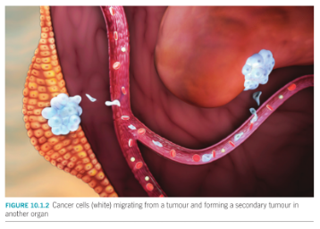
Metastasis: occurs when cancer cells break away from the original tumor, travel through the blood and lymph vessels and form secondary tumors at other locations
Cancer Treatments:
include chemotherapy, radiation therapy and surgery to remove tumors.
Chemotherapy:
involves administering drugs that are cytotoxic to cells that multiply rapidly. Although these drugs have improved, they are not specific enough to not damage healthy cells that also divide rapidly such as bone marrow and hair follicle cells. Radiation therapy kills cells by damaging their DNA. Damage to surrounding tissue is inevitable. Surgery can be effective, but it can be difficult to ensure that all malignant cells are removed.
Cancer Vaccines:
Stimulate the immune system to attack cancer cells. Preventative; directed against viruses that cause cancer (HPV and HBV). These introduce specific antigens into the body. Therapeutic; given to people who already have cancer to trigger an immune response against specific cancer antigens. Personalized; developed for an individual using their own cancer cells or immune cells to improve recognition by the immune system. Can be tumor cells that have been removed from the patient, altered in the lab to make them more visible and then injected back into the patient.
Genetically Modified T-Cell Therapy:
Cytotoxic T cells are removed from patient, modified to recognize specific cancer cell antigens, and returned to the patient where they target and destroy cancer cells.
Monoclonal Antibody Therapy:
Monoclonal antibodies (mAbs) are produced by a single clone of a B lymphocyte that is grown in culture in a laboratory to produce a large volume of the same clone. Are all identical and specific to the same antigen. Used to target specific antigens on tumour cells. Also used to target cells of the immune system and direct the immune response in a way that helps destroy tumour cells.
Production of Monoclonal Antibodies:
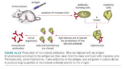
Mice are injected with a specific antigen for cancer cell.
The mice’s B lymphocytes produce specific antibodies.
These B cells are isolated from spleens of mice.
The B cells are fused with myeloma cells (from a plasma cell tumour which is an immortal cell line that continually undergo cell division without mutating as they age) to create a hybridoma.
The hybridoma makes many copies of the specific antibody (mAbs) which are then harvested.
Benefits of the Process (Production of Monoclonal Antibodies)
A hybridoma has the capability of continually dividing without mutating as it ages (‘immortal’). Being that it is fused with a specific B cell that produces antibody of interest, we now have a cell line that can continually produce the same antibody.
These antibodies are termed monoclonal antibodies because they are produced by clones of the same hybrid cell and are thus identical.
Types of Monoclonal Antibodies:
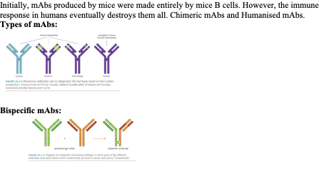
Initially, mAbs produced by mice were made entirely by mice B cells. However, the immune response in humans eventually destroys them all. Chimeric mAbs and Humanised mAbs.
Types of mAbs:
Bispecific mAbs:
Autoimmune Disease:
Failure of recognizing self, which leads to an adaptive immune response directed towards self-antigens. Autoimmune diseases tend to be inherited and more common in females.
- Cytotoxic T lymphocytes attack tissues directly.
- B lymphocytes secrete antibodies.
- Mast cells release histamines = inflammation in affected tissues.
Examples:
- Haemolytic anaemia – autoantibodies directed against red blood cells.
- Type 1 diabetes mellitus – T cells attack and destroy insulin-producing pancreas cells.
- Rheumatoid arthritis – antigen-antibody complexes are deposited in joint tissue e.g. knees and hands.
- Multiple sclerosis – myelin layer on neurons is attacked.
Treatments: Traditionally been managed with anti-inflammatory and immunosuppressive drugs, which reduce inflammation and suppress immune response. Autoimmune diseases are typically chronic and long-term use of such medicated has undesirable side effects. Drugs that suppress the immune system have the potential to result in cancer or infection.
Monoclonal Antibody Therapy:
mAb therapy complements traditional immunosuppressive medications used to treat autoimmune conditions.
Examples include:
- Cytokine inhibitors (Tumor necrosis factor alpha inhibitors and interleukin inhibitors) that reduce inflammatory response.
- B and T lymphocyte inhibitors that reduce immune cell activity and can be used to treat autoimmune diseases and some cancers.