The Skull
 Call Kai
 Learn
 Practice Test
 Spaced Repetition
 Match
 Flashcards
 Knowt Play
1/70

There's no tags or description
Looks like no tags are added yet.
71 Terms
Skull Structure
22 bones→all but 6 paired
structures between individual bones→immobile fibrous joints
Skull Function
protects
brain
receptor systems associated with special senses
nasal cavities→smell
orbits→vision
ears→hearing+balance
oral cavity→taste
Mandible
lower jaw
combines with cranium to form skull
articulation→neurocranium at temporomandibular joint
temporal bone
mandible

Cranium
top portion of skull
combines with mandible to form skull
divisions
neurocranium
-houses brainface
surrounds:
-orbital
-nasal
-oral cavities

Bones of Neurocranium List
frontal
parietal
occipital
temporal
sphenoid
ethmoid

Calvarium
superior portion of neurocranium→roof of skull
upper portions of
frontal bones
occipital bones
parietal bones

Floor of Neurocranium
made up of all bones in the neurocranium
occipital
temporal
sphenoid
frontal
ethmoid
houses neural cavity

Frontal Bone
forehead+roof of orbit
paired when born→fused to single bone around age 2
articulation→posteriorly with parietal bones (paired)

Parietal Bones
paired
posterior to frontal bone
anterior to occipital bone

Occipital Bone
single bone
posterior to parietal bones

Temporal Bone
paired
articulation
parietal
occipital
mandible
sphenoid
zygomatic

Sphenoid Bone
complex bone
looks small in lateral view→much larger in back wall of orbits
see better extent in inferior view

Face Bones List
maxilla
nasals
lacrimal
zygomatic
ethmoid
inferior nasal concha
vomer
palatine
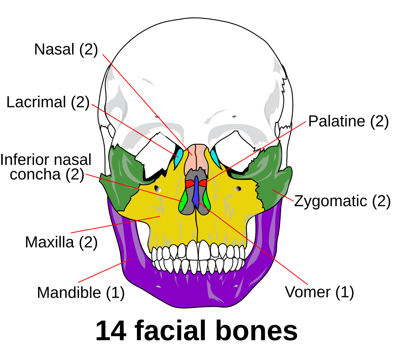
Maxilla
paired
forms
upper jaw
-houses teeth of upper jawportion of hard palate
floor of orbit
majority of face inferior to orbit

Nasal Bones
paired
small
form bridge of nose
articulation
maxilla
frontal

Lacrimal Bones
paired
articulate with maxilla
inside orbit

Zygomatic Bones
paired
forms portion of zygomatic arch
-with temporal bone
“cheek bone”
forms lateral wall of orbit

Ethmoid
see in nasal cavity→medial wall of orbits
deepest bone of skull
contains
superior nasal concha
middle nasal concha
perpendicular plate
-bony nasal septumcrista galli (interior)
cribiform plate (inferior)
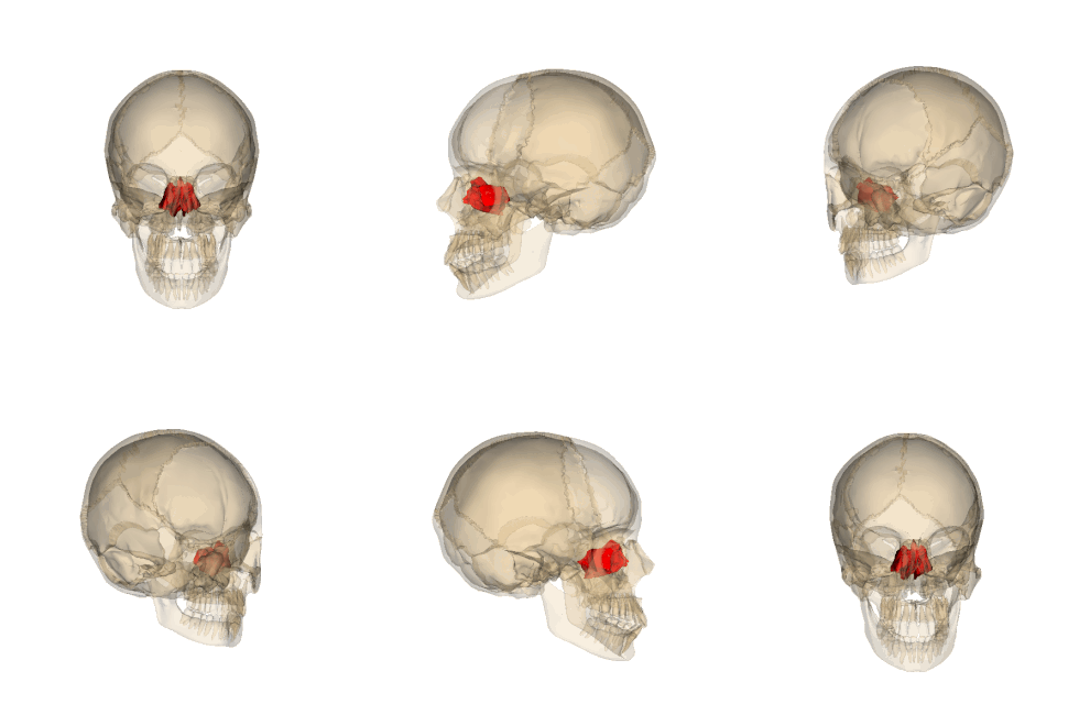
Inferior Nasal Concha
not part of ethmoid
in nasal cavity
paired
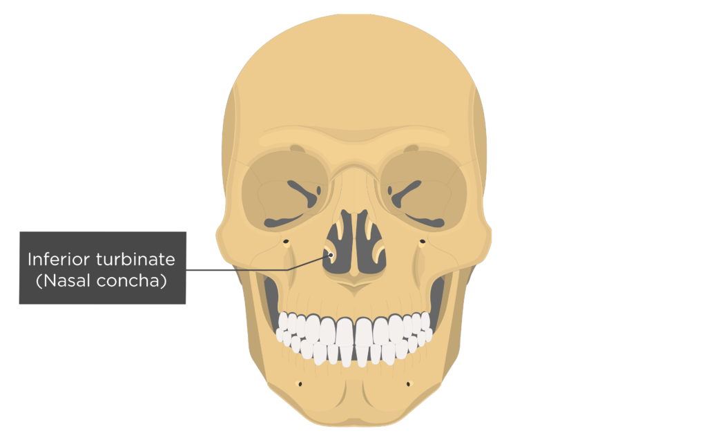
Vomer
pink
single bone
in nasal cavity
forms part of nasal septum with ethmoid
-bony septum

Palatine Bone
paired
posterior portion of hard palate

Mandibular Condyle of the Mandible
articulates with temporal bone→temporomandibular joint
insertion point→lateral pterygoid (both heads)

Coronoid Process of the Mandible
insertion point→temporalis

Body of the Mandible
houses roots of teeth
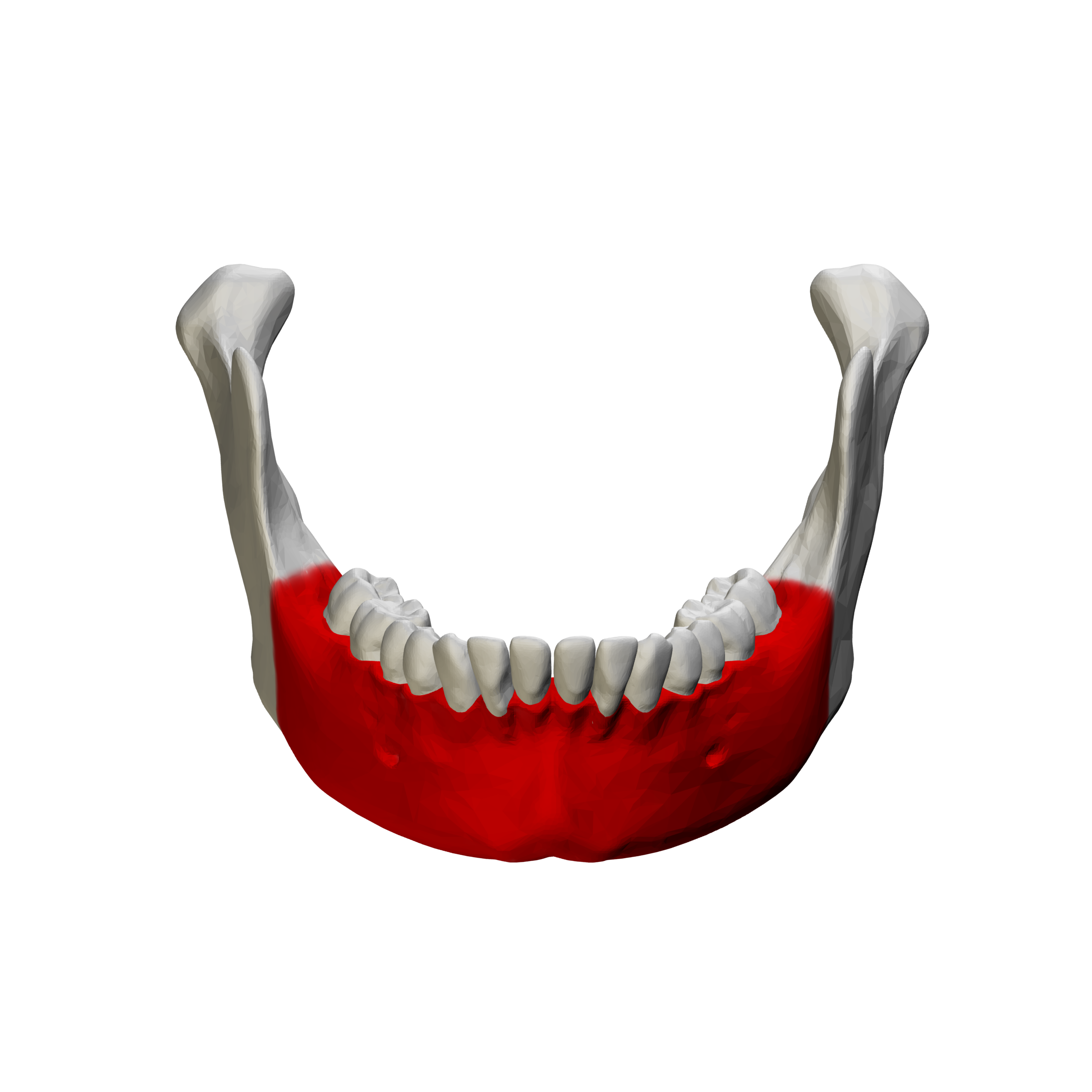
Ramus of the Mandible
vertical process
contains
condyle
coronoid process
insertion→masseter

Mental Protuberance of the Mandible
“mental→the thinker”
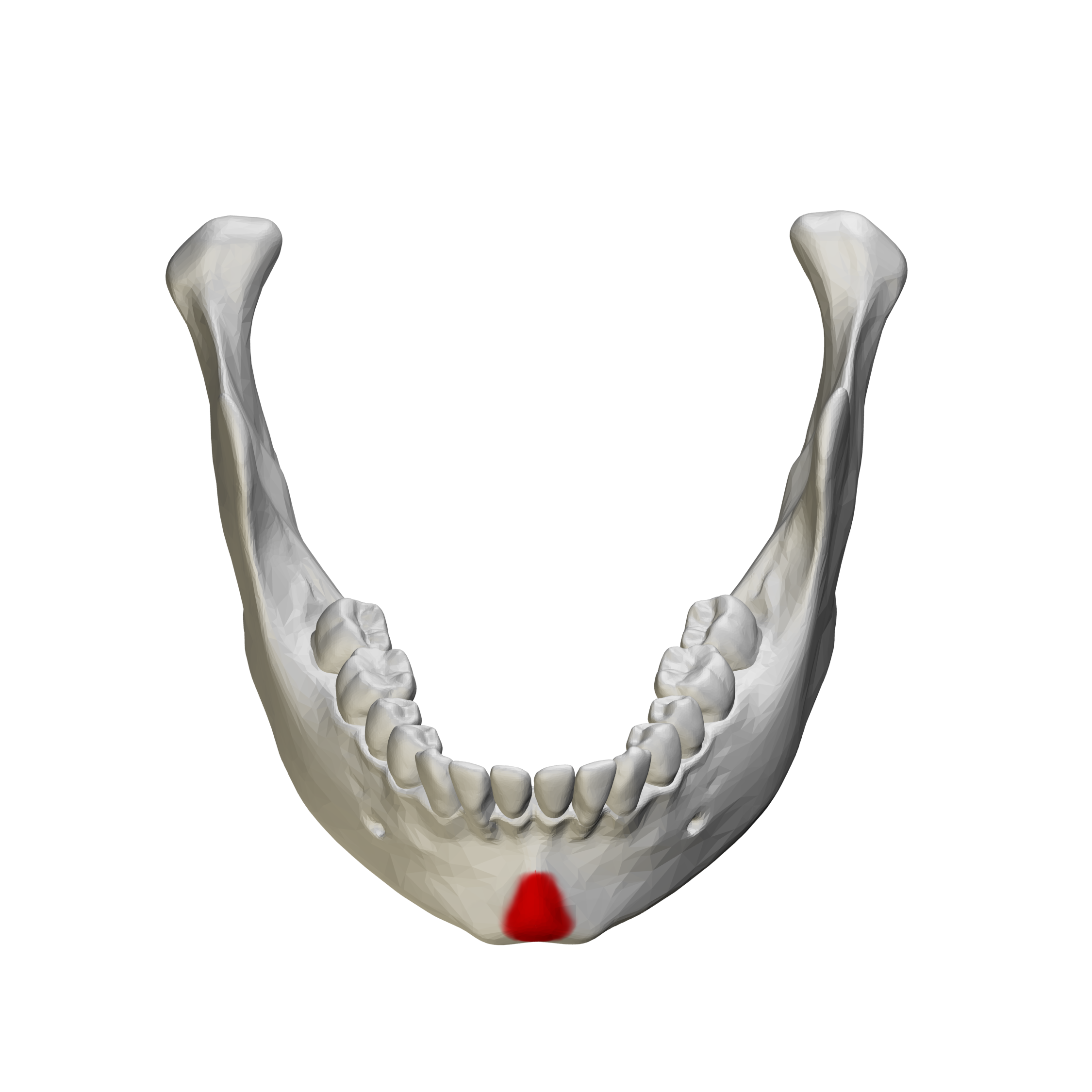
Mental Foramen of the Mandible
houses mental branch of mandibular N

Mandibular Foramen of the Mandible
on internal surface of ramus
houses inferior alveolar N

Foramen Magnum of Occipital Bone
transmits
spinal cord
blood vessels
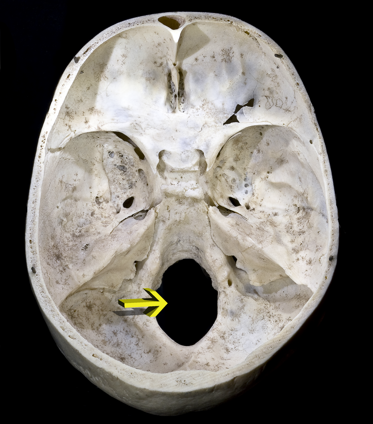
Occipital Condyles of Occipital Bone
articulate with atlas

Protuberance+Lines of Occipital Bone
protuberance→external occipital protuberance
nuchal lines
superior
inferior
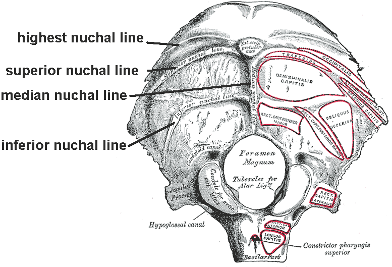
Hypoglossal Canal of Occipital Bone
transmits hypoglossal N

Zygomatic Process of Temporal Bone
forms portion of zygomatic arch
zygomatic process of temporal bone
temporal process of zygomatic bone

Glenoid Fossa of Temporal Bone
depression
articulation point with mandibular condyle→form temporomandibular joint

Mastoid Process of Temporal Bone
attachment
SCM
digastric
longissimus
splenius capitis

Styloid Process of Temporal Bone
origin
styloglossus
stylohyoid

Stylomastoid Foramen of Temporal Bone
transmits terminal branch of CN VII (facial N)

Carotid Foramen of Temporal Bone
runs through carotid canal
transmits internal carotid A

Foramen Lacerum of Temporal Bone
transmits greater petrosal N

Jugular Foramen of Temporal Bone
transmits
internal jugular V
glossopharyngeal N (CN IX)
vagus N (CN x)
accessory N (CN XI)

Internal Acoustic Meatus of Temporal Bone
transmits to middle+inner ear cavities
facial N (CN VII)
vestibulocochlear N (CN VIII)

External Acoustic Meatus of Temporal Bone
anterior to mastoid process
posterior to temporomandibular joint

Sella Turcica+Hypophyseal Fossa of Sphenoid Bone
sella turcica→ “saddle”
contains depression→hypophysial fossa
-houses pituitary gland
Greater+Lesser Wings of Sphenoid
located on posterior wall of orbit

Optic Canals of Sphenoid
transmit
optic N (CN II)
opthalmic A

Superior Orbital Fissure of Sphenoid
separates greater+lesser wings of sphenoid
transmits
oculomotor N (CN III)
trochlear N (CN IV)
abducens N (CN VI)
opthalmic division of trigeminal N

Foramen Rotundum of Sphenoid
transmits maxillary division of trigeminal N

Foramen Ovale of Sphenoid
transmits mandibular divison of trigeminal N

Foramen Spinosum of Sphenoid
transmits middle meningeal A

Medial Pterygoid Plate of Sphenoid
insertion→medial pterygoid
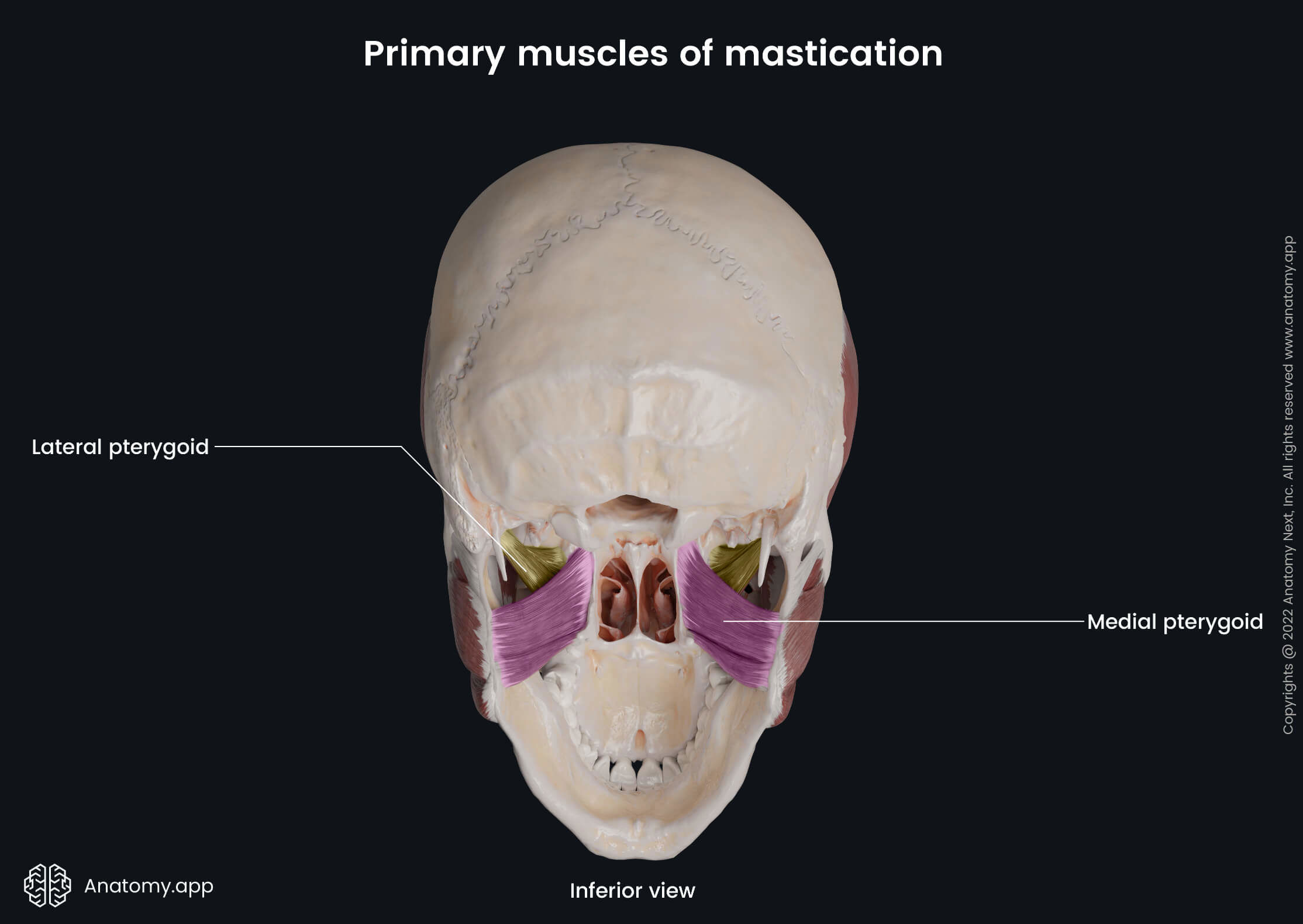
Lateral Pterygoid Plate of Sphenoid
origin
medial pterygoid
both heads of lateral pterygoid

Crista Galli of Ethmoid
raised crest
anchors fold of dura mater
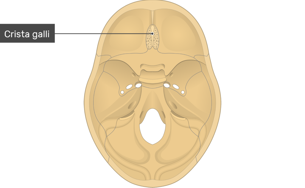
Cribiform Plate of Ethmoid
flanks crista galli
pierced with tiny holes→transmit olfactory N (CN I)

Superior+Middle Nasal Concha of Ethmoid
superior nasal concha→high up in nose→not visible
middle nasal concha

Perpendicular Plate of Ethmoid
articulates with vomer→forms bony nasal septum

Nasolacrimal Canal of Maxilla
transverses maxilla from the orbit→drains into nasal cavity
clinical→why nose runs when crying

Lacrimal Bone
smallest bone of the face
fragile
forms lacrimal groove with the maxilla
-portions of lacrimal apparatus

Sagittal Suture
between L+R parietal bones

Coronal Suture
between frontal and L+R parietal bones

Anterior Fontanelle
aka: bregmatic fontanelle
open area
between L+R frontals and parietal bones
-in fetuses+infants
covered with membrane→area of no bone
-”soft spot”
clinical→allows for flexible head to pass through birth canal
usually gone by age 2

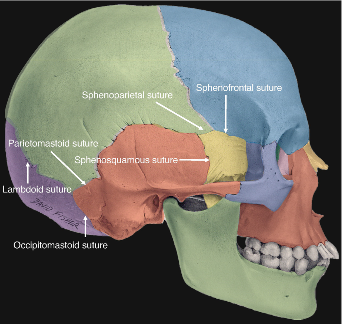
Lambdoid Suture
between L+R parietal bones+occipital bone

Posterior Fontanelle
in fetuses+infants
opening between L+R parietal bones and occipital bones
membrane over opening→area of no ossification

Occipitomastoid Suture
between L+R temporal bones and occipital bone
posterior to mastoid process

Squamous Suture
between parietal+temporal bones

Parietomastoid Suture
between parietal bones+portion of temporal bone
superior to mastoid process

Anterior Temporal Sutures
sphenoparietal
sphenosquamous
zygomaticofrontal
Facial Sutures
frontonasal
zygomaticomaxillary

Bony Orbit
composed of 7 bones
frontal
zygomatic
maxilla
lacrimal
ethmoid
sphenoid
palatine

Paranasal Sinuses
mucosa-lined air-filled spaces
lighten skull
enhance resonance of voice
warm+humidify air
found in
frontal
sphenoid
ethmoid
maxillary
Hyoid Bone (General)
bone in neck→level of C3
small+u-shaped
doesn’t articulate with other bones→suspended by ligaments+muscles below mandible
attachment point for 10 muscles

Hyoid Bone Structures
greater horn
lesser horn
body
