Extremity Venous Pathology
 Call Kai
 Learn
 Practice Test
 Spaced Repetition
 Match
 Flashcards
 Knowt Play
1/106

There's no tags or description
Looks like no tags are added yet.
107 Terms
A 56yr old female presents with mild calf pain that is caused by dorsiflexion of the foot. The symptoms began after she returned from vacation in France. Yesterday she noticed a sudden red color change and edema, localized to the lower leg. These clinical findings are most suggestive of:
A. cellulitis
B. pseudoaneurysm
C. chronic insufficiency
D. acute DVT
D. acute DVT
The patient noticed the problem after a vacation in France = long
airplane ride = venous stasis. The sudden onset of additional
symptoms indicates propagation of a clot in the calf.
Which of the following is a sign or symptom of chronic DVT?
A. Redness
B. Positive Homan's Sign
C. Warmth
D. Brawny Discoloration
D. Brawny Discoloration
Hyperpigmentation refers to brawny discoloration of the distal 1/3 of the calf. It is caused by cell debris and waste accumulating in the tissues outside the veins. Homan's sign refers to calf pain with quick dorsiflexion of the foot. This can be a sign of a DVT with the calf pain relating to the inability to pump out the blood when the muscle is quickly contracted. Swelling, warmth and redness are all related to venous stasis in the vessels below the area of DVT.

What is the most accurate sonographic indicator for the presence of DVT?
A. loss of augmentation response
B. flow abnormalities found with Color Doppler
C. flow abnormalities found with PW Doppler
D. lack of compressibility
D. lack of compressibility
Bilateral leg and foot edema are present. What is the most common cause for this?
A. Lymphedema
B. Chronic insufficiency in deep system
C. Chronic insufficiency in the superficial system
D. Congestive heart failure
D. Congestive heart failure
Cardiac and systemic problems lead to bilateral swelling of the lower extremities, including the feet. Lymphedema can cause bilateral leg and foot swelling, but it is usually unilateral and less common than CHF. Chronic venous disease does not normally cause swelling of the feet and is most commonly unilateral.
Which of the following causes of thrombosis is associated with anatomic variations of the thoracic outlet?
A. Paget - Schroetter Syndrome
B. indwelling catheters
C. radiation therapy treatments to the chest
D. shoulder surgery
A. Paget - Schroetter Syndrome
Paget - Schroetter Syndrome AKA spontaneous effort thrombosis:
Most common cause of axillary/subclavian DVT in the ambulatory patient population
Related to the presence of a cervical rib and thoracic outlet syndrome
Higher incidence in the dominant arm
Indwelling catheters are a common cause of upper extremity thrombosis in patients that are ill and require intravenous medication/treatment
Which of the following are common risk factors for varicose vein formation?
A. diabetes and smoking
B. post-phlebitic syndrome and high cholesterol
C. smoking and high cholesterol
D. pregnancy and standing for long periods of time
D. pregnancy and standing for long periods of time
Smoking, high cholesterol and diabetes are risk factors for atherosclerotic disease. Post-phlebitic syndrome, pregnancy, and standing for long periods of time.
If greater saphenous venous flow velocity is approximately 20cm/s and appears continuous, which other vessel should be evaluated for a cause for the flow abnormality?
A. short saphenous vein
B. profunda vein
C. internal iliac vein
D. femoral and popliteal vein
D. femoral and popliteal vein
Venous perforators normally send flow from the superficial system into the deep system. If the deep system of the thigh/popliteal fossa has occlusive thrombus, the majority of flow in the GSV will be rerouted to the groin to enter the common femoral vein. The increased flow will increase the velocity in the GSV and cause a continuous flow pattern.

A thrombus is identified in the soleal veins of the calf. The thrombus will propagate into what calf veins first?
A. posterior tibial and peroneal
B. peroneal, posterior tibial and anterior tibial
C. anterior tibial
D. great saphenous
A. posterior tibial and peroneal
When answering questions regarding clot propagation, consider what vessel is proximal to the clot. When answering questions regarding distal flow changes due to a proximal clot, consider what vessel is emptying blood into the vessel with the clot.
The soleal vessels drain into the peroneal and posterior tibial veins of the calf. A clot in the soleal veins will first extend into the posterior tibial and peroneal veins.
Which of the following is a cause for deep venous HTN?
A. Congestive heart failure
B. Klippel Trenaunay Webber Syndrome
C. Diffuse atherosclerotic disease
D. Aortic coarctation
A. Congestive heart failure
Venous HTN is described as increased venous volume that causes fluid to leak into interstitial spaces between tissues. Related to thrombus formation, venous insufficiency, CHF, pregnancy
A 65yr old female presents with left leg swelling for over one year. Mild hyperpigmentation is present in the distal calf and ankle area. These clinical findings are most suggestive of:
A. Chronic Insufficiency
B. Acute DVT
C. Superficial Phlebitis
D. Cellulitis
A. Chronic Insufficiency
Chronic swelling and brown discoloration are characteristics of venous stasis. This is most commonly associated with chronic insufficiency. Acute DVT would result in acute swelling with a red discoloration.
Which of the following describes symptoms of a patient with lymphedema?
A. Unilateral non-pitting edema that is unaffected by leg elevation
B. Warm, red calf with significant swelling
C. Bilateral calf swelling with severe pitting edema
D. Unilateral left leg swelling that is relieved by elevating the legs for 30 minutes
A. Unilateral non-pitting edema that is unaffected by leg elevation
Lymphedema does not normally cause skin discoloration, while acute DVT can be associated with warmth and redness. Lymphedema causes non-pitting edema, DVT causes pitting edema. Elevating the leg will have no effect on the swelling, but can reduce swelling associated with DVT. Lymphedema is typically unilateral. DVT formation can affect one or both legs.

The most common location of valvular insufficiency is:
A. femoral and profunda femoral vein junction
B. short saphenous vein
C. calf vein confluence
D. great saphenous and common femoral vein junction
D. great saphenous and common femoral vein junction
The most common location of valvular insufficiency is at the SFJ (junction of great saphenous and common femoral vein junction).
Which of the following causes flow reversal in venous perforators?
A. superficial thrombosis
B. deep vein thrombosis
C. compression stockings
D. combination of superficial and deep thrombosis
B. deep vein thrombosis
DVT in the deep system forces any remaining flow into areas of lower pressure, the perforators. Flow exits the deep system through the perforators to try to "escape" into the patent superficial system.

The images of the neck demonstrate which of the following?
A. Acute DVT of the SVC
B. Acute DVT of the IJV
C. Chronic DVT of the lJV
D. Large neck mass anterior to CCA
B. Acute DVT of the IJV
Note the engorged vein with mildly hyperechoic debris within the lumen. The acute thrombosis is causing complete obstruction of the IV. Chronic thrombosis would not present as an engorged vein and the thrombus would be excessively echogenic with potential recanalization.
NORMAL IJV - FLATTENED, FLEXIBLE

Which of the following carries an increased risk of developing varicose veins?
A. Wearing compression stockings
B. Sitting with legs elevated for long periods of time
C. Oral contraceptive use
D. Wearing flat shoes instead of high heels
C. Oral contraceptive use
Oral contraceptive use will increase the risk of developing varicose veins. Family history, pregnancy and obesity also increase the risk.
The other three choices would reduce the risk of developing varicose veins.

The attached picture is most suggestive of what acute vascular disorder?
A. Peripheral arterial disease
B. Congestive heart failure
C. Unilateral DVT
D. Bilateral DVT
C. Unilateral DVT
Significant edema is evident in the subcutaneous tissues. This is most commonly associated with Acute DVT. Peripheral arterial disease is a chronic disorder. CHF or bilateral DVT would cause bilateral swelling.

The image is a transverse view of the distal popliteal fossa. Which of the following would not be a recommended treatment?
A. No medication, just a warm compress
B. Lovenox injection
C. Coumadin therapy
D. IV Heparin
A. No medication, just a warm compress
DVT and superficial thrombosis are present. DVT is always treated aggressively with protocols using IV Heparin, Lovenox injection and Coumadin/Warfarin therapy. If only superficial thrombosis of the small saphenous vein is present, a warm compress and an aspirin would usually be sufficient treatment.

The image attached demonstrates which of the following?
A. Greenfield filter in a normal vein
B. Chronic venous thrombosis with recanalization
C. Chronic thrombosis with Greenfield filter
D. Venous dissection with multiple intimal flaps
B. Chronic venous thrombosis with recanalization
Note the fibrous strands of the thrombus as it becomes denser and atrophies within the lumen. The anechoic areas are thrombus free areas which allow some flow through the vessel.
Which of the following Doppler findings would indicate significant venous hypertension?
A. Absence of flow in the deep veins on deep expiration
B. Increased systolic velocities with decreased diastolic velocities
C. Triphasic flow patterns in the lower extremity deep veins
D. Phasic flow patterns in the lower extremity deep veins
C. Triphasic flow patterns in the lower extremity deep veins
Significant pulsatility in the deep veins of the lower extremity indicates increased venous pressures (venous HTN). CHF, electrolyte imbalance and kidney dysfunction can lead to venous HTN. If the flow pattern normally seen in the VC and hepatic veins is seen in the legs, venous HTN is present.
Triphasic flow means the flow has 3 phases of movement in a single cardiac cycle. (The Doppler tracing crosses the baseline 3 times in a cardiac cycle.) The term is not limited to describing arterial flow.
Continuous flow patterns in the veins can be a sign of chronic obstruction proximally.

Which of the following is an expected clinical finding in a patient with a venous stasis ulcer?
A. dry and crusty
B. deep crater in the skin
C. severe pain
D. located at the medial malleolus
D. located at the medial malleolus
Venous ulcers are usually mildly painful, oozing, shallow ulcers located at the medial malleolus.
Arterial ulcers are usually severely painful, dry and crusty, deep ulcers located at the toes or heel.

A Greenfield filter is used to prevent which of the following?
A. Cardiac tamponade
B. Blue toe syndrome
C. DVT
D. Pulmonary embolism
D. Pulmonary embolism
A greenfield filter is inserted into the abdominal portion of the IVC (below the level of the renal veins) to catch embolic material before it reaches the heart and lungs. It does not prevent the formation of DVT, but it does protect the patient from complications related to embolism from a DVT.

A patient presents with bilateral distention of the internal jugular veins and arm swelling. The lower extremities are normal on physical examination.
These findings are most suggestive of:
A. Congestive heart failure
B. Interrupted IVC
C. Subclavian steal syndrome
D. SVC Syndrome
D. SVC Syndrome
SVC syndrome refers to the progressive obstruction of the SVC which causes venous dilatation of the upper extremities and neck. Doppler evaluation of the bilateral internal jugular veins will demonstrate continuous flow patterns. Respiratory phasicity and cardiac pulsatility will be lost in the bilateral IJVs due to the SVC obstruction.
Lymphedema is commonly related to:
A. a history of radiation therapy or cancer
B. Leriche syndrome
C. peripheral arterial disease
D. a pulmonary embolism
A. a history of radiation therapy or cancer
Lymphedema occurs due to an obstructed lymphatic system. It can be caused by many things, but is commonly seen with infection, injury, trauma, or cancer that affects the lymphatic system. Radiation therapy or removal of lymph nodes related to cancer can also cause lymphedema.
How is Heparin administered to a patient with DVT?
A. orally
B. subcutaneous injection
C. intra-arterial
D. intravenous
D. intravenous
DVT treatment can include 5-10 day dose of intravenous Heparin to prevent clot extension, but it does not destroy current thrombus.
Which of the following lower extremity vessels would most likely be occluded in a patient with Phlegmasia Alba Dolens?
A. femoral vein, deep calf veins, popliteal vein
B. common femoral artery
C. deep veins, superficial veins and common femoral artery
D. saphenous veins only
A. femoral vein, deep calf veins, popliteal vein
Phlegmasia Alba Dolens refers to a condition where all deep venous vessels are occluded in the extremity causing reduced arterial inflow. The superficial veins are usually patent. The leg has a milky white appearance due to severe outflow obstruction and markedly reducing arterial inflow. Leads to ischemia in entire extremity with severe pain.
A patient presents for a venous insufficiency exam, but is unable to stand for the exam. How will this affect your exam?
A. The segmental pressures will be falsely elevated.
B. The PPG tracings will be increased in amplitude.
C. Venous reflux may not be demonstrated in veins where it is actually present.
D. No change as long as they can move their feet to perform the heel raises.
C. Venous reflux may not be demonstrated in veins where it is actually present.
Standing increases hydrostatic pressure to better demonstrate the reflux. If the patient is supine, a false negative exam could be obtained because the augmentation response will be diminished without hydrostatic pressure. A seated patient will have bent knees and hip joints which can inhibit/reduce the flow response to augmentation.
The Valsalva maneuver is most useful when trying to assess which of the following?
A. median arcuate syndrome
B. Venous insufficiency
C. ECA/ICA differentiation
D. DVT
B. Venous insufficiency
In the lower extremities, the Valsalva maneuver increases intra-abdominal pressure and inhibits normal venous return to the heart. If the venous valves are incompetent, flow reversal will be seen while the maneuver is applied.
_______ cause primary varicose veins, while ________ cause secondary varicose veins.
A. incompetent valves, incompetent perforators
B. elevated deep venous pressures, elevated arterial pressures
C. incompetent valves, elevated deep venous pressures
D. elevated deep venous pressures, incompetent valves
C. incompetent valves, elevated deep venous pressures
Incompetent valves cause primary varicose veins, while elevated deep venous pressures in a diseased deep system (hx of DVT or abnormal valves) cause secondary varicose veins. Primary varicose veins are much more common than secondary.
Which of the following is the most common cause of venous stasis ulcer formation on the distal medial calf?
A. insufficiency in the paratibial perforators and the posterior arch vein
B. insufficiency in the posterior tibial perforators and the posterior arch vein
C. insufficiency in the distal thigh perforators and the great saphenous vein
D. insufficiency in the proximal thigh perforators and the great saphenous vein
B. insufficiency in the posterior tibial perforators and the posterior arch vein
Insufficiency in the posterior tibial perforators and the posterior arch vein most commonly leads to venous stasis ulcer formation on the distal medial calf.
A patient presents with a wet ulcer located on the medial calf just above the ankle. What vessel should be evaluated first as the most likely primary cause of the ulcer?
A. posterior accessory saphenous vein
B. posterior tibial vein
C. small saphenous vein
D. posterior tibial artery
A. posterior accessory saphenous vein
Insufficiency of the posterior arch vein (posterior accessory saphenous vein) and perforators is most commonly related to venous ulcer formation. The PTV are part of the deep venous system and do not typically demonstrate reflux. The SSV courses up the back of the calf, not near the ankle. A wet, oozing ulcer near the medial malleolus indicates a venous stasis ulcer.
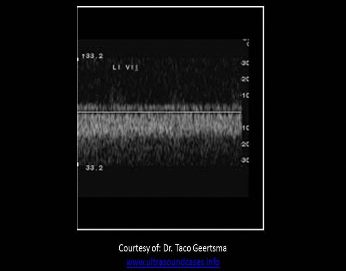
You perform a bilateral venous Doppler due to bilateral swelling and pain in the legs. If both external iliac veins look like the tracing on the image, which of the following should be reported on your technologist worksheet?
A. Congestive heart failure is likely
B. IVC obstruction is suspected
C. May-Thurner syndrome is likely
D. Severe tricuspid regurgitation is suspected
B. IVC obstruction is suspected
Bilateral continuous waveforms from the iliac veins indicate a proximal obstruction that affects them both. An IVC obstruction is suspected. CHF and tricuspid regurgitation cause cardiac pulsatility in both extremities. May Thurner syndrome leads to thrombosis of the left common iliac vein. Only the left leg would demonstrate continuous flow in the external iliac vein.
A clot in the gastrocnemius veins will first propagate into what vessel(s)?
A. anterior tibial veins
B. popliteal vein
C. peroneal veins
D. deep femoral vein
B. popliteal vein
When answering questions regarding clot propagation, consider what vessel is proximal to the clot. When answering questions regarding distal flow changes due to a proximal clot, consider what vessel is emptying blood into the vessel with the clot.
The gastrocnemius veins empty their blood into the popliteal vein in most patients. Gastrocnemius thrombosis will first extend into popliteal vein.
Which of the following patients has the lowest risk of upper extremity venous thrombosis?
A. hemophilia
B. recent shoulder surgery
C. polycythemia vera
D. central venous catheter for chemotherapy
A. hemophilia
Polycythemia vera (Hypercoagulability), central venous catheter (trauma), and recent surgery (trauma/stasis) will increase the risk of thrombus formation.
Hemophilia refers to a condition causing abnormally thin blood and would be a contraindication to clot formation.
A patient presents with gangrene of the foot and toes. The most likely cause of these symptoms is:
A. coarctation of the aorta
B. a 5cm aortic aneurysm with mural thrombus
C. chronic occlusion of the popliteal artery
D. extensive deep and superficial vein thrombosis
D. extensive deep and superficial vein thrombosis
Venous gangrene usually involves the foot and all toes, while arterial gangrene usually involves one or two toes.
During a color evaluation of the common femoral vein, the patient is asked to perform the Valsalva maneuver. The blue color in the CFV changes to red for approximately 3 seconds before returning to blue. What do these color Doppler findings indicate?
A. The CFV has minimal venous insufficiency.
B. Superficial venous insufficiency is present.
C. There is most likely an occlusive DVT in the superficial femoral vein causing the venous turbulence proximally.
D. A significant level of insufficiency is present and should be further evaluated with PW or CW Doppler techniques.
D. A significant level of insufficiency is present and should be further evaluated with PW or CW Doppler techniques.
0.5 seconds or less of flow reversal is considered normal in the superficial veins.
1 sec or less of flow reversal is considered normal in the deep veins.
3 seconds or greater of flow reversal indicates significant venous reflux.
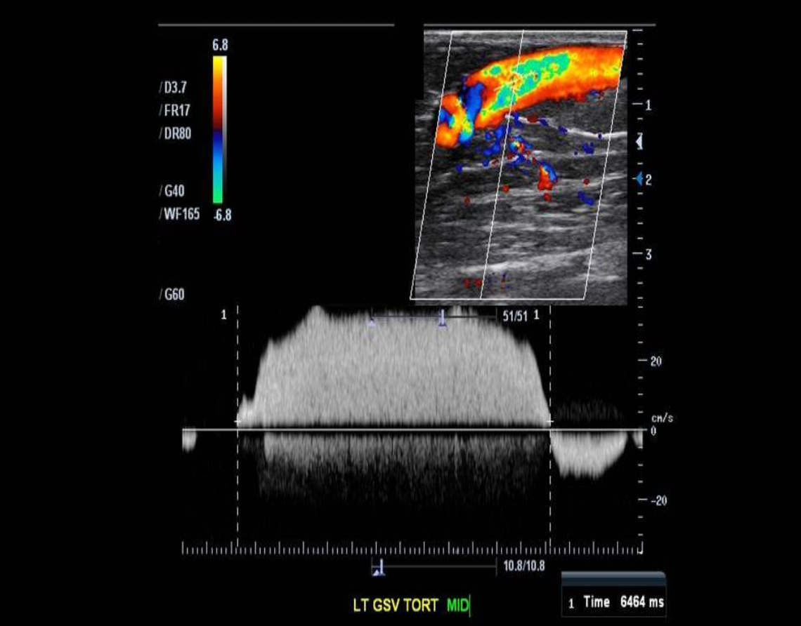
Which of the following would be contraindicated in this patient?
A. thermal ablation
B. compression stockings
C. Coumadin therapy
D. daily exercise
C. Coumadin therapy
Venous reflux is usually treated with compression stockings, thermal or laser ablation and lifestyle changes that increase the venous flow to the heart. (exercise, elevating legs when sitting, avoid high heel shoes) Coumadin therapy would thin the blood causing further dilatation of the damaged veins.
Post-phlebitic Syndrome:
A. will usually resolve within 2 weeks after treatment for DVT
B. refers to black discoloration of the toes caused by embolism from a AAA
C. refers to the hair loss and thickened toenails that occur with chronic DVT
D. refers to valve damage in the veins after thrombus formation
D. refers to valve damage in the veins after thrombus formation
Post-phlebitic Syndrome refers to valve damage in the veins after thrombus formation. Varicosities form and insufficiency is present.
A venous insufficiency exam is performed for a patient with multiple varicosities. The deep system is found to be competent, but there is insufficiency present in the great saphenous vein. Which of the following statements regarding the findings is true?
A. Venous insufficiency is normal in the superficial veins when varicosities are present.
B. The multiple varicosities are primary varicosities.
C. The multiple varicosities are primary and secondary varicosities
D. The multiple varicosities are secondary varicosities.
B. The multiple varicosities are primary varicosities.
Varicosities with superficial venous insufficiency related to deep venous system disease = secondary varicosities
Varicosities with incompetent valves causing superficial venous insufficiency and normal deep system = primary varicosities.
Which of the following is an expected complication caused by a Baker cyst?
A. Atherosclerosis formation in the popliteal artery
B. Thrombosis of the small saphenous vein due to valvular insufficiency
C. Aneurysm formation in the popliteal artery
D. Thrombosis of the popliteal vein due to extrinsic compression
D. Thrombosis of the popliteal vein due to extrinsic compression
A Baker cyst can cause pain and swelling in the popliteal fossa. The extrinsic compression of the popliteal vein by the cyst can lead to a continuous flow pattern, blood stasis and DVT formation. The arterial walls are muscular and can prevent the effects of extrinsic compression on the popliteal artery.
The most significant complication seen with DVT is _______
A. Varicose veins
B. Cardiac Tamponade
C. Incompetent valves
D. Pulmonary embolism
D. Pulmonary embolism
Varicose vein formation is the most common complication of DVT and pulmonary embolism is the most significant complication of DVT. A pulmonary embolism occurs when a piece of thrombus breaks off and travels to the lungs. This occludes the bronchial artery and leads to the inability of the lungs to replace the oxygen in the blood. 15% of all cases of sudden death are PE related.

A patient presents for an exam due to a history of Lemierre Syndrome.
What vessels should be evaluated for associated findings?
A. Bilateral radial and ulnar veins
B. Bilateral internal jugular veins
C. Superficial veins of the lower extremity
D. Deep veins of the lower extremity
B. Bilateral internal jugular veins
Lemierre Syndrome refers to thrombophlebitis of the jugular veins with oropharyngeal infection, such as pharyngitis/tonsillitis.
Duplex evaluation can demonstrate thrombus within the jugular vein or other neck or facial veins.
A patient presents with a script for a "vascular ultrasound" due to a history of Paget Schroetter syndrome. What type of exam should be performed for this patient?
A. lower extremity venous Doppler exam
B. upper extremity venous Doppler exam
C. arterial PPG of the toes
D. segmental pressures on the lower extremities
B. upper extremity venous Doppler exam
Paget Schroetter syndrome refers to the formation of upper extremity DVT due to excessive motion. It is usually associated with thoracic outlet syndrome.
Which of the following is a common infection that causes skin discoloration in patients with venous insufficiency?
A. staphylococcus epidermidis
B. staphylococcus aureus
C. arteritis
D. stasis dermatitis
D. stasis dermatitis
Stasis dermatitis is a common inflammatory skin disease. It is usually the earliest cutaneous sequela of chronic venous insufficiency with venous hypertension and causes discoloration of the ankle and calf.
Which of the following describes abnormal lower extremity venous flow response to distal augmentation in a superficial vein?
A. Augmentation causes increased flow toward heart followed by flow reversal >0.5sec toward the feet
B. Augmentation causes increased flow toward feet followed by at least 3 seconds of flow reversal toward the feet
C. Augmentation causes increased flow toward heart followed by flow normalization
D. Augmentation causes increased flow toward feet followed by flow reversal >0.5 sec toward the heart
A. Augmentation causes increased flow toward heart followed by flow reversal >0.5sec toward the feet
<0.5 sec flow reversal with distal augmentation is considered within normal limits. Reflux is diagnosed when flow reversal exceeds 0.5sec.
>3 sec flow reversal is considered severe venous reflux. The best answer is Augmentation causes increased flow toward heart followed by flow reversal >0.5sec toward the feet.

Patients with lower extremity venous insufficiency may describe pain
A. that is relieved by placing legs in a dependent position
B. that is relieved by elevating the legs
C. that is relieved by walking
D. that is relieved by lying down
B. that is relieved by elevating the legs
The dependent position helps blood to reach the lower legs more quickly. Walking and elevating the legs would increase the flow toward the heart and reduce blood pooling in the calves. With venous insufficiency, walking will increase the arterial flow into an already inefficient venous system. This can lead to increased pooling in the lower leg with swelling and pain.
Most lower extremity thrombosis begins in the _______
A. femoral vein in the adductor canal
B. soleal sinuses
C. tibial veins
D. peroneal vein
B. soleal sinuses
Most lower extremity thrombosis begins in the soleal sinuses and propagates into the PTA and/or peroneal veins.
________ is a nonspecific sign of DVT. It is defined as pain
experienced upon quick dorsiflexion of the foot.
A. Allen Sign
B. Asher Sign
C. Henderson Sign
D. Homan Sign
D. Homan Sign
Homan sign is a nonspecific sign of DVT. It is defined as pain experienced upon quick dorsiflexion of the foot. Asher sign refers to enlargement of the spleen that extends across the midline, anterior to the abdominal aorta. The Allen Test is used to determine ulnar artery patency related to graft availability for removing the radial artery.
The mid left femoral vein appears to be occluded by hypoechoic, non-compressible thrombus. The popliteal vein is completely compressible.
Which of the following Doppler waveform characteristics is expected from the popliteal vein?
A. normal response to distal augmentation of calf muscle
B. loss of respiratory phasicity
C. decreased response to distal augmentation of calf muscle
D. increased pulsatility
B. loss of respiratory phasicity
The flow response to the distal augmentation would most likely be decreased but NEVER augment a patient's calf when an acute DVT is diagnosed. The best answer is loss of respiratory phasicity because continuous flow is expected with the proximal obstruction.
_______ is the "gold standard" exam technique for diagnosis of
venous disease in the lower extremities.
A. Arteriography
B. Venography
C. Doppler Ultrasound
D. MR Angiography (MRA)
C. Doppler Ultrasound
Doppler ultrasound has surpassed venography as the gold standard for evaluation of the veins in the lower extremities.
Sclerotherapy:
A. refers to the treatment of veins using a radiofrequency catheter
B. involves the injection of saline into superficial varicosities
C. is used to treat incompetent deep veins
D. refers to the treatment of deep venous reflux with compression stockings
B. involves the injection of saline into superficial varicosities
Sclerotherapy refers to the injection of saline or other sclerosing agent that damages the vein and causes fibrosis to occur. Ablation can be used to avoid surgical intervention in some cases, catheter based radiofrequency application can occlude the vessels leading to overall atrophy of the vessels.
Which of the following portions of the leg would be swollen with chronic insufficiency?
A. calf only
B. ankle and calf, but not the foot
C. foot and ankle, but not the calf
D. calf, foot, ankle
B. ankle and calf, but not the foot
Swelling of the feet normally does not occur with venous disease
(systemic or cardiac disease, yes)
Venous perforators with a diameter of ______ or greater will usually demonstrate reflux
A. 6mm
B. 4mm
C. 2mm
D. 2cm
B. 4mm
Normal perforator diameter is 2mm. Venous perforators with a diameter of 4mm or greater will usually demonstrate reflux.
Which of the following is a long term treatment for DVT?
A. Lovenox
B. Heparin
C. venous ligation
D. Warfarin
D. Warfarin
Treatment for Acute DVT:
Subcutaneous shot of Lovenox immediately
Heparin administered for 5-10 days to stop the progression of the clot, but does not have clot lysing properties to decrease the clot that has already formed
After Heparin dosage completed and recanalization has occurred, Warfarin is administered to prevent future clot formation
First DVT occurrence - Warfarin prescribed for 3 months
Recurrent DVT - Warfarin prescribed for 6+ months
If a patient is diagnosed with a pulmonary embolism, what should be the next step in their patient care?
A. IV Heparin
B. lower extremity venous Doppler exam
C. pulmonary angiography
D. V/Q scan
A. IV Heparin
If the patient has been diagnosed with a PE, additional exams may or may not be required to locate the source of the embolism. Immediate IV Heparin therapy should be administered
A patient has a prior history of popliteal DVT and currently complains of the ncreasing appearance of small varicosities on the calf. Which statement below best describes his condition?
A. There is most likely a Baker's cyst in the popliteal fossa causing extrinsic compression.
B. The patient is suffering from secondary varicose vein formation.
C. There is most likely a clot in the gastrocnemius veins of the calf.
D. The patient most likely has a recurrent acute DVT.
B. The patient is suffering from secondary varicose vein formation.
Primary varicosities form due to abnormal valves in the superficial
venous system.
Secondary varicosities form if there is an associated problem in the deep system, such as previous DVT.

The image attached demonstrates which of the following?
A. Reflux time, post augmentation
B. Velocity of venous reflux
C. Normal augmentation response time
D. Time of venous augmentation
A. Reflux time, post augmentation
The flow reversal is quantified by the time reflux is present following augmentation. <0.5 sec of reflux is normal in the superficial system with >3 sec of reflux indicates severe insufficiency.

A patient presents with persistent calf pain after a recent biking accident and spending a few days on bed rest. After visual inspection of the right leg, what type of vascular exam should be performed?
A. Lower extremity venous Doppler
B. Segmental pressure exam
C. Lower extremity arterial Doppler
D. Soft tissue exam to rule out hematoma formation
A. Lower extremity venous Doppler
The significant difference in size demonstrates fluid accumulation in the swollen leg. The recent trauma and few days of stasis while on bed rest most likely caused a DVT to form in the leg.

What is demonstrated by the images from a lower extremity venous Doppler exam?
A. AV fistula
B. pseudoaneurysm
C. incorrect gain settings on color Doppler, normal spectral waveform from the CFV
D. Klippel-Trenaunay Webber Syndrome
A. AV fistula
Note the pulsatility in the venous waveform which is consistent with the presence of arterial inflow proximally in the vessel. The mosaic color pattern indicates turbulence at the connection with the artery.
Which of the following is a sonographic finding of acute DVT?
A. Partial compressibility of the vein
B. Valvular insufficiency
C. Flattened, collapsed vessel with increased echogenicity of the wall
D. Engorged vein with increased echogenicity in the lumen
D. Engorged vein with increased echogenicity in the lumen
Chronic thrombus will demonstrate as echogenic material lining a partially collapsed vein. Acute thrombus causes the vein to grow in size and appear engorged with blood/debris.
NORMAL IJV - Flattened, flexible shape


The complications demonstrated on the image are most suggestive of what vascular abnormality?
A. Acute superficial vein thrombosis
B. Peripheral arterial disease
C. Acute deep vein thrombosis
D. Infrarenal aortic aneurysm
C. Acute deep vein thrombosis
Note the hypoechoic and anechoic areas of tissue due to increased fluid accumulation in the tissues. Peripheral edema is a sign of DVT. The venous outflow is occluded and fluid in the veins is "pushed" into the surrounding tissues. Acute superficial thrombosis would not typically lead to peripheral edema if the deep system is unaffected. If the superficial vein is chronically occluded, mild edema may develop. Peripheral arterial disease causes dry skin and toenails due to a lack of blood supply.
Which of the following is related to chronic DVT?
A. Dry ulceration
B. Swelling and erythema
C. Engorged, non-compressible vein
D. Wet ulceration
D. Wet ulceration
Ulcerations take time to form. They are a sign of chronic venous disease. Venous ulcers are wet/oozy and mildly painful. Arterial ulcers more painful and dry.
A patient presents 3 days after left hip surgery for a DVT evaluation. There is severe edema from the hip to the ankle and the leg has a mild cyanotic appearance. You identify acute thrombus from the common femoral vein through the calf veins. The great saphenous vein is also occluded by thrombus. What is the diagnosis for this condition?
A. Phlegmasia Alba Dolens
B. Budd Chiari syndrome
C. Phlegmasia Cerulea Dolens
D. Acute arterial occlusion
C. Phlegmasia Cerulea Dolens
Phlegmasia Cerulea Dolens refers to extensive DVT occluding deep system and superficial system. Arterial inflow is significantly affected. Severe limb edema and mottled skin are noted with cyanosis due to high concentration of deoxygenated blood. Tip for remembering the difference between Cerulea Dolens and Alba Dolens, "C"erulea causes "C"yanosis of the leg, while Alba Dolens results in pallor.

A patient presents for a lower extremity venous Doppler due to leg pain in the groin and upper thigh after a recent car accident. After viewing these findings in the common femoral vein, what should you do next on this patient's exam?
A. evaluate the common femoral artery for signs of an AV fistula
B. evaluate the common femoral artery for a pseudoaneurysm
C. perform the color and Doppler evaluation of the superficial femoral vein to rule out thrombus
D. perform a blood pressure on the thigh
A. evaluate the common femoral artery for signs of an AV fistula
Note the pulsatility in the venous waveform which is consistent with the presence of arterial inflow proximally in the vessel. The mosaic color pattern indicates turbulence at the connection with the artery.
Localized redness and warmth of the distal calf is most commonly associated with:
A. Acute arterial occlusion of the popliteal artery
B. Anterior compartment syndrome
C. Cellulitis
D. Acute DVT formation in the popliteal vein
C. Cellulitis
Redness and warmth are most commonly seen with cellulitis but can be a symptom of acute DVT.
A patient presents for an upper extremity venous Doppler evaluation due to bilateral arm and face swelling. There are also varicosities visible on the upper chest wall. These symptoms are most consistent with:
A. occlusive thrombus in the internal jugular vein
B. congestive heart failure and venous stasis
C. occlusive thrombus in the superior vena cava
D. Nutcracker syndrome
C. occlusive thrombus in the superior vena cava
Chest wall varices, facial and bilateral arm swelling are indicative of SVC thrombosis. IJV thrombosis can be related to facial swelling but the arm would not be affected. CHF usually causes swelling in the legs.

What vessel is demonstrated as thrombosed on the image?
A. basilic vein
B. short saphenous vein
C. cephalic vein
D. great saphenous vein
D. great saphenous vein
Note the body marker indicator on the image along with the location of the vein just beneath the subcutaneous fat and anterior to the muscular fascia. The lack of a corresponding artery also indicates a superficial vein.
Which of the following conditions is usually related to cardiac pulsatility in the leg veins?
A. DVT
B. Bradycardia
C. Congestive heart failure
D. Tachycardia
C. Congestive heart failure
As the heart moves in the chest it causes the ripple effect in the venous system. Normally, the upper IVC and Hepatic veins demonstrate this ripple effect as flow reversal during cardiac systole. The "ripples" of cardiac pressure/motion usually dissipate as they reach the mid abdomen area. In patients with CHF, the venous blood volume is increased throughout the system. The cardiac pulsatility will extend further into the lower extremities due to the increased blood volume.

Which of the following will be administered to a patient with extensive DVT to cause lysis of the clot?
A. Heparin or Coumadin
B. Heparin
C. Coumadin
D. Streptokinase
D. Streptokinase
Heparin, Coumadin and Warfarin are medications that reduce the viscosity of blood to prevent propagation of the clot but they have no clot lysing properties. Streptokinase and Urokinase are used to dissolve the thrombus in cases with significant thrombosis of the venous system.
What is the most common lower extremity venous disorder?
A. May Thurner syndrome
B. superficial thrombophlebitis
C. venous insufficiency
D. DVT
C. venous insufficiency
Venous insufficiency is the most common lower extremity venous disorder.
A patient presents with intermittent left lower leg swelling for several months. The popliteal vein demonstrates a collapsed appearance with echogenic, thickened walls. These findings are most consistent with:
A. acute femoral DVT
B. vasculitis of the popliteal vein
C. chronic popliteal DVT
D. proximal venous stenosis
C. chronic popliteal DVT
Chronic DVT will become echogenic as it becomes more fibrous while attached to the vein wall. The fibrous material limits the pliability of the walls leading to a semi-collapsed appearance. Acute femoral DVT would cause a dilated popliteal vein.
Which of the following correctly describes DVT in the lower extremity?
A. Recanalization of a thrombus usually occurs around 5 days after formation occurred.
B. Chronic thrombus will demonstrate complete coaptation of the venous walls upon compression.
C. Acute thrombus will be completely rigid upon compression.
D. Heparin is used to treat a DVT due to its clot lysing properties.
A. Recanalization of a thrombus usually occurs around 5 days after formation occurred.
Recanalization of a thrombus usually occurs around 5 days after formation occurred. Heparin is used to treat a DVT because it thins the blood and prevents further propagation of the clot. It does not have clot lysing properties. Acute thrombus will be somewhat pliable upon compression. Chronic thrombus will be non-pliable upon compression.
When performing an upper extremity venous Doppler evaluation for thrombosis, which of the following veins should be evaluated on the exam?
A. axillary, brachial and deep forearm veins
B. subclavian, axillary, cephalic, basilic and brachial veins
C. cephalic, basilic and brachial veins
D. axillary, cephalic, basilic and brachial veins
B. subclavian, axillary, cephalic, basilic and brachial veins
When performing an upper extremity venous Doppler evaluation for thrombosis, the subclavian, axillary, cephalic, basilic and brachial veins should be evaluated on the exam. The radial and ulnar veins are not typically evaluated at length, similar to the calf veins on a lower extremity exam.
Unilateral swelling of the lower extremity would be an expected clinical finding with:
A. Left popliteal DVT
B. Thrombus in the IVC
C. Congestive heart failure
D. Chronic kidney disease
A. Left popliteal DVT
Bilateral swelling is associated with renal disease, CHF, and IVC thrombosis. Left popliteal DVT would cause unilateral swelling of the left leg.
Virchow's triad of symptoms includes blood stasis, recent trauma and:
A. Hyperhydration
B. Polycythemia vera
C. Anemia
D. Hyperpigmentation
B. Polycythemia vera
Virchow's Triad:
Blood Stasis
Trauma
Hypercoagulability. Polycythemia vera refers to abnormally thickened blood.
Hyperpigmentation is related to chronic vascular disease.
Hyperhydration and anemia would lead to thinner blood and less risk of thrombus formation.

You perform a bilateral venous Doppler due to bilateral swelling and pain in the legs. Both common femoral vein waveforms look like the tracing on the image. These findings are most consistent with:
A. May Thurner syndrome
B. severe tricuspid regurgitation
C. peripheral arterial disease
D. IVC obstruction
B. severe tricuspid regurgitation
Cardiac pulsatility in the lower extremity veins can be related to venous HTN. This is caused by CHF, renal dysfunction and severe tricuspid regurgitation. May Thurner syndrome only affects the left leg due to compression of the left iliac vein. IVC obstruction would cause continuous flow in the lower extremities.
The most common complication of lower extremity DVT is:
A. valvular incompetence
B. phlegmasia cerulea alba
C. pulmonary embolism
D. phlegmasia cerulea dolens
A. valvular incompetence
The presence of DVT damages venous valvular structure and motion and venous insufficiency is the most common sequela see with DVT.
Pulmonary embolism is the most critical complication of a DVT.
Phlegmasia alba dolens is an extensive iliofemoral thrombosis, usually due to stasis of uterine blood with recent pelvic surgery or pregnancy. It presents as painful swelling and pallor of the entire leg (milk leg).
Phlegmasia cerulea dolens is an uncommon severe form of DVT which results from extensive thrombotic occlusion of the veins of an extremity. It presents as painful "blue" swollen leg. Milk leg becomes Blue leg if untreated.
Which of the following statements is true regarding a partial venous obstruction?
A. May cause continuous flow
B. May cause phasic flow
C. Usually results in significant flow reversal during Valsalva maneuver
D. May cause increased volume of flow seen with distal augmentation
A. May cause continuous flow
Partial DVT will decrease the flexibility of the venous wall causing a more continuous/less phasic flow pattern. Insufficiency is assessed using the Valsalva maneuver. The maneuver does not provide pertinent information related to the diagnosis of a partial DVT.
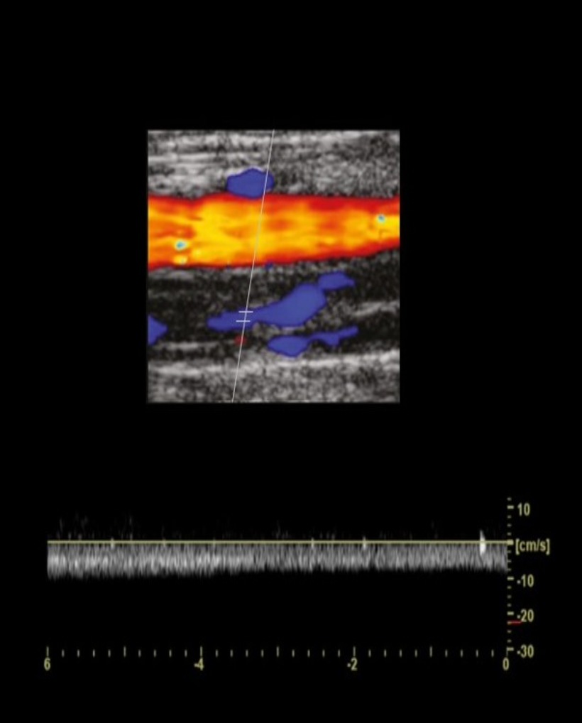
A patient presents for a lower extremity venous Doppler exam due to a history of May-Thurner Syndrome. What vessel should you closely evaluate for related findings?
A. left iliac vein
B. right iliac vein
C. bilateral popliteal veins
D. bilateral distal femoral veins
A. left iliac vein
May - Thurner Syndrome refers to increased incidence of left DVT due to extrinsic compression by RIGHT iliac artery.
Venous gangrene usually involves ______, while arterial gangrene usually involves ______
A. all the toes and foot, just one or two toes
B. the foot but no toes, the toes but not the foot
C. just one or two toes, all the toes and foot
D. right foot, left foot
A. all the toes and foot, just one or two toes

Which of the following cardiac abnormalities will have no effect on venous flow characteristics in the lower extremities?
A. significant tricuspid regurgitation
B. premature ventricular contractions
C. severe tricuspid stenosis
D. congestive heart failure
B. premature ventricular contractions
Premature ventricular contractions are a type of cardiac arrhythmia. A PVC will cause smaller ectopic contractions before a larger compensatory contraction. This abnormality alone will not affect venous flow characteristics in the extremities. The other choices lead to a backlog of flow in the IVC. When the IVC/iliac veins are dilated and engorged with blood, the cardiac pulsatility is transmitted further away from the heart due to the ripple effect.
Which of the following correctly describes the effects of walking on the deep venous pressure in the lower extremity in a patient with post phlebitic syndrome?
A. Walking causes a gradual decrease in pressure during the first 1-2 minutes of exercise that quickly returns to pre-exercise levels once the patient stops walking.
B. Walking causes a significant drop in deep venous pressure that takes several minutes to return to pre-exercise levels once the patient stops walking.
C. Walking has little to no effect on the deep venous pressure, only the arterial pressure is affected by exercise.
D. Walking causes an immediate, quick drop in pressure in the deep veins at the onset of exercise that quickly returns to pre-exercise levels once the patient stops walking.
D. Walking causes an immediate, quick drop in pressure in the deep veins at the onset of exercise that quickly returns to pre-exercise levels once the patient stops walking.
Valvular insufficiency in the deep system commonly occurs after an episode of DVT and is a key characteristic of post-phlebitic syndrome.
Normal deep veins will only allow flow toward the heart. As the leg veins empty and pressure quickly drops with muscle contraction, the arterial inflow is the only source of blood to refill the veins and pressure rises slowly upon stopping. In order to refill the veins and increase deep venous pressure to pre-exercise levels, the patient will need to stand for several minutes (>5min). Venous refill time is very fast if the deep veins contain incompetent valves. The pressure will drop quickly at the start of exercise but will quickly return to pre-exercise levels upon standing. The deep veins are refilled by arterial inflow and retrograde flow in the veins.
Which of the following changes in flow can be seen with a Baker cyst?
A. continuous flow in the popliteal vein
B. pulsatile flow in the popliteal vein
C. pulsatile flow in the superficial femoral vein
D. continuous flow in the popliteal artery
A. continuous flow in the popliteal vein
A Baker cyst can cause pain and swelling in the popliteal fossa. The extrinsic compression of the popliteal vein by the cyst can lead to a continuous flow pattern, blood stasis and DVT formation. The arterial walls are muscular and can prevent the effects of extrinsic compression on the popliteal artery.
Which of the following describes chronic lower extremity DVT?
A. echogenic vein walls
B. exaggerated respiratory phasicity
C. pulsatile flow pattern
D. vein is completely non-compressible
A. echogenic vein walls
Chronic thrombus formation can cause the vein walls to appear thickened and echogenic. The wall changes diminish the venous response to respiration. It can also lead to partial compressibility of the vein and continuous flow in the vessel (loss of respiratory phasicity).
It is not possible to evaluate and document valvular insufficiency with only:
A. Color Doppler Evaluation
B. Plethysmography
C. 2D real time imaging
D. PW Doppler evaluation
C. 2D real time imaging
The black and white 2D images are not capable of demonstrating blood flow characteristics. Venous valve movement may be assessed in 2D but this does not help quantify valvular incompetence.
Which of the following are common symptoms of chronic venous disease?
A. pallor and paresthesia
B. pitting edema and ulceration on great toe
C. varicosities and pallor
D. brawny discoloration and pitting edema
D. brawny discoloration and pitting edema
Toe ulceration, pallor and paresthesia are signs of arterial disease.
Varicosities, brawny discoloration and pitting edema are signs of chronic venous disease.
Which of the following is related to the appearance of Rouleaux formation on the image?
A. Thoracic outlet syndrome
B. DVT
C. Raynaud Syndrome
D. Intracranial collateral flow
B. DVT
Proximal to a DVT the flow may be very slow and visible on 2D imaging; the RBCs appear lined up or stacked in rolls similar to coin rolls from your bank which is called the Rouleaux Formation
A patient presents for a lower extremity venous Doppler to rule out venous insufficiency. Which vessels should be completely evaluated for the exam?
A. iliac, femoral and great saphenous veins
B. great and short saphenous veins
C. femoral and popliteal veins
D. femoral, popliteal, short saphenous and great saphenous veins
D. femoral, popliteal, short saphenous and great saphenous veins
The deep veins should be evaluated for thrombus before the reflux exam is performed. The deep veins should also be evaluated for reflux along with the superficial veins.
A patient in the ER has just been diagnosed with acute DVT of the politeal vein. What would be the physician's preferred treatment plan?
A. Immediate IV treatment with Heparin with 90 days of Warfarin medication once released from the hospital
B. Insert a valvulotome into the popliteal vein and recanalize the lumen.
C. Perform sclerotherapy to resolve the clot.
D. Begin the use of compression stockings and perform a follow up exam in 30 days
A. Immediate IV treatment with Heparin with 90 days of Warfarin medication once released from the hospital
Acute DVT is usually initially treated with IV Heparin followed by at least 90 days of Warfarin/Coumadin therapy once the patient is out of the hospital.
Which of the following risk factors has the highest incidence of lower extremity thrombus formation?
A. Recent hip surgery
B. Polycythemia vera
C. Rotator cuff repair
D. Diabetes
A. Recent hip surgery
Patients who have undergone any type of lower extremity surgery are at the greatest risk of DVT. The 3 factors of Virchow's triad are present in these patients. Usually these patients are confined to a bed for several days/weeks which leads to stasis. There are significant limitations with using blood thinners immediately following surgery because it would prevent the healing of the surgery and could lead to hemorrhage.
Proximal to a DVT in the femoral vein, the flow appears very slow and visible on 2D imaging. The red blood cells appear lined up or stacked in rolls. What is this phenomenon called?
A. May-Thurner Syndrome
B. Phlegmasia cerulea dolens
C. Raynaud phenomenon
D. Rouleaux Formation
D. Rouleaux Formation
Proximal to a DVT in the superficial femoral vein, the flow appears very slow and visible on 2D imaging. The red blood cells appear lined up or stacked in rolls. This phenomenon is called Rouleaux formation.

While scanning the popliteal artery, you notice a thrombus in the popliteal vein. Which vein(s) should be evaluated next for ascending propagation of the clot?
A. GSV
B. common femoral vein
C. distal femoral vein
D. ATA and tibioperoneal trunk
C. distal femoral vein
If a clot is identified in the popliteal vein, the distal femoral vein should be evaluated next for propagation of the thrombus.
Venous ulcers are normally located on the _____, while arterial ulcers are normally found on the _____
A. toes and heel, medial calf/ankle
B. heel, toes
C. calf, thigh
D. medial calf/ankle, toes and heel
D. medial calf/ankle, toes and heel


This image demonstrates which of the following?
A. Chronic DVT
B. Partial DVT
C. Acute DVT
D. Superficial venous thrombosis
C. Acute DVT
Note the engorged vein with mildly echogenic material within the lumen. The vein appears completely obstructed by acute thrombus.

Which of the following is not an expected finding related to the abnormality displayed on the image?
A. dilated posterior arch vein
B. varicose veins in the calf
C. dilated paratibial perforators
D. spontaneous flow in the distal great saphenous vein
D. spontaneous flow in the distal great saphenous vein
Severe GSV reflux would most likely lead to the formation of varicosities distally. The paratibial perforators connect the GSV to the PTVs. The GSV pressures will be increased and this will dilate the perforators. The posterior accessory saphenous vein (posterior arch vein) empties into the GSV below the knee. If the GSV is dilated, the accessory vein will also dilate. Flow in the superficial veins is not usually spontaneous. As the pressure in the GSV increases, there will be no flow toward the heart without augmentation.
A varicose vein is usually:
A. a dilated vessel of the perforator system.
B. a dilated tributary of the greater saphenous vein.
C. a dilated lesser saphenous vein.
D. a dilated tributary of the deep system.
B. a dilated tributary of the greater saphenous vein.
The greater saphenous vein and associated tributaries are most commonly associated with varicosity formation.
Which of the following carries an increased risk of pulmonary embolism?
A. renal vein thrombosis
B. common femoral artery thrombosis
C. thrombus accumulation in an aortic aneurysm
D. portal vein thrombosis
A. renal vein thrombosis
A clot in the systemic venous system can break loose and travel to the lungs. The renal veins drain into the IVC. The portal system does not directly communicate with the systemic veins, so a clot in the portal vein cannot lead to pulmonary embolism. An embolus in the arterial system will only travel until it becomes lodged in a smaller distal artery and cause ischemia.
Which of the following describes an abnormal response to proximal augmentation?
A. Flow reversal begins at the onset of the augmentation maneuver
B. No flow reversal occurs with proximal augmentation, but will occur with distal augmentation
C. Flow reversal begins upon release of the augmentation maneuver
D. Long venous refill time
A. Flow reversal begins at the onset of the augmentation maneuver
If the proximal thigh is squeezed while evaluating the distal GSV, flow reversal will occur as soon as you start to squeeze. The Valsalva maneuver can also be used for proximal augmentation. Flow reversal begins at the start of the Valsalva strain.

What is the most common cause of upper extremity DVT in the normal ambulatory patient population?
A. radiation therapy treatments to the chest
B. indwelling catheters
C. Paget - Schroetter Syndrome
D. shoulder surgery
C. Paget - Schroetter Syndrome
Paget - Schroetter Syndrome AKA spontaneous effort thrombosis:
Most common cause of axillary/subclavian DVT in the ambulatory patient population
Related to the presence of a cervical rib and thoracic outlet syndrome
Higher incidence in the dominant arm
Indwelling catheters are a common cause of upper extremity thrombosis in patients that are ill and require intravenous medication/treatment.
Warfarin is administered to a patient:
A. to prevent thrombus propagation
B. to assist in atheroma lysis and recanalization
C. to increase platelet aggregation
D. to assist in clot lysis and recanalization
A. to prevent thrombus propagation
Warfarin is administered to a patient to prevent thrombus propagation and reduce platelet aggregation. Streptokinase is used for clot lysis in phlegmasia patients. Increased platelet aggregation would cause clot propagation.
A patient presents with a swollen left leg from hip to ankle and the right leg is normal.
A lower extremity venous duplex exam shows compressible vessels bilaterally.
Doppler demonstrates normal flow in the right leg, but there is loss of phasicity of flow and decreased augmentation of the left common femoral vein and proximal femoral vein.
These findings are most suggestive of:
A. left ovarian mass or left external iliac vein thrombosis
B. right external iliac thrombosis or IVC thrombosis
C. bilateral external or internal iliac vein thrombosis
D. IVC thrombosis
A. left ovarian mass or left external iliac vein thrombosis
A left ovarian mass could compress the iliac vein to cause the swelling. Left external iliac vein thrombosis could also cause unilateral swelling of the entire leg.
IVC thrombosis or bilateral external iliac thrombosis would cause flow abnormalities in both extremities.
Internal iliac thrombosis will not affect the venous outflow of the legs.