EMT M6 - L1 - Respiratory Emergencies - (Quiz 3)
 Call Kai
 Learn
 Practice Test
 Spaced Repetition
 Match
 Flashcards
 Knowt Play
1/115

There's no tags or description
Looks like no tags are added yet.
116 Terms
What is Dyspnea?
Shortness of breath
What are the 3 stages of dyspnea?
– Respiratory Distress: Patient still ventilating but with difficulty
– Respiratory Failure: Poor or inadequate ventilation
– Respiratory Arrest: No ventilation
What is Respiratory Arrest?
No ventilation
What is Respiratory Failure?
Poor or inadequate ventilation
What is Respiratory Distress?
Patient still ventilating but with difficulty
What are the general causes of shortness of breath?
Issue of distribution with:
Ventilation
Respiration (The exchange of oxygen for CO2 in the lungs and in the cells of the body)
Oxygenation (The loading of oxygen onto hemoglobin)
What can the failure of ventilation, oxygenation, or respiration lead to?
Tissue hypoxia (lack of oxygen) and death.
What are the signs and symptoms of inadequate breathing?
– Anxiety or altered mental status in conjunction with abnormal respirations
– Respiration rate too fast, too slow, or irregular
– Poor skin signs (pale, cyanotic, cool, clammy / diaphoretic)
– Pale mucus membranes
– Patient can only speak of few words between breaths
– Accessory muscle use
– Unequal or inadequate chest expansion
– Excessive coughing
– Abnormal positioning (tripod, sniffing)
– Pursed lips or nasal flaring
– Abnormal or decreased breath sounds

What is this an example of?
Cyanosis
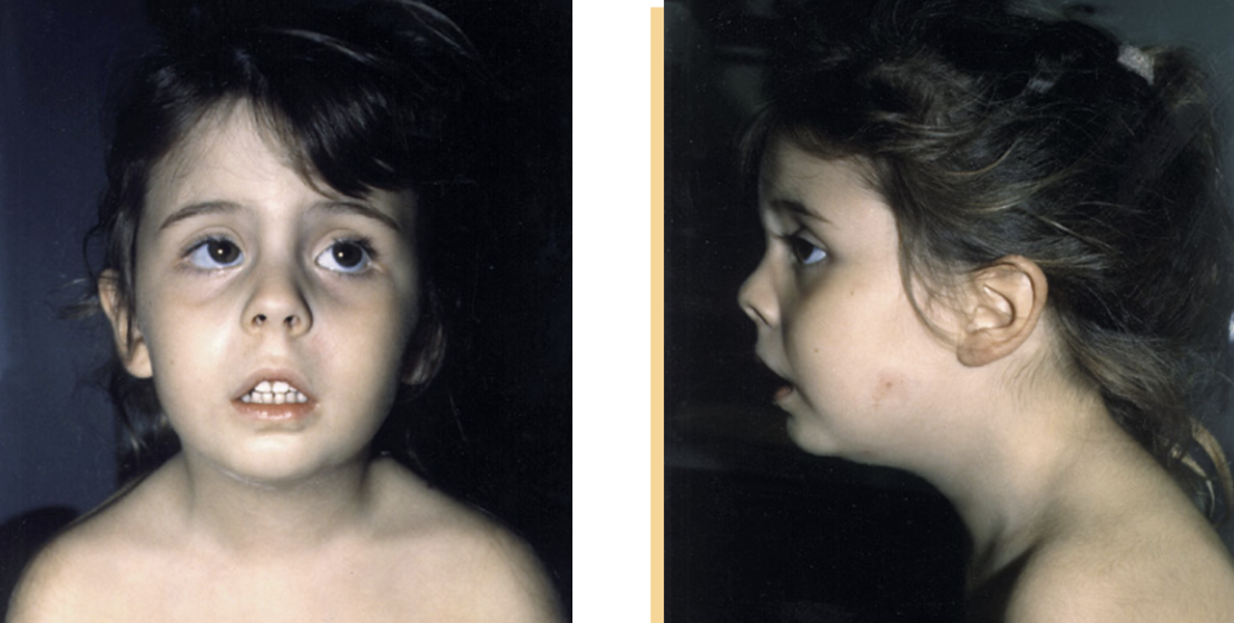
What is this an example of?
The Sniffing Position.
A position taken naturally when someone is having trouble breathing. Common in kids.

What is this an example of?
Tripod position
Pursed lip breathing
Accessory muscle use
He’s putting his weight on his knees and leaning forward to try and breath easier. He’s using his neck to help him breath, as you can see from the retraction around his clavicles.
Barrel chest (the shape of his chest in the photo).
What is barrel chest a characteristic of?
COPD (Chronic Obstructive Pulmonary Disease, a progressive group of lung conditions—including emphysema and chronic bronchitis—that block airflow and make breathing difficult).
What are the types of abnormal breath sounds?
wheezing
crackles
rhonchi (low, coarse breath sounds)
stridor (high pitched airway sound)
What are crackles? (Abnormal breathing sounds)
– Crackling, clicking, or rattling sound
– Heard on inspiration
– Caused by popping open of small airways and alveoli
***** Usually means there is fluid in the lungs
***** Lower airway sounds, usually we use stethoscope sounds to hear
What is wheezing? (Abnormal breathing sounds)
– Whistling, musical in character
– Wheezes are higher pitched
– Indicative of narrowing of airways
***** Lower airway sounds, usually we use stethoscope sounds to hear
What is rhonchi? (Abnormal breathing sounds)
– Lower, coarse breath sound
– Can be from mucus and thick secretions in upper airways
What is Stridor? (Abnormal breathing sounds)
– High pitched upper airway sound
– Indicative of airway obstruction
– Can be heard without stethoscope
What are the Common Causes of Dyspnea?
• Upper or lower airway infection*
• Acute pulmonary edema
• COPD
• Asthma
• Allergies or Anaphylaxis
• Spontaneous pneumothorax
• Pleural effusion
• Prolonged seizures
• Airway obstruction
• Pulmonary embolism
• Hyperventilation syndrome
• Environmental / industrial exposure, CO poisoning
• Cystic Fibrosis
What is Acute Pulmonary Edema?
Fluid accumulation in the air spaces and interstitial tissue of the lungs. Gas exchange is inhibited because fluid increases the distance between the lung capillaries and alveolar wall.
Becomes and issue of external respiration.
Inhibited meaning?
Held back, restrained, or not confident enough to speak, act, or express feelings freely.
What is the common cause of Acute Pulmonary Edema?
Left sided heart failure.
Blood starts to back up behind the heart when it is not able to let out blood as fast as the blood is coming back in, leading to an increase in the blood pressure in the lungs, (pulmonary hypertension), and causing acute pulmonary edema.
What is pulmonary hypertension?
An increase in the blood pressure in the lungs
When do flash pulmonary edemas occur?
When an acute event, such as a myocardial infarction, suddenly disrupts function of left ventricle and causes a very rapid onset of dyspnea.
What should you do if a person is drowning?
Use pressure to push fluid of of their lungs and into their bloodstream.
The opposite of what happens during an acute pulmonary edema.
What are some other causes of acute pulmonary edema?
– Hypertensive crisis (if your blood pressure is too high generally)
– Injury to lung
– Aspiration of fluid (or if you’re drowning)
– Drowning or near drowning
– Severe infection or inflammation
What are the signs and symptoms of Acute Pulmonary Edema?
– Severe shortness of breath
– Poor skin signs
– Paroxysmal nocturnal dyspnea
– Orthopnea (Shortness of breath that occurs when laying flat, and results in patient sleeping upright or with head elevated)
– Pink frothy sputum (Not to be confused with hemoptysis from trauma. End inspiratory crackles (heard in the lungs))
What is Orthopnea?
Shortness of breath that occurs when laying flat, and results in patient sleeping upright or with head elevated
If you have a patient that wakes up suddenly out of breath, suspect that there may be fluid in their lungs.
If you’re considering pulmonary edema, should you lie your patients supine or prone?
No, that can cause Orthopnea.
What is sputum?
A mixture of thick mucus, saliva, and cellular debris that is coughed up from the lower respiratory tract, including the lungs and bronchial tubes
What is hemoptysis?
Coughing up blood or blood-stained mucus from the lower respiratory tract (the larynx, trachea, bronchi, or lungs)
What do some patients with pulmonary edema due to CHF exhibit?
Wheezing, coughing, and dyspnea know as cardiac asthma.
What is CHF?
Congestive heart failure.
A chronic condition where the heart muscle cannot pump blood effectively enough to meet the body's normal needs, causing blood and fluid to back up into the lungs, legs, and other tissues.
Should you treat cardiac asthma like true asthma?
No, although the signs and symptoms mimic true asthma, they are treated differently.
Should you request ALS for patient with acute pulmonary edema?
Yes.
ALS intercept should be requested and/or patient should be rapidly transported.
Acute Pulmonary Edema Treatment:
In patients with suspected acute pulmonary edema and respiratory distress, should EMTs begin positive pressure ventilations even if patient is breathing?
Yes.
– Positive pressure will aid gas exchange by forcing fluid out of the air spaces
– CPAP should be used if indicated by protocol
– BVM may be necessary based on patient condition
Acute Pulmonary Edema Treatment:
Should patients be placed upright in Fowler’s position?
Yes to aid breathing.
Supine position should be avoided if at all possible.
What is CAD?
Coronary artery disease (CAD). A common heart condition caused by atherosclerosis, where cholesterol, fat, and calcium build up as plaque inside the heart's major blood vessels, narrowing or blocking oxygen-rich blood flow.
Can patients with CAD may be prescribed nitro for CHF as well as angina?
Yes.
– EMTs should assist as allowed by local protocols
– ALS providers also carry nitro for CHF exacerbations
What is angina?
AKA Angina pectoris is chest pain, pressure, heaviness, or squeezing discomfort that happens when the heart muscle does not get enough oxygen-rich blood.
What is Chronic Obstructive Pulmonary Disease (COPD)?
A chronic and progressive inflammatory lung disease that results in the obstruction of airflow in the lungs.
“What is commonly referred to as ‘lung disease’.“
What patients most commonly have COPD (Chronic Obstructive Pulmonary Disease)?
Patients who smoke cigarettes, or who have direct lung and airway damage from repeated infections or inhalation of toxic agents.
Amount and duration of smoking correlates with severity of COPD.
What are the two forms of COPD (Chronic Obstructive Pulmonary Disease)?
– Chronic bronchitis (ongoing irritation of the trachea and bronchi results in the production of excess mucus that obstructs small airways and alveoli)
– Emphysema: dilation and destruction of the walls of the alveoli from chronic stretching (Alveoli stretch due to the inability to easily expel gases through inflamed airways).
What is Chronic bronchitis?
A type of COPD (Chronic Obstructive Pulmonary Disease)
Ongoing irritation of the trachea and bronchi that results in the production of excess mucus that obstructs small airways and alveoli.
What is Emphysema?
A type of COPD (Chronic Obstructive Pulmonary Disease)
The dilation and destruction of the walls of the alveoli from chronic stretching. (Alveoli stretch due to the inability to easily expel gases through inflamed airways).
Do most COPD patients have elements of both chronic bronchitis and emphysema?
Yes, although one may play a more significant role in their disease.

What is this an image of?
Lungs without COPD vs. with.
With, the:
airways are smaller making it harder to move air in and out
Alveoli have less surface area, making it harder to have adequate gas exchange, and the alveoli start to flow into one another and expand outwards, which is not good for surface area.
Bronchioles are narrower and misshapen and clogged with mucus
What are the signs and symptoms of Chronic Obstructive Pulmonary
Disease (COPD)?
– Dyspnea
– Chronic cough and sputum production
– Long expiratory phase of breathing
– Wheezing or decreased breath sounds
– Pursed lip breathing
– Barrel chest
– Poor skin signs
What is the expected oxygen saturation of Chronic Obstructive Pulmonary
Disease (COPD) patients?
Lower O2 saturation (88-92%) may be present in patients with COPD
Baseline oxygen saturations differ and you should ask your patients if they know their baseline O2 saturation
Level of distress should be your primary indicator for oxygen administration
PATIENTS IN DISTRESS SHOULD STILL RECEIVE OXYGEN REGARDLESS OF O2 SATURATIONS!
What can advanced COPD patients suffer from due to pulmonary hypertension?
Right sided heart failure (cor pulmonale).
– Vasoconstriction and destruction of arterioles in lungs as a result of chronic hypoxia increases vascular resistance and causes right side of heart to have to push harder
– In these patients, you may see JVD and peripheral edema due to heart failure
Is there a cure for COPD?
No, but symptoms can be treated and progression of the disease can be delayed.
What are some methods of managing COPD?
– Smoking cessation (the process of stopping tobacco use)
– Exercise
– Bronchodilators
– Corticosteroids
– Supplemental oxygen
What is acute exacerbation of COPD?
The sudden onset or worsening of respiratory symptoms over several hours or days.
EMS if commonly called for these cases.
What are some prehospital BLS treatment of COPD exacerbation?
– Assisting the patient with their metered dose inhaler
– Applying supplemental oxygen and assisting ventilations as necessary
– CPAP (if allowed by protocol)
– Albuterol via nebulizer (if allowed by protocol) (Usually an ALS intervention)
– Requesting ALS intercept and/or rapid transport
What is Asthma?
Inflammatory disease of the airways that is characterized by spasm of bronchioles
What is the acute exacerbation of asthma often referred to?
An ‘asthma attack’
What are the signs and symptoms of acute exacerbation?
– Dyspnea
– Expiratory wheezes
– Chest tightness
– Accessory muscle use
– Coughing
What are asthma patients typically prescribed?
Bronchodilators and corticosteroids
What are corticosteroids?
Human-made or synthetic drugs that mimic cortisol, a natural hormone produced by your body's adrenal glands.
Why should you ask patients with asthma about previous episodes?
To understand the severity of a patient’s disease
– Patients with asthma will likely have had previous episodes of asthma exacerbation
– If patient has been previously intubated for asthma, then EMT should expect that the current episode may be just as severe
What is Status asthmaticus?
An acute exacerbation of asthma that does not respond to standard treatments.
Patients with this condition require rapid transport and ALS intercept
What is Anaphylaxis?
A severe allergic reaction characterized by airway swelling and systemic vasodilation.
How long does it take for Anaphylaxis to occur?
Typically occurs within 30 minutes of exposure to an allergen
What should you do when a patient is suffering from anaphylaxis?
Epinephrine via autoinjector should be immediately administered.
What is Pneumothorax?
A collapsed lung.
The partial or complete accumulation of air in the pleural space
What is the thorax/your thoracic cavity?
Your chest/everything above your diaphragm.
What is Spontaneous Pneumothorax?
A pneumothorax caused by a medical condition rather than trauma.
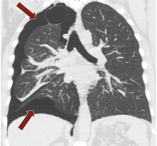
What is this an example of?
Spontaneous Pneumothorax.
What usually keeps your lungs inflated?
A vacuum in pleural space.
Thus if air leaks into pleural space due to disruption to lung surface then the vacuum is lost and the lung collapses.
What are the signs and symptoms of Spontaneous Pneumothorax?
– Sharp, stabbing, pleuritic chest pain
– Dyspnea
– Diminished breath sounds on affected side
– Hyperresonance to percussion on affected side (not commonly assessed by EMTs)
In which patients does Spontaneous Pneumothorax typically occur?
In young adults (20-30 y/o) and patients with chronic lung conditions like COPD.
– Tall, thin males are more susceptible than the rest of the population
– Patients with Marfan Syndrome
– Patients with COPD or asthma may rupture a weakened portion of their lung while coughing
What does pleuritic chest pain mean?
Chest pain that seems to be related to breathing.
What can Spontaneous pneumothorax progress to?
Tension pneumothorax.
When does Tension Pneumothorax occur?
When pneumothorax leads to significant impairment of respiration and blood circulation. Occurs due to buildup of air in pleural cavity that compresses the affected lung and causes heart and other mediastinal tissues to shift to opposite side.
What are some indicators that a pneumothorax has progressed to tension pneumothorax?
Severely labored breathing,
tachycardia,
hypotension
Other signs:
Chest pain,
decreasing O2 saturation,
diminished lung sounds on affected side,
altered mental status.
Other late signs:
JVD
tracheal deviation
What should you do if tension pneumothorax is suspected?
Call for an ALS intercept, and manage ABCs while initiating rapid transport to hospital.
ALS may be able to decompress the pleural cavity and allow lung to re-expand
What are some causes of Pleural Effusion?
Inflammation
Irritation
Infection
Congestive heart failure
Cancer
When does Pleural Effusion happen?
Occurs when fluid accumulates in the pleural cavity and compresses one or both lungs.
Fluid buildup is usually gradual, but dyspnea is often sudden in onset.
Pleural Effusion: What should you hear?
In area of fluid buildup, there is often dullness to percussion and decreased breath sounds.
Pleural Effusion: What position should you have the patient do?
Sit up.
This can usually help them feel better, but the fluid in their lungs can only be drained in the hospital, so getting them there is really all you can do.
What is Pleural Effusion?
The gradual build up of fluid in the lungs.
What are tonic-clonic seizures?
“Generalized grand mal seizure”
A type of generalized seizure that consists of two phases:
– Tonic phase: patient loses consciousness and their skeletal muscles stiffen
– Clonic phase: patient’s muscles begin jerky, rhythmic contractions
What happens to a patient’s breathing and airway during a tonic-clonic seizure?
The patient’s breathing will be impaired
Their airway may become compromised from biting of the tongue or sputum
Patients can also turn cyanotic, when they are not moving enough air.
How long do most tonic-clonic seizures last?
Less than a few minutes before a patient regains consciousness and regular breathing is quickly restored.
What are Status epilepticus?
A seizure of greater than five minutes or multiple seizures within five minutes without the patient returning to normal.
What do patient in status epilepticus need?
Aggressive airway management
Ventilatory assistance (because their ability to breathe will be compromised for an extended period of time)
***** Personal safety of EMT should be taken into account
What should you not do to a patients in status epilepticus?
Do not place anything in a patient’s mouth during a seizure, as it could result in an airway obstruction
What should you do to patients in status epilepticus after managing airway and breathing?
Rapidly transport the patient and call for an ALS intercept. ALS can also potentially terminate the seizure by administering a benzodiazepine.
What is an embolus?
A mass (solid, liquid, or gas) that travels from its point of origin and lodges to a distant site, obstructing blood flow.
What is a Pulmonary Embolism (PE)?
A condition that occurs when an embolus lodges in the pulmonary artery or one of its branches.
Typically caused by a blood clot that formed in veins of the legs or
pelvis and travelled to through the heart to the pulmonary artery
The severity is influenced by how much of the pulmonary circulation is blocked
What is deep vein thrombosis?
A serious medical condition where a blood clot (thrombus) forms in a deep vein, most commonly in the lower leg, thigh, or pelvis.
Where do emboli caused by blood pooling in the heart go? What can they cause?
To the brain.
They can cause a stroke.
What are some risk factors for pulmonary embolism?
Prolonged immobilization
“The main reason that blood will pool in the legs”
After a long flight maybe
Being bed-ridden after a surgery
Increased ability of blood to coagulate
Can be caused by:
Cancer
Heart disease
Smoking
Estrogen based contraception pills
Pregnancy
Obesity due to hormonal reasons
Damage to vessel wall
Recent surgery maybe
Previous Deep Vein Thrombosis (DVT)
What are some common signs and symptoms of pulmonary embolism?
– Dyspnea (typically rapid onset within seconds or minutes)
– Tachypnea
– Pleuritic chest pain
– Crackles or other abnormal lung sounds
– Cough
– Hemoptysis (coughing up blood)
– Tachycardia
What can a large embolis cause?
A Pulmonary Embolism that leads to rapid circulatory collapse and sudden death.
What can you, as an EMT, do in the case of a Pulmonary Embolism?
Get them to the hospital as fast as possible.
There is nothing you can do to dislodge the emboli.
What is Hyperventilation?
Over breathing to the point where the level of arterial CO2 falls below normal.
Our bodies often uses hyperventilation to compensate for acidosis by blowing off CO2 and correct the blood’s pH.
What life-threatening illnesses can hyperventilation be an indicator of?
Asprin overdose
Severe infection
Diabetic patient in diabetic ketoacidosis (DKA) due to high blood sugar
What should you do if you have a patient that is hyperventilating and you do not know why?
Take their blood sugar and monitor it super closely
Consider taking them to the hospital super fast
What is Hyperventilation Syndrome also referred to?
“Panic Attack”
What are the symptoms of Hyperventilation Syndromes?
– Anxiety (can be the cause and a resulting symptom)
– Dizziness
– Numbness and tingling in hands and feet
– Dyspnea despite rapid breathing
**** Usually these symptoms are due to blowing off too much CO2, resulting in alkalosis