BIOCHEM501 Unit 2
 Call Kai
 Learn
Learn Practice Test
 Spaced Repetition
 Match
Match Flashcards
 Knowt Play
1/61

Name | Mastery | Learn | Test | Matching | Spaced | Call with Kai | Chat |
|---|
No analytics yet
Send a link to your students to track their progress
62 Terms
basic structure of nucleotides
composed of a purine or pyrimidine base, a pentose, and a phosphate

pyrimidine
single-ring nitrogenous base; C, T, and U

purine
double-ring nitrogenous base; A and G

adenine
purine; pairs with thymine (DNA) and uracil (RNA)

guanine
purine; pairs with cytosine

cytosine
pyrimidine; pairs with guanine

thymine
pyrimidine (DNA); pairs with adenine

uracil
pyrimidine (RNA); pairs with adenine

ribose
pentose for RNA; -OH on C2
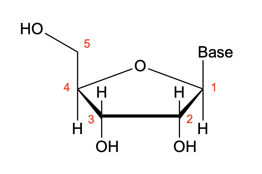
deoxyribose
pentose for DNA; no -OH on C2

glycosidic bond (N-glycosidic linkage)
the connection between the nitrogenous base and the pentose; between the 1’ position on the pentose and an N on the nitrogenous base

nucleoside
a nitrogenous base connected to a pentose

nucleotide
nucleosides with phosphoryl group(s) attached via ester linkage
nucleoside monophosphate
nucleoside with one phosphoryl group attached; what we see in the final nucleic acid polymer

nucleoside triphosphate
nucleoside with three phosphoryl groups attached; serve as high-energy building blocks used by cells to synthesize the nucleic acid polymer

phosphodiester bond
backbone of nucleic acid structure; between the 5’ and 3’ end of pentose

phosphodiester bond mechanism
base activation of 3’ OH
nucleophilic attach of 3’ OH on an “activated” 5’ phosphate (LG attached to drive rxn foreward)

cleavage of phosphodiester bonds
base activation of H2O molecule
nucleophilic attack on phosphodiester bond

Watson-Crick-Franklin (WCF) H-bonding
occurs between bases on strands oriented anti-parallel to one another; produce double-helix
hydrogen bond donors in the bases
adenine: NH2
cytosine: NH2
guanine: N1H, NH2
thymine: N3H

hydrogen bond acceptors in the bases
adenine: N1
cytosine: N3, O
guanine: O
thymine: O on C4, O on C2

WCF base pairs
adenine forms two H bonds with thymine
guanine forms three H bonds with cytosine
anti-anti conformation

B-form double-stranded DNA double-helix
most common
right-handed helix; 10.4 bp per turn
strands are anti-parallel
sugar-phosphate backbone on the outside
nucleobases on the inside
asymmetrical

base-stacking interactions (dsDNA helix)
van der Waals interactions between hydrophobic nucleobase faces (sterics)
π-stacking (electronic effects)
reenforce the individual A/T and C/G H-bonding interactions to stabilize the dsDNA helix
major groove
allows for sequence-specific interactions to occur from the outside without unwinding the strands

preferred RNA nucleotide conformer
C3’-endo conformer; the 2’ position of the sugar contains an OH, and the C2-endo conformation creates steric problems with the nucleobase

preferred DNA nucleotide conformer
C2’-endo conformer; the 2’ position is an H, so there are no steric issues, however, both conformations are still possible

B-form dsDNA conformation
C2’-endo conformation; places phosphates farther apart from one another
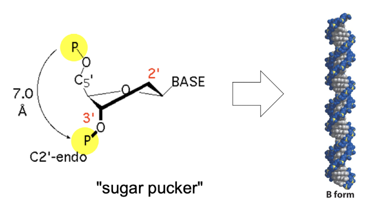
A-form dsDNA conformation
C3’-endo conformation; more tightly spaced backbone

Hoogsteen base pairs
syn/ani conformation
possible A/T, C/G, or G/G base pairs
far less common than WCF
allows 3-4 strands in a helix
found in damaged DNA
RNA primary (1°) structure
linear sequence of nucleotides linked together by phosphodiester bonds in a single strand

RNA secondary (2°) structure
Watson-Crick and Hoogsteen pairs, formed within a single-stranded RNA molecule as it folds back on itself

RNA tertiary (3°) structure
3D arrangement of folded RNA molecule, formed by long-range interactions between 2° structure elements like stems and loops

why is DNA the primary form of information storage?
RNA is more susceptible to degradation (2’ OH can lead to breakdown of the chain)
easier access to information via the major groove
when does DNA need to be separated into strands?
DNA is denatured during genome replication
DNA melts at the 5’ end of a gene before it is expressed
which base pairs form stronger interactions?
cytosine and guanine
Tm
temperature at which the helix is half double-stranded, half single-stranded (50% denatured)
how is Tm related to dsDNA stability?
stable helix = high Tm
unstable helix = low Tm

what determines the melting temperature of dsDNA?
relative A/T and G/C content; more C/G bp make it harder to separate strands

why is knowing Tm useful?
tells us what temperature we need to go above to cause dsDNA strands to denature (or what temp to go below to get two strands to anneal)
topology
describes the 3D shape of dsDNA; how it twists and arranges itself
Lk = Tw + Wr
linking number; cannot be changed by deforming the structure
Lk = Tw + Wr
twists; the number of times each of the curves rotates around the central axis C of the double helix

Lk = Tw + Wr
writhe; the number of times the intact B-form helix twists about itself

can topology be changed?
no; any configuration with the same Lk value is the same topography
Lk0
natural twist of relaxed B-form dsDNA
equal to (# of bp) / 10.4
what determines observed topology?
for a given DNA topology Lk, the Tw will be ~Lk0 and Wr will account for the difference between Lk and Lk0
ΔLk = Lk - Lk0
difference between the “natural twist” and a actual topology
ΔLk < 0
DNA is under wound and negative supercoiling will result
ΔLk > 0
DNA is overwound and positive supercoiling will result
ΔLk = 0
DNA is relaxed
DNA topoisomerase
enzymes that relieve torsional strain and prevent tangling in DNA by creating temporary single- or double-strand breaks in the sugar-phosphate backbone
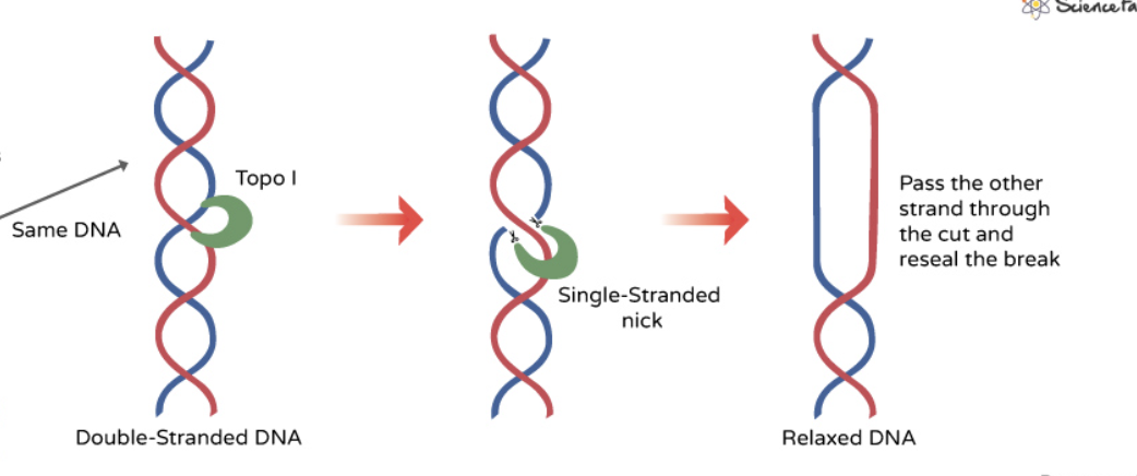
type I topoisomerase mechanism
cut one strand of the double helix
attach Tyr to the DNA phosphate backbone
pass unbroken strand through the break, or rotate broken strand around intact one to release tension
seal the cut back together
type II topoisomerase mechanism
cut both strands of the double helix
pull unbroken double-stranded section of DNA through the gap
seal cut back together

histones
small, highly alkaline proteins found in eukaryotic and archaeal cell nuclei that package and order DNA
nucleosome
DNA + histone nucleoprotein
30nm fiber
higher-order structure of DNA and histone proteins formed by the tight folding and compaction of nucleosome arrays
histone methylation
adding methyl groups to histone proteins to bind chromatin and turn off gene transcription; makes DNA less accessible

histone acetylation
adding an acetyl group to histone proteins to relax chromatin and turn on gene transcription; makes DNA more accessible

heterochromatin proteins
bind across methylated histones to promote chromatin compaction
euchromatin
accessible; transcription can occur; marked by histone acetylation
heterochromatin
inaccessible; transcription cannot occur; marked by histone methylation and binding of heterochromatin proteins