Anatomy Lab Practical 1
 Call Kai
 Learn
Learn Practice Test
 Spaced Repetition
 Match
Match Flashcards
 Knowt Play
1/126

There's no tags or description
Looks like no tags are added yet.
Name | Mastery | Learn | Test | Matching | Spaced | Call with Kai | Chat |
|---|
No analytics yet
Send a link to your students to track their progress
127 Terms
Prophase
Chromosomes are visible and look like dark tangled threads.

Metaphase
Chromosomes are lined up in one line across by the middle of the cell.

Anaphase
Chromosomes in two groups, moving toward opposite ends of the cell.

Telophase
Nuclei reforming around chromosomes that are located at opposite ends of the cell.

M-Phase
Mitosis + Cytokinesis → One cell becomes two
Interphase
Lightly stained area in cell.

Karyokinesis (Mitosis) vs. Cytokinesis
Karyokinesis (Mitosis): Division of the nucleus, separated duplicated chromosomes into two nuclei, stages of prophase, metaphase, anaphase, and telophase, results in two genetically equivalent nuclei.
Cytokinesis: Division of the cytoplasm, separates the cell into two daughter cells, occurs after or around telophase, results in two separate daughter cells.
Simple Squamous Epithelial Tissue
One thin layer of flat cells, nuclei are dark, clear cell boundaries.

Stratified Squamous Epithelial Tissue
Many cell layers, flat cells on the top surface, dark nuclei.

Simple Cuboidal Epithelial Tissue

One layer of boxy or cubed cells, dark nuclei are in the center of each cell, cells form tube or ring around an opening.

Simple Columnar Epithelial Tissue
One layer of tall and narrow cells in columns, nuclei are narrow and elongated, cells are tightly packed, goblet cells are scattered around column (lighter).
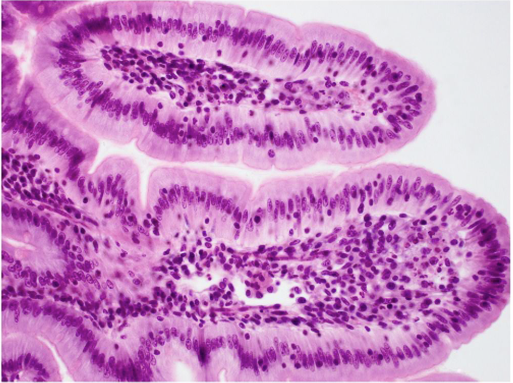
Pseudo-stratified Stratified Ciliated Columnar Epithelial Tissue

Hair like cells at the top of the surface, Looks like multi cell layer but is actually only one layer (all cells touch the basement membrane), cells are tall.

Transitional Epithelial Tissue

Multiple layers of cells, cubital toward bottom, top surface are more large and rounded, cells change shape/look like tissue can stretch.

Areolar Connective Tissue
Loose messy arrangement/fibers in many different directions, open space, Collagen fibers → thicker, usually pink/lightly stained, wavy fibers. Elastic fibers → thinner, darker fibers that may look wavy or branching. Fibroblast nuclei → small, dark, elongated/oval spots scattered throughout.

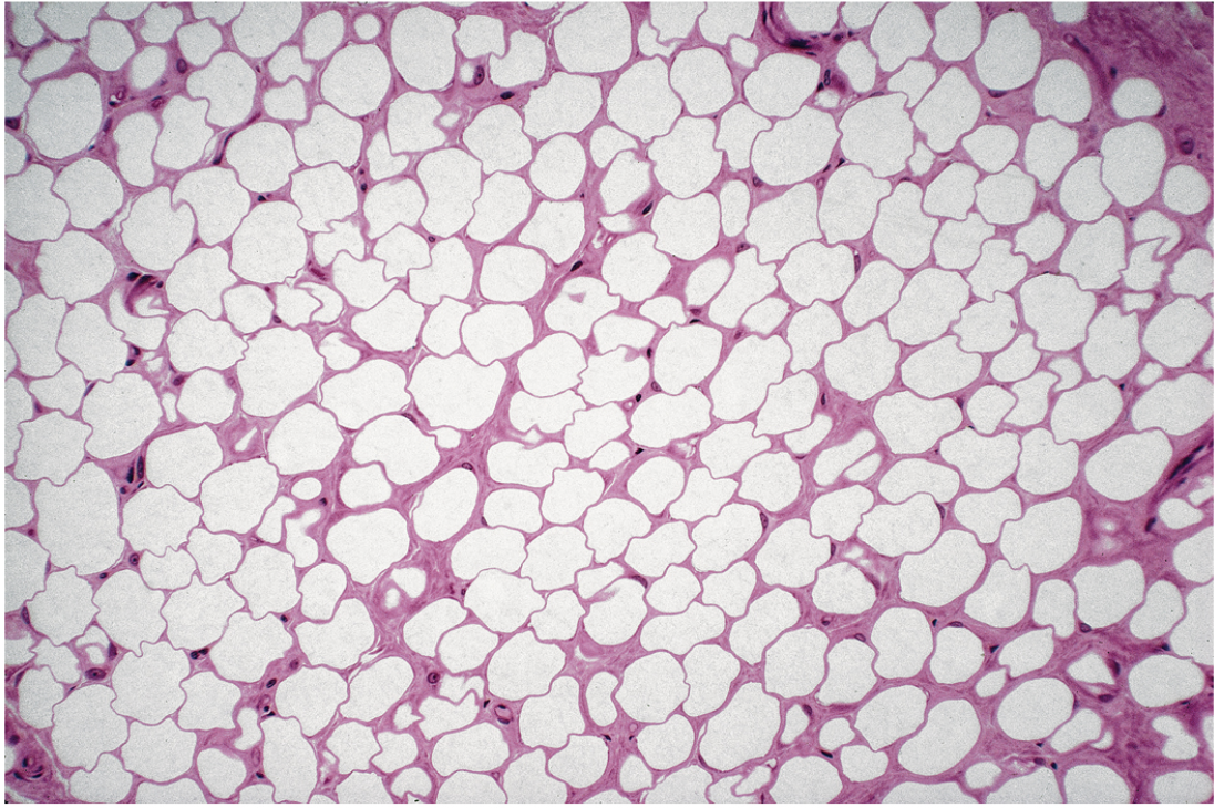
Adipose Connective Tissue
Large empty looking white cells, fat cells, nucleus is pushed to the edge, little visible material between cells, each cell is called an adipocyte.
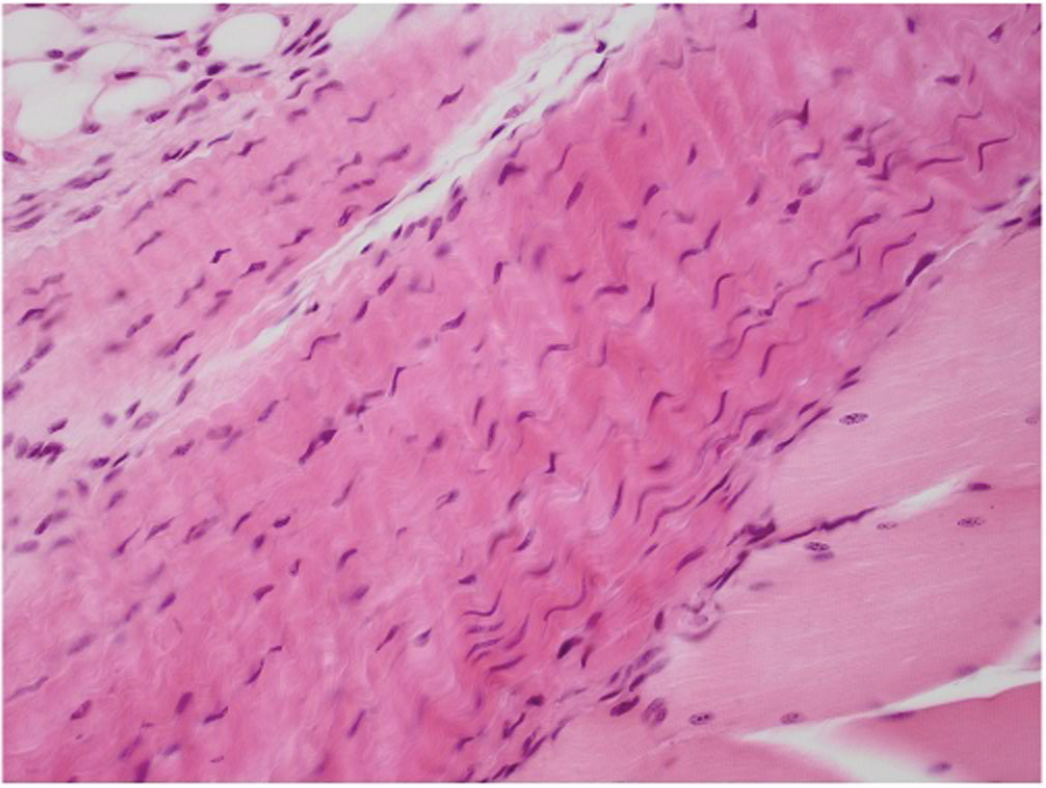
Dense Regular (Fibrous) Connective Tissue
Thick fibers packed tightly together, organized same direction fibers, Fibroblast nuclei between fibers appear thin and long, very little open space.
Dense Irregular (Loose) Connective Tissue
Thick packed fibers, run in many directions, Fibroblast nuclei between fibers appear small and dark, tangled, disorganized.

Cartilage Connective Tissue
Cells in little pockets. Chondrocytes in Lacunae.

Hyaline Cartilage Connective Tissue
Lacunae are rounded spaces or "holes" in the tissue Chondrocytes are round cells/nuclei found inside the lacunae. The background often looks relatively uniform, large, and smooth (matrix).

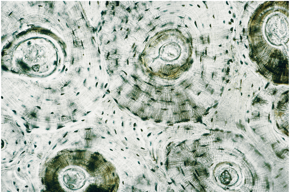
Bone (Compact Ground) Connective Tissue
Ring like organization. Osteons are circular structures that look like bullseyes. Concentric lamellae are rings of bone tissue surrounding the center of each osteon. Lacunae are tiny dark spaces between the rings where osteocytes are located. The tissue appears very dense and organized, with little open space
Blood (Vascular) Connective Tissue
Lots of RBCs and few WBCs. Red blood cells (erythrocytes) are small, round, pink/red cells. Usually no visible nucleus in mature human RBCs. White blood cells (leukocytes) are much larger and less numerous than RBCs. Have clearly visible, dark-staining nuclei. Platelets are tiny cell fragments scattered between RBCs. Plasma is the light-colored fluid background surrounding the cells.

Skeletal Muscle Tissue
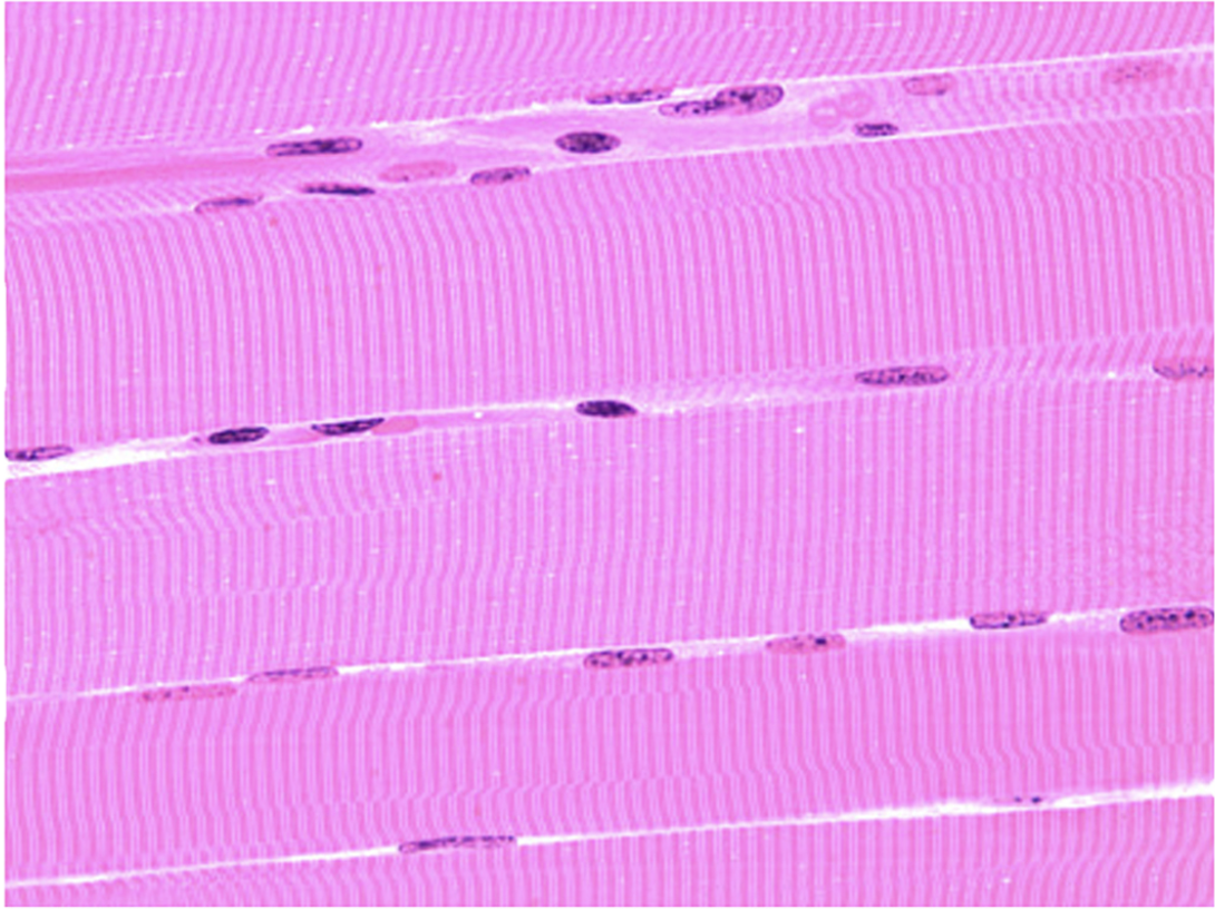
Stripes and many nuclei. Long, cylindrical muscle fibers that run parallel to each other. Striations are alternating light and dark bands running across the fibers. Multiple nuclei in each muscle fiber. Nuclei are usually located near the edge (periphery) of the fibers. Fibers are unbranched and look like long strands.

Cardiac Muscle Tissue
Striations are alternating light and dark bands across the cells. Cardiac muscle cells often branch and connect with neighboring cells. Usually a single nucleus located near the center of each cell. Intercalated discs are dark lines crossing the muscle fibers. These are a major clue for cardiac muscle.

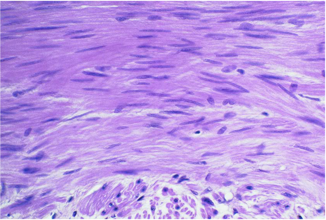
Smooth Muscle Tissue
No stripes, Spindle-shaped cells wide in the middle and tapered at both ends. One central nucleus in each cell, usually elongated or cigar-shaped. No visible striations — this is the biggest clue. Cells are arranged in sheets or layers, often running in the same general direction.
Multipolar Neuron Nervous Tissue
Visible axon and many dendrites, round nucleus in center of cell body.

Thick Skin Integument Tissue
Skin where you get calluses and do lots of things, harder to break, ex: on palm of hands and soles of feet. Easily see 5 layers, no hair follicles or sebaceous glands.

Thin Skin Integument Tissue
Skin on the rest of your body on normal and delicate areas. Four layers, lacking the lucidum. Contains hair follicles, sweat glands, and sebaceous glands.

Stratum Basale or Germinative
Layer of epidermis right along the basement membrane/locks directly onto the dermis. Single row of dark cells.

Stratum Spinosum
Thick multi layered region located right above the stratum basale. Prickly star spine like look to the cells. Thinner in thin skin.

Stratum Granulosum
Appears as the dark ribbon. Granular give it the dark color.

Stratum Lucidum
Glassy strip directly above the granulosum layer. No separate nuclei or organelles, almost opaque looking, homogenous layer. Absent in thin skin.
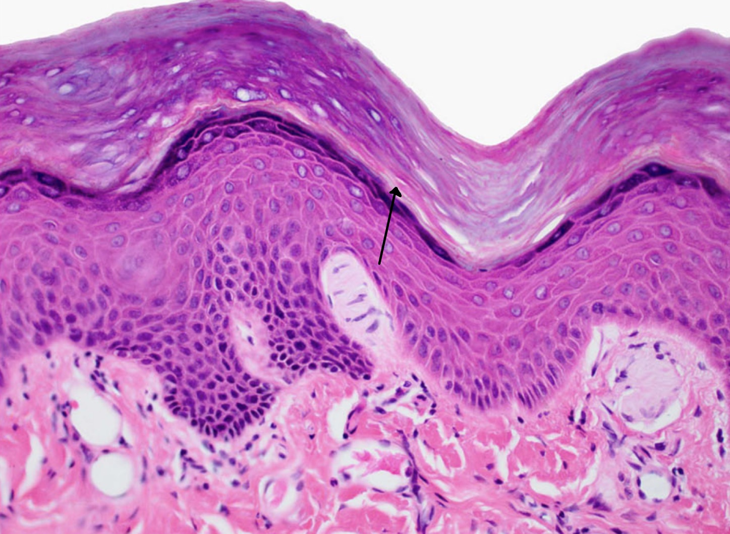
Stratum Corneum
Outermost layer of the skin flakey appearance, much thicker in thick skin. No nuclei.

Dermis
Irregular connective tissue scattering dark nuclei.

Dermis Papillary Layer
The finger-like extensions of the dermis pushing up into the epidermis.

Subcutaneous Layer

Contains fat cells, hair follicles, and sweat glands, is located below the dermis.

Hair Follicle
Stretched out at an angle.

Sebaceous Glands
Attached to hair follicle.

Sweat Glands
Pack of cells.

Occipital Bone

Pariental Bone

Frontal Bone

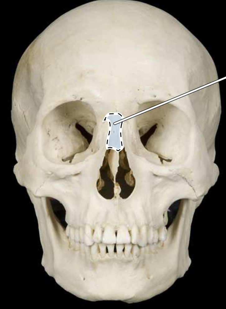
Nasal Bone
Maxilla Bone

Mandible Bone

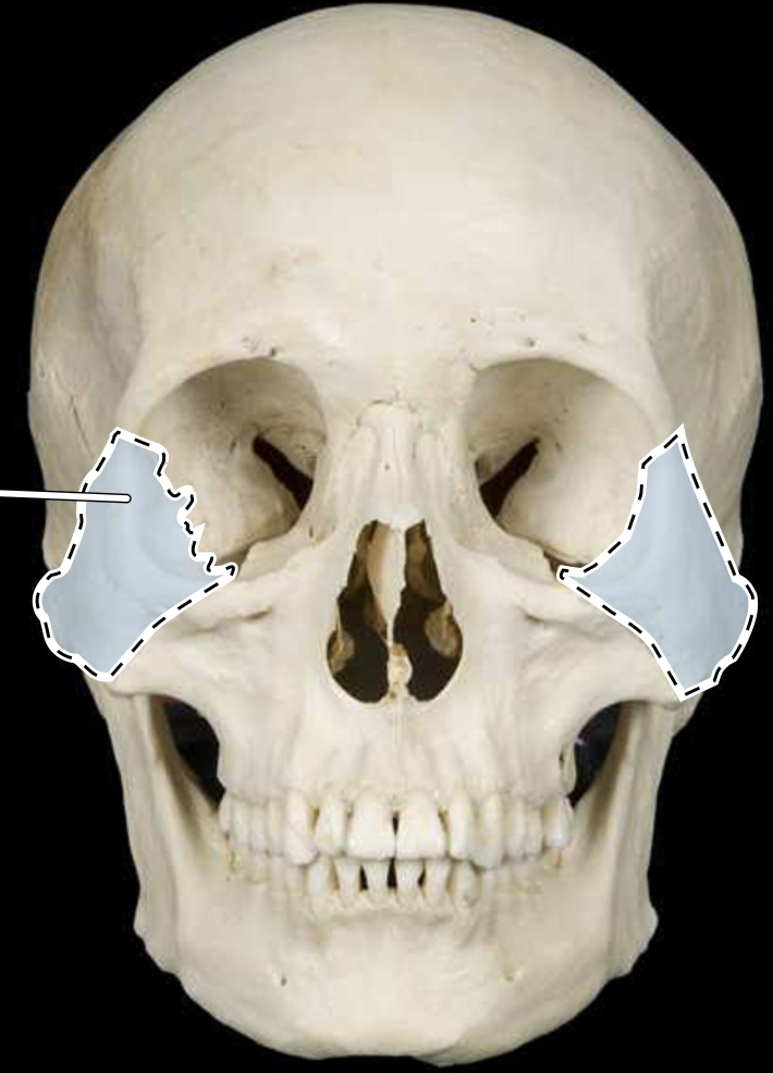
Zygomatic/Malar Bone
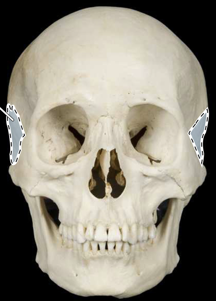
Temporal Bone
Sphenoid Bone

Ethmoid Bone
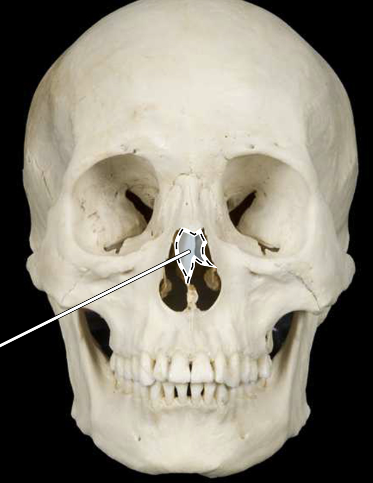
Lacrimal Bone

Vomer Bone

Palatine Bone

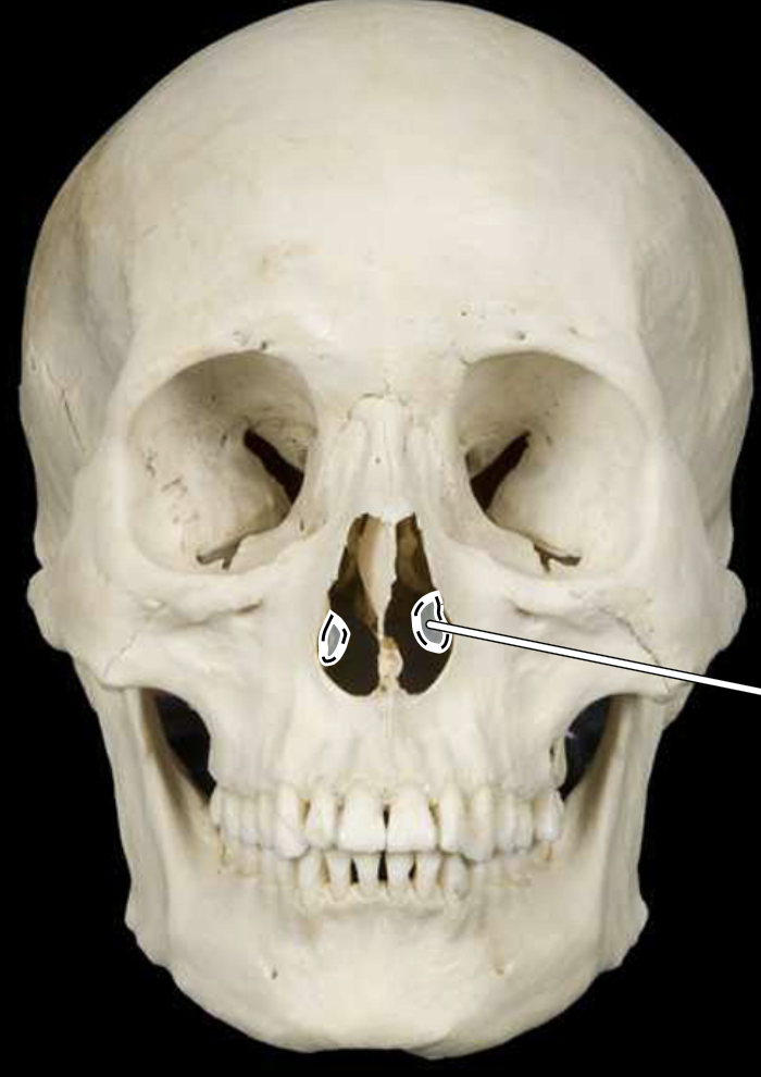
Conchae Bone
Cervical Vertebral Column

Atlas Bone
C1

Axis Bone
C2

Thoracic Vertebral Column

Lumbar Vertebral Column
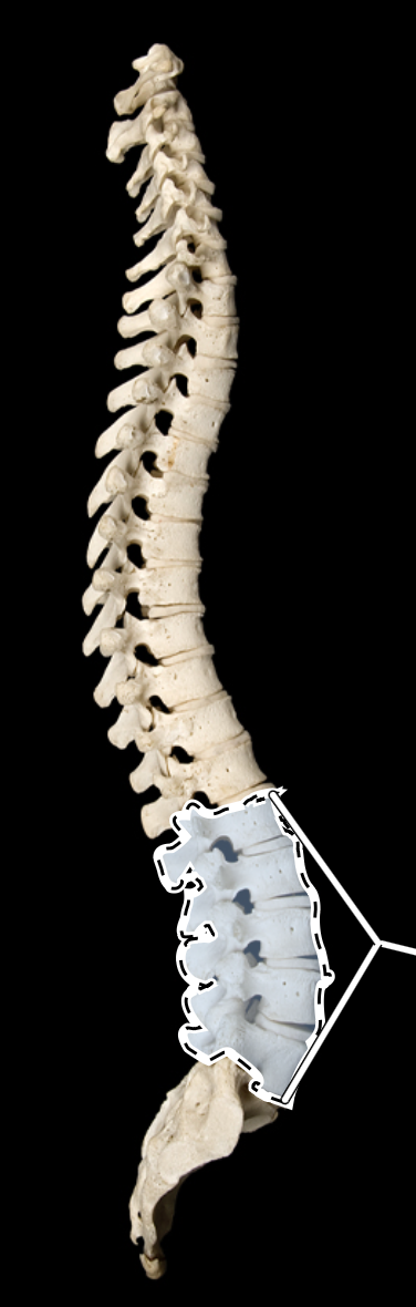
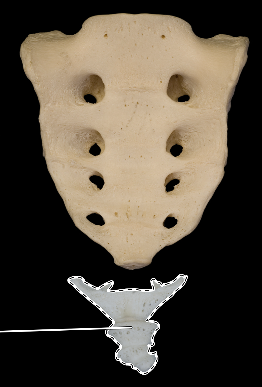
Sacrum

Coccygeal
Vertebra Body

Vertebral Foramen/Spinal Canal

Spinous Process

Transverse Process

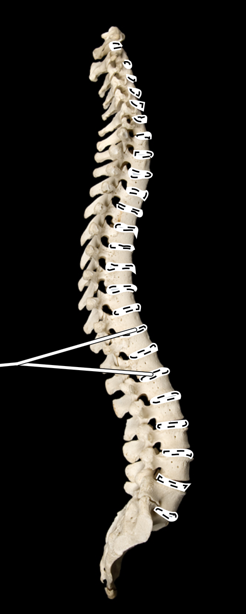
Intervertebral Discs/Fibrocartilage
Sternum Body/Gladiolus
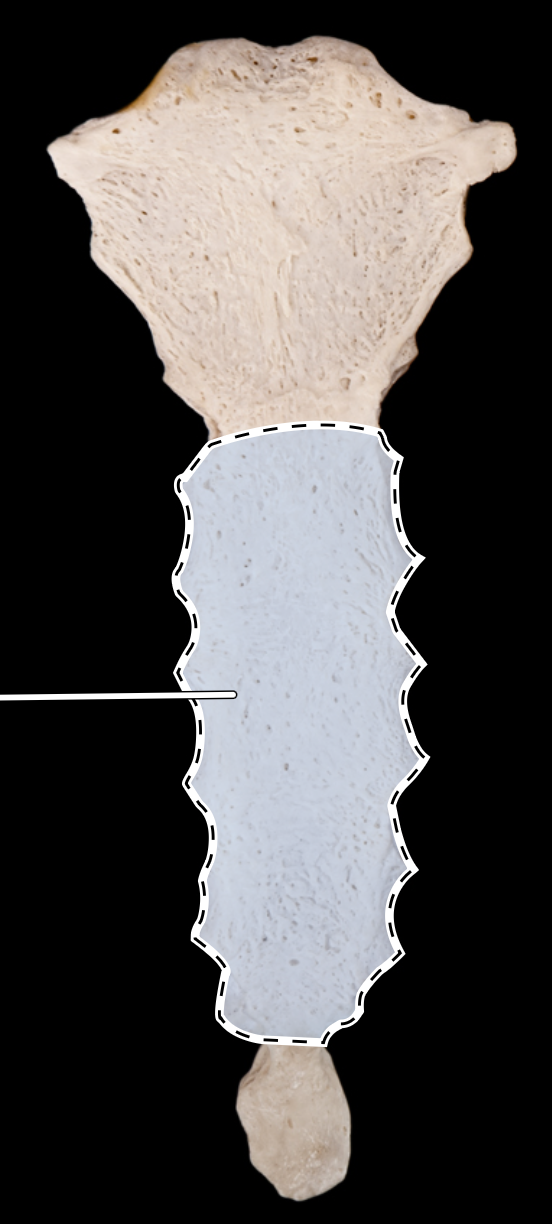
Manubrium

Xyphoid

Ribs
12 pairs
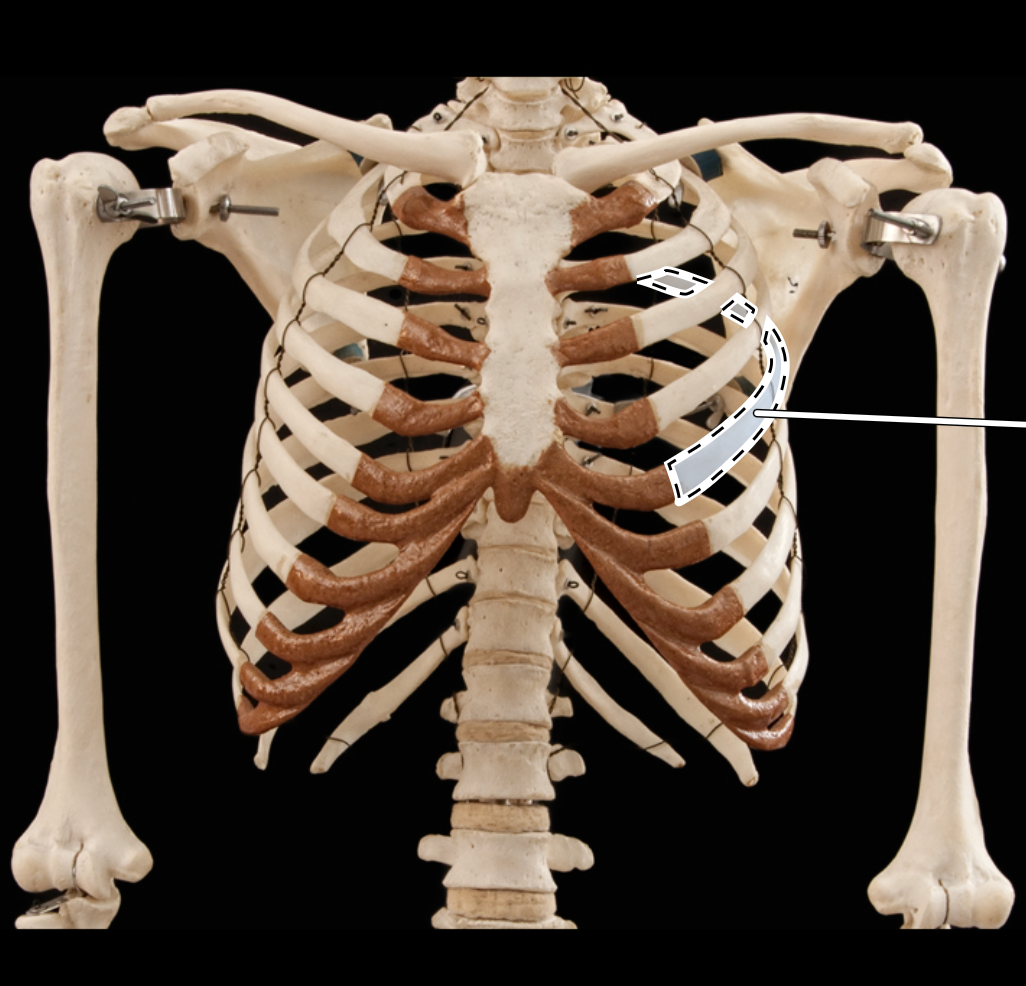
Floating Ribs
Pair 11 and 12

Costal Cartilage

Incisive foramen

Optic foramen
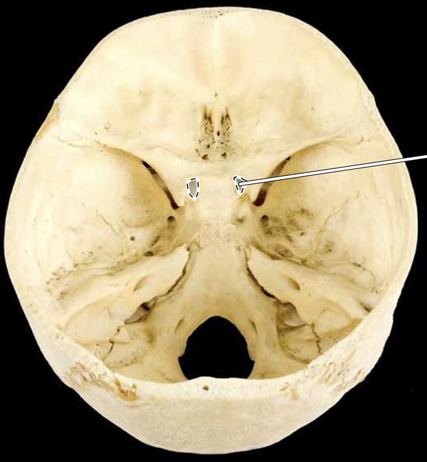
Alveolar process of mandible
Hold the lower teeth in place.

Incisors

Palatine process of maxilla

Alveolar process of maxilla

Inferior nasal concha
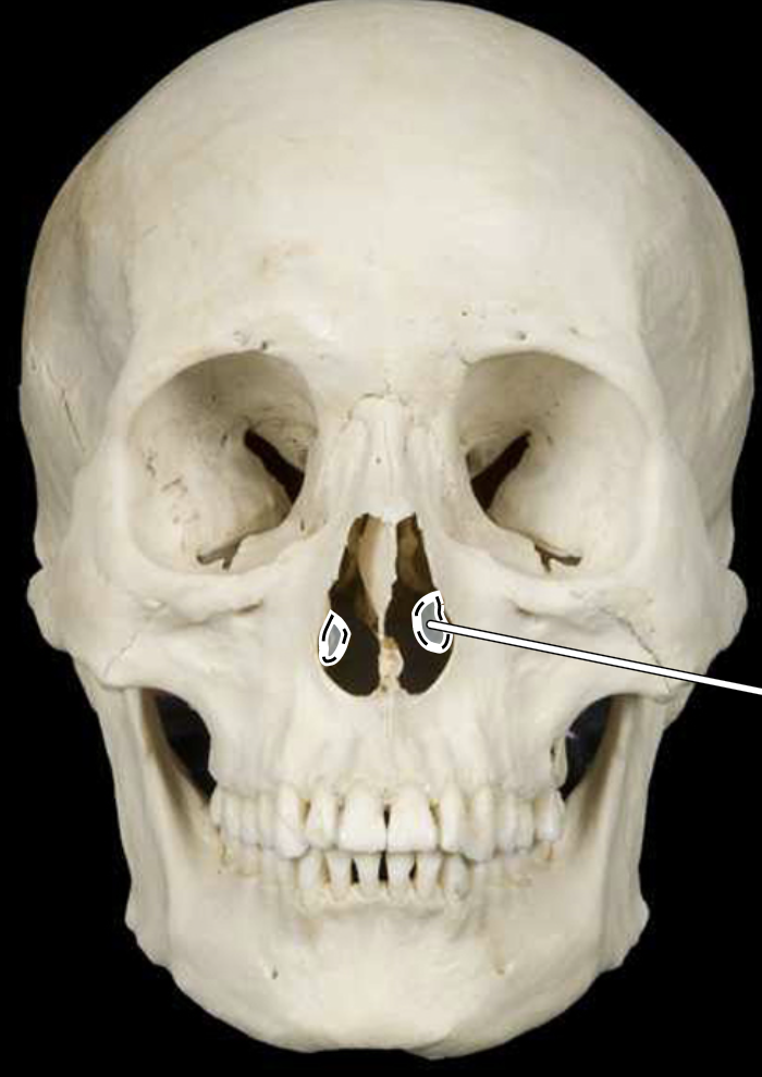
Angle of mandible

Infraorbital foramen

Premolars

Canines

Lambdoidal suture
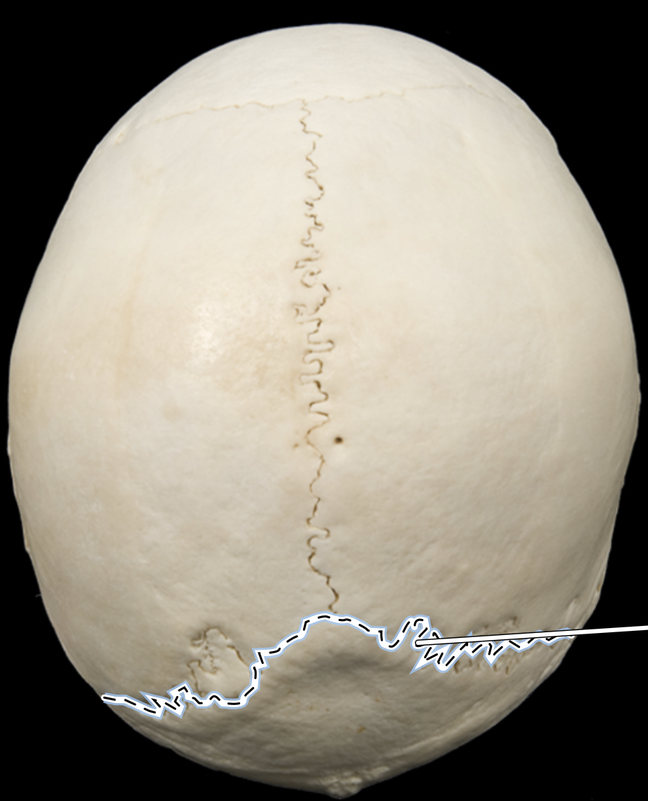
Sella turcica

Coronoid process of mandible

Cribriform plate of ethmoid

Crista galli

Lesser wing of sphenoid

External auditory meatus

Mandibular condyle

Styloid process of temporal

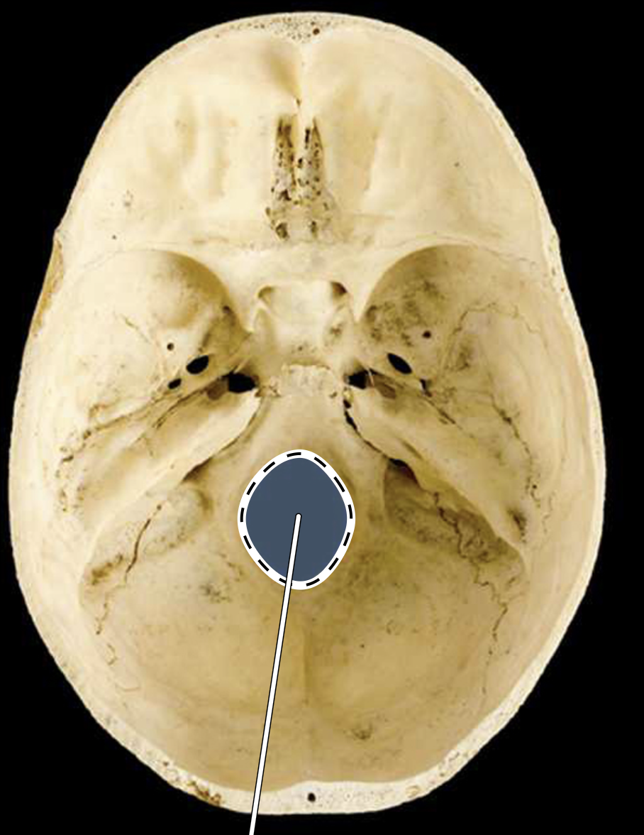
Foramen magnum
Mandibular fossa

Foramen ovale

Mandibular symphysis

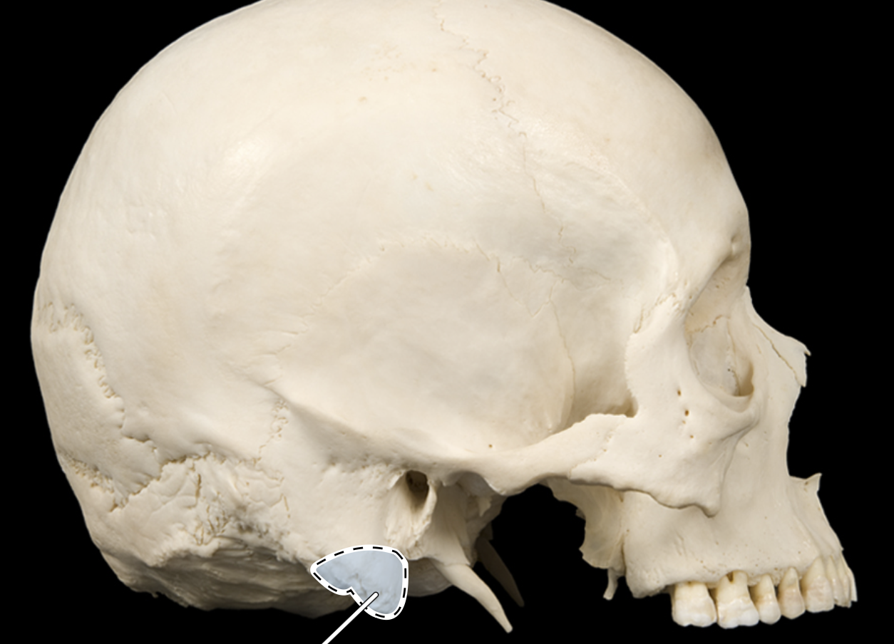
Mastoid process
Glabella

Supraorbital foramen

Molars

Greater wing of sphenoid

Mental foramen
