Lecture 19 - Cancer Genetics and Gene Therapy
 Call Kai
 Learn
 Practice Test
 Spaced Repetition
 Match
 Flashcards
 Knowt Play
1/105

There's no tags or description
Looks like no tags are added yet.
106 Terms
Cancer
A collection of diseases characterized by abnormal and uncontrolled cell growth
Carcinomas
Cancer arising in the epithelial tissue of the skin or of the lining of the internal organs
usually form solid tumors
Sarcomas
Cancers that arise in the bones and connective tissues such as fat and muscles
Lymphomas
Hematopoietic cancers that start in the cells that are part of the body's immune system (lymphoid tissue)
Leukemias
Hematopoietic cancers that begin in the bone marrow
result in a high number of abnormal white blood cells
Myelomas
Hematopoietic cancers involving white blood cells (plasma cells) responsible for producing antibodies (B-cells)
Gliomas
Cancer of brain cells

Symptoms of cancer


Leading cause of cancer death in both sexes (US, 2018)
Lung and bronchus — 83,550 deaths (26%) in males and 70,500 (25%) in females

Second leading cancer deaths by sex (US, 2018)
Prostate in males (29,430; 9%) and breast in females (40,920; 14%)

Tumor vs cancer
Cancer is uncontrolled cell division almost anywhere in the body
a tumor is when that uncontrolled growth occurs in solid tissue such as an organ, muscle or bone
Cancers that do not form tumors
Hematopoietic cancers
How tumors spread
Tumors may spread to surrounding tissues through the blood and lymph systems

Cancer and growth control
Cancer arises from a loss of normal growth control

Density-dependent inhibition in cancer
Cancer cells ignore density-dependent inhibition
continue to multiply until nutrients are exhausted

Growth/death balance in cancer
The rate of new cell growth and of old cell death are disrupted

Two ways cell growth is disrupted
Uncontrolled cell growth
loss of the cell's ability to undergo cell suicide (apoptosis)
Cancer as a genetic disease
Cancer results from a disruption of one or more genes
as genes switch “on and off” they determine when and how fast a cell grows and divides, when it stops dividing, and when it dies
Fraction of cells undergoing genetic mutations
5–10%
Main cause of cancer
The cumulative effect of somatic mutations from carcinogenic chemicals, radiation, and some viruses

Initiation phase
Genetic alteration causes the cell to acquire or lose specific functional aspects
i.e., attitude to proliferate or modifications of apoptotic mechanisms

Promotion phase
The initiated clone is amplified

Progression phase
Through further mutations the cell acquires metastatic potential
Characteristics of cancer cells
Undifferentiated (dysplasia)
less adherent
loss of cell cycle control
tissue invasion and metastasis
escaping apoptosis
insensitivity to antigrowth factors
increased mutation rate
induction of angiogenesis


Three aspects of lost cell cycle control
Length of telomere
chemical signals within and outside the cell
failed checkpoints

Angiogenesis
The physiological process through which new blood vessels form from pre-existing vessels
formed in the earlier stage of vasculogenesis
Atrophy
Morphologic change from failure to exercise
Hypertrophy
Morphologic change from repeated exercise (increase in cell size)
Hyperplasia
Morphologic change involving an increase in cell number, as in a callus
Dysplasia
Abnormal proliferation of cells

Metastasis
The development of secondary malignant growths at a distance from a primary site of cancer
advanced cancer

How metastasis happens
Cancer cells break away from the original (primary) tumor
travel through the blood or lymph system
form a new tumor in other organs or tissues

Apoptosis
Programmed cell death (PCD)
cells die in a controlled, regulated fashion
activation of initiator enzymes (caspases) that activate effector caspases to cleave cellular substrates

Features of apoptotic cells
Cytoplasmic and nuclear condensation
DNA damage
formation of apoptotic bodies
maintenance of an intact plasma membrane
exposure of surface molecules

Events during apoptosis
Caspases are produced
break down cellular components (cytoskeleton and nuclear proteins like DNA repair enzymes)
caspases also activate other enzymes such as nucleases that begin to break down nuclear DNA

Major players in apoptosis
Caspases
adaptor proteins
TNF
the TNFR family
anti-apoptotic genes (BCL-2 and BCL-XL)
pro-apoptotic genes (BAD, BAX, BIM)

Death-inducing signaling complex (DISC)
A multi-protein complex formed by members of the "death receptor" family of apoptosis-inducing cellular receptors

Example of DISC formation
FasR forms the DISC upon trimerization as a result of its ligand (FasL) binding

Fas ligands
Also called cytotoxicity-dependent APO-1-associated proteins
they physically associate with APO-1, AKA the Fas receptor or CD95

Composition of the DISC
The death receptor, FADD, and caspase 8
it transduces a downstream signal cascade resulting in apoptosis

Caspases in inflammation, apoptosis initiation, execution of apoptosis, keratinocyte differentiation
inflammation: Caspase 1, 4, 5
apoptosis initiation: Caspase 2, 8, 9, 10
execution of apoptosis: Caspase 3, 6, 7
keratinocyte differentiation: Caspase 14
Why apoptosis is needed
Keeping cell numbers relatively constant through cell death and division
replacing cells when they malfunction or become infected
stopping the spread of viral infection

Advantage of apoptosis
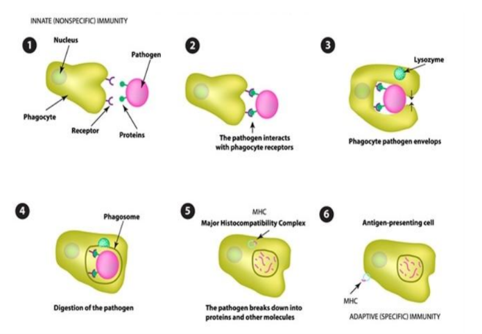
packages the dead cell into small blisters
easily digested by immune cells through phagocytosis

Phagocytosis
phagocytes engulf large prokaryotic cells such as bacteria, or eukaryotic cells such as yeast or dead cells (>0.5 µm)
kill them or remove small particles from the circulation
Excess apoptosis
Takes place during neurodegenerative disease

Deficient apoptosis
Takes place during cancer and autoimmune disorders

Germline mutations
Responsible for 5–10% of cancer cases
also called familial cancer
inherited and present in all cells from birth

Acquired mutations
sporadic cancer or somatic mutations
a series of mutations that develop during a person's lifetime
not present in every cell and not passed from parent to child

Causes of acquired mutations
Bad habits
tobacco and alcohol consumption, over-exposure to UV radiation, and toxins and chemicals

Four normal regulatory genes that may promote cancer
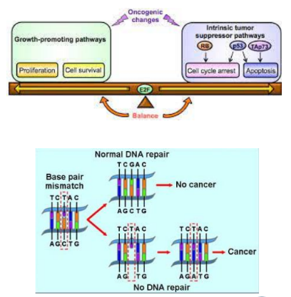
Growth promoting (proto-oncogenes)
growth inhibiting (tumor suppressor genes)
genes that regulate apoptosis (BCL2, p53 and MYC)
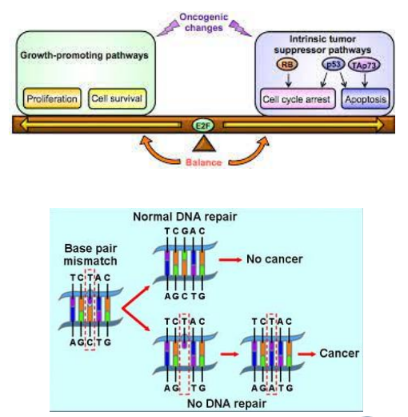
DNA repair genes
Proto-oncogene
A normal gene
when altered by mutation, becomes an oncogene that can contribute to cancer
these genes are often mutated or expressed at high levels

Functions of proto-oncogenes
Some provide signals that lead to cell division
others regulate programmed cell death

What proto-oncogenes code for
Proteins that help regulate cell growth and differentiation
often involved in signal transduction and execution of mitogenic signals through their protein product
Tumor suppressor genes
Normal genes that…
slow down cell division
repair DNA mistakes
tell cells when to die by apoptosis
Med1
A DNA repair gene that codes for one of the key enzymes involved in repairing DNA

Point mutations activating oncogenes
Deletions or insertions that lead to a hyperactive gene product
deletions/insertions in promoter region of a proto-oncogene leading to increased transcription
Gene amplification
An event leading to extra chromosomal copies of a proto-oncogene

Chromosomal translocation (oncogene activation)
Relocation of a proto-oncogene to a new chromosomal site → higher expression
fusion between a proto-oncogene and a second gene → fusion protein with oncogenic activity

Conversion of Proto-oncogene → oncogene reason 1: coding region mutation
Mutation of the coding region → protein higher physiological activity
e.g. HTLV (human T-lymphotropic virus), which can cause T-cell leukemia and lymphoma
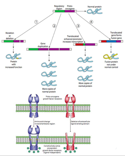
Conversion of Proto-oncogene → oncogene reason 2: gene duplication
Multiple copies of the gene are each expressed normally → making many more copies of the protein

Conversion reason 3: regulatory mutation
Mutation of the regulatory region, or translocation of a stronger enhancer or promoter to the proto-oncogene → leads to enhanced transcription and therefore more protein

Conversion reason 4: translocation in line with coding region
Translocation of another gene in line with part of the coding region puts the proto-oncogene under the control of modifiers of the translocated gene → leading to over-activity
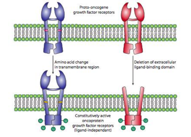
Two ways a mitogen receptor becomes an oncogene
Point mutation leading to an amino acid change in the transmembrane region (left), or truncation of the ligand-binding domain

p53 gene
A cancer suppressor gene located on chromosome 17
one of the most frequently mutated genes in human cancer

Consequence of p53 mutation
The mutant p53 protein no longer effectively binds DNA
cannot act as the "stop signal" for cell division → over-proliferation of cells

What tumor suppressor genes encode
Negative regulator proteins that keep the cell from undergoing uncontrolled division
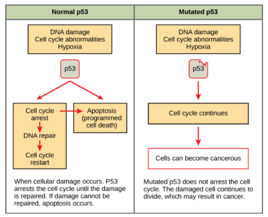
p53 and the G1 checkpoint
Without fully functional p53 → G1 checkpoint of interphase is severely compromised
the cell proceeds directly from G1 → S, creating two daughter cells that inherit the mutated p53 gene
Sources of DNA damage
Reactive oxygen species (ROS) inside cells
environmental agents such as UV light, pesticides, atmospheric carcinogens, and mycotoxins in food
Number of known human DNA repair genes
130, acting through distinct pathways of repair
Consequence of an error in DNA repair
It may attach a proto-oncogene to part of a different gene
producing a "combo" protein with unregulated activity
Ras protein
Activated by mutation in 20–30% of cancers
p53 protein mutation frequency
Mutated or deleted in 50% of cancers
BRCA1
Found in increased number in 20% of breast cancers
Abl kinase and Src kinase
Abl kinase: Activated by abnormal chromosomes in chronic myelogenous leukemia
Src kinase: Activated by mutations in 2–5% of cancers
Types of chemical carcinogen
Polyaromatic hydrocarbons
aromatic amines/amides and solvents
N-nitroso compounds
carbamates
halogenated compounds
mycotoxins
heavy metals (Hg, Cd, Ni, Cr)
radioisotopes
anticancer drugs
asbestos
preservatives such as sodium nitrate that produce nitrosamine
pesticide and herbicide residue
Viral mechanism: mutation
A classic example is point mutation of the RAS oncogene, giving a small GTPase with less activity in tumors and resultant stimulation of adenylyl cyclase activity
Viral mechanism: promoter or enhancer insertion
Insertion of a viral promoter or enhancer region near a gene activates it
Viral mechanism: chromosomal translocation
A piece of one chromosome is split off and joined to another — classic examples are Burkitt's lymphoma and the Philadelphia chromosome
Viral mechanism: gene amplification
Abnormal multiplication of a gene results in many copies, which can occur with oncogenes and with genes involved in tumor drug resistance

Telomerase
An enzyme that extends the telomeres of chromosomes
“the end replication problem”

End replication problem
Because DNA replication is asymmetric along double strands, the RNA primer sequence at the 3′-hydroxyl end cannot be replaced by DNA polymerase I, (no 3′-OH primer group for the polymerase to extend)
causing loss of 30–200 nucleotides with each replication and cell division

Telomeres in normal cells
Telomeres shorten at each cell division
when they reduce below a critical length the cell dies

Telomeres in cancer cells
Shortening of telomeres is prevented by telomerase
telomerase inhibitors may have anti-cancer activity
Factors influencing longevity
Genetics, the environment, and lifestyle
Cellular functions tied to longevity
DNA repair, maintenance of telomeres, and protection of cells from damage caused by free radicals
genes associated with blood lipid levels, inflammation, and the cardiovascular and immune systems also contribute by reducing risk of heart disease, stroke and insulin resistance
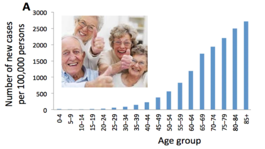
Why cancer develops in older people
prolonged exposure to carcinogens (sunlight, radiation, environmental chemicals and substances in food)
mutations also occur from random errors when a cell's DNA is copied before it divides
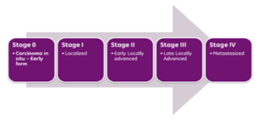
Cancer staging
The process of determining the extent to which a cancer has developed by growing and spreading
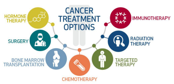
Chemotherapy
Drugs are used to kill cancer cells
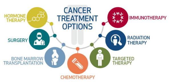
Surgery (cancer treatment)
Cancer tissues are removed by a surgical procedure

Radiation therapy
X-rays are used to kill cancer cells
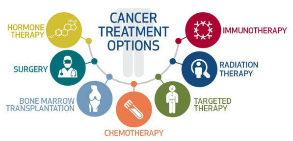
Targeted therapy
Treatment using different types of gene therapy

Immunotherapy
Treating cancer using monoclonal antibodies

Transplant (cancer treatment)
Replacing cancer cells with healthy cells, such as bone marrow
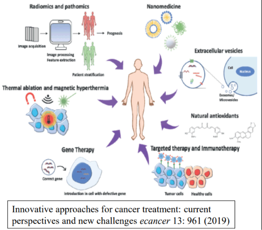
Gene therapy
Replacement of a deficient gene in the form of a protein or an enzyme, or correction of an abnormal gene

Gene therapy approach 1: immunotherapy
Uses genetically modified cells and viral particles to stimulate the immune system to destroy cancer cells

Gene therapy approach 2: oncolytic viro-therapy
Uses viral particles that replicate within the cancer cell to cause cell death
an emerging modality that shows great promise, particularly with metastatic cancers

Gene therapy approach 3: gene transfer
Introduces new genes into a cancerous cell or the surrounding tissue to cause cell death or slow the growth of cancer

In-vivo gene therapy
The therapeutic gene is introduced directly into the body of the patient (e.g. muscle, liver)
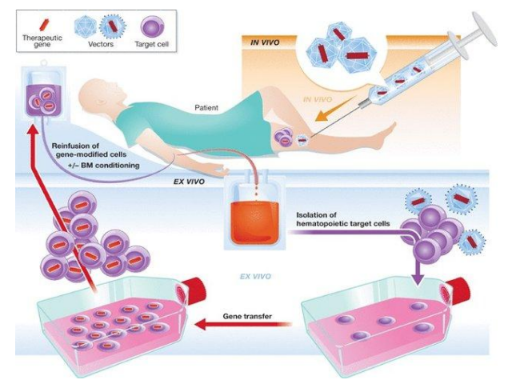
Ex-vivo gene therapy
Patient cells are first isolated from the body → genetically modified outside the body → reintroduced into the patient

Stable transfection
AKA permanent transfection
the transferred DNA is integrated into chromosomal DNA and the genetics of recipient cells is permanently changed