B&C Psych Lecture 6 From brain to cognition
 Call Kai
 Learn
 Practice Test
 Spaced Repetition
 Match
 Flashcards
 Knowt Play
1/56

There's no tags or description
Looks like no tags are added yet.
57 Terms
What is distributed processing, and why do cognitive functions depend on networks of brain regions?
Distributed processing means that different aspects of information are processed by interconnected neurons and brain regions rather than by one isolated area.
For example, an object's colour, shape, movement and sound may be processed in different regions.
These regions communicate and integrate information to produce coherent perception, cognition and behaviour.
Memory cue: Like an orchestra, different brain regions have specialised roles but must work together.
Connection to previous lectures: Distributed neural coding concerns populations of neurons; distributed brain processing emphasises interactions between brain regions.
How does the information-processing approach explain the transformation of sensory input into behaviour?
The brain can be understood as an information-processing system:
Input → Processing → Output
Input (stimulus): Sensory systems receive information from the environment.
Processing (cognition): Brain networks interpret the information and select an appropriate response.
Output (response): Motor systems produce an observable action.
This is a simplified model. In reality, processing also involves interactions and feedback between brain regions.

What are the four cerebral lobes, and what are their main cognitive functions?
Occipital lobe: Visual perception and processing.
Temporal lobe: Auditory processing, object identification (what information) and long-term memory.
Parietal lobe: Somatosensory processing and spatial/action-related (where/how) information.
Frontal lobe: Cognitive control, planning, working memory and action organisation.
Important distinction: Posterior cortical regions are strongly involved in perception, while frontal regions are particularly important for using information to guide behaviour.
Memory cue: Occipital = see; Temporal = hear/identify; Parietal = feel/locate; Frontal = plan/act.

How do the dorsal and ventral visual processing pathways differ in their functions?
Visual information is processed through two interacting pathways:
Dorsal pathway (where/how): Processes spatial information and visual information used to guide actions.
Ventral pathway (what): Processes object identity and characteristics, helping recognise what is being perceived.
Both pathways contribute to interpreting visual stimuli and guiding behaviour.
Memory cue: Dorsal = where/how to act; Ventral = what it is.

How is a visual stimulus processed and used to select an appropriate behavioural response?
Visual input: Information travels from the retina, through the thalamus, to the occipital cortex for visual processing.
Perceptual processing: Temporal and parietal association regions process relevant stimulus characteristics through the ventral (what) and dorsal (where/how) pathways.
Cognitive control: Prefrontal regions integrate perceptual information with goals, motivations and task rules to select an appropriate response.
Example: Seeing an arrow pointing right and deciding to press the right-hand button.
Key distinction: Perceptual regions process stimulus information; prefrontal regions help determine what action should be taken.
How does the brain transform a selected response into an actual movement?
After the appropriate response has been selected:
Motor planning: The premotor cortex and supplementary motor area (SMA) help organise the intended action.
Motor execution: The primary motor cortex sends motor commands that travel towards the spinal cord.
Movement coordination: The basal ganglia and cerebellum contribute to selecting, regulating and coordinating movement.
Response: Motor signals reach the relevant muscles (effectors), producing movement.
Example: Activating the muscles needed to press the right-hand button.
Memory cue: Plan → Execute → Coordinate → Move.
Why is transforming sensory information into behaviour not simply a one-way processing sequence?
Although information can be described as flowing from sensory input to motor output, brain processing also involves feedback and interactions between regions.
Feedback provides information about an ongoing or completed action, allowing the nervous system to monitor and adjust behaviour.
For example, sensory feedback from a button press helps the brain evaluate the movement.
Key principle: Behaviour depends on continuous communication between perceptual, control and motor systems, not a rigid one-way chain.

What is the role of the prefrontal cortex (PFC) in cognitive control, and what makes it particularly important in humans?
The prefrontal cortex (PFC) supports cognitive control: regulating thoughts and behaviour according to goals, task rules and situational demands.
Task complexity: PFC involvement generally increases when tasks require more planning, flexibility or control.
Human development:
The PFC is particularly large in humans.
It matures relatively late during development.
Behavioural significance: Supports flexible, goal-directed behaviour, contributing to functioning in complex social and technological environments.
Key idea: The PFC helps determine how information should be used to guide behaviour.
What are the three major surfaces of the prefrontal cortex, and what functions are they associated with?
Lateral PFC
Primarily associated with the cognitive aspects of executive functioning.
Supports goal-directed cognitive control.
Medial PFC
Associated with emotional and social regulation of behaviour.
Orbital PFC
Also associated with emotional and social regulation of behaviour.
Memory cue: Lateral = cognitive control; Medial + Orbital = emotional/social regulation.


How does the spatial oculomotor delayed-response task investigate the role of the dorsolateral PFC (dlPFC) in working memory?
The task tests whether the dlPFC remains active while spatial information must be remembered to guide a later action.
Procedure:
Fixation: Participant fixates on a cross.
Cue: A visual stimulus briefly appears at another location.
Delay: The cue disappears, but the participant must maintain fixation and remember its location.
Response: Once the fixation cross disappears, the participant moves their eyes towards the remembered location.
Measurements:
Monkeys: Single-cell recordings.
Humans: fMRI.
Key principle: The delay period separates remembering the location from seeing the stimulus and performing the movement.

What did Curtis & D'Esposito (2003) observe in the dlPFC during the delayed-response task, and what did this demonstrate?
Findings:
Macaque monkeys: Single-cell recordings showed sustained dlPFC neuronal activity during the delay period.
Humans: fMRI showed dlPFC activation during the delay period.
Interpretation:
The dlPFC remained active even though:
The visual cue was no longer present.
The participant had not yet performed the eye movement.
This demonstrates that the dlPFC participates in working-memory-related processes during the maintenance of task-relevant information.
Important limitation: Delay-period activity alone does not prove that the dlPFC directly stores the remembered location.

According to Curtis & D'Esposito (2003), how do the prefrontal and posterior cortices contribute differently to working memory?
Curtis & D'Esposito proposed a distinction between controlling information and maintaining its representation.
- Posterior cortical regions — Maintenance
Maintain representations of information, such as spatial locations and object identities.
- Prefrontal cortex — Control
Controls and manipulates these representations according to behavioural goals.
Influences posterior cortical activity to guide appropriate responses.
Example: During the delayed eye-movement task, posterior regions may maintain the remembered location while the dlPFC controls how that information is used to guide the response.
Memory cue: Posterior cortex = what is represented; PFC = what to do with it.

How can the dlPFC influence posterior brain representations and guide behaviour through other cortical regions?
The dlPFC can exert goal-directed control by influencing other cortical regions involved in maintaining information and producing responses.
Parietal cortex: Helps represent spatial information.
The dlPFC can influence these representations directly.
Frontal eye fields (FEF): Involved in controlling eye movements.
Provide a pathway through which prefrontal control can guide spatially directed eye movements.
Broca's region: Involved in speech production.
Can contribute to prefrontal control of verbal information.
Connection to predictive coding: Both involve top-down influences from higher-level regions to posterior processing areas. In predictive coding, feedback predictions help determine prediction errors.
Key idea: The PFC coordinates goal-directed processing by interacting with specialised brain regions rather than performing every cognitive function itself.

What is working memory, and what are its main characteristics?
Working memory (WM) is the temporary, active maintenance of information needed for ongoing cognitive activities.
Main characteristics:
Temporary: Holds information for current tasks.
Active: Maintains information for use in cognitive processing, rather than simply storing it passively.
Limited capacity: Only a restricted amount of information can be maintained.
Vulnerable to interference: Distractions can disrupt maintained information.
Multiple information types: Can maintain verbal, auditory, visual and spatial information.
Multiple sources: Information can come from current perception or long-term memory.
Key idea: WM allows information to remain available while we use it to think, reason or act.
What are the three components of Baddeley & Hitch's (1974) working-memory model, and what does each do?
The model distinguishes one control system and two temporary-storage systems.
1. Central executive — Control
Directs attention and coordinates working-memory activities.
2. Phonological loop — Verbal/auditory storage
Temporarily maintains verbal and sound-based information.
3. Visuospatial sketchpad — Visual/spatial storage
Temporarily maintains visual images and spatial information.
The phonological loop and visuospatial sketchpad are called slave systems because they operate under central-executive control.
Memory cue: Executive = controls; Loop = sounds; Sketchpad = images and space.

What are the main functions of the central executive in Baddeley & Hitch's working-memory model?
The central executive is the attentional control system that coordinates working-memory processes.
Main functions:
Recruitment: Activates the relevant storage systems.
Selective attention: Focuses on relevant information while ignoring distractions.
Attention distribution: Allocates attention between tasks or storage systems.
Coordination: Organises complex cognitive activities, such as mental arithmetic.
Long-term memory activation: Temporarily activates relevant information retrieved from long-term memory.
Key distinction: The central executive primarily controls how information is processed, rather than serving as a temporary-storage system itself.
What is the function of the phonological loop, and how does it maintain information?
The phonological loop temporarily maintains verbal and auditory information in working memory.
Information type: Speech, words and sounds.
Maintenance mechanism: Silent/internal rehearsal keeps phonological information active.
Capacity: Limited; information can be lost without continued maintenance.
Example: Mentally repeating a telephone number until you can enter it.
Memory cue: Phonological = sound; Loop = repeating information.
What is the function of the visuospatial sketchpad, and what kinds of information does it maintain?
The visuospatial sketchpad temporarily maintains visual and spatial information.
Visual information: Mental images, shapes and appearances.
Spatial information: Locations and spatial relationships.
Information sources: Can use current perceptual input or representations retrieved from long-term memory.
Capacity: Limited.
Example: Mentally picturing a cat sitting in a tree and imagining where the cat is positioned.
Memory cue: Visuo = what something looks like; Spatial = where it is.
What are the four working-memory subcomponents described in Baddeley's (2003) anatomical mapping, and what does each do?
Phonological loop:
Articulatory rehearsal (AR)
Maintains verbal/phonological information through rehearsal.
Phonological store (PS)
Temporarily represents phonological information.
Visuospatial sketchpad:
Inner scribe (IS)
Maintains visuospatial information.
Visual cache (VC)
Temporarily represents visual information.
Key distinction: Rehearsal/maintenance processes help keep information active, while storage components represent the information.

How did Baddeley (2003) tentatively relate working-memory functions to different brain regions and hemispheres?
Baddeley's proposed anatomical mapping suggests:
Left hemisphere: Predominantly associated with phonological working-memory functions.
Right hemisphere: Predominantly associated with visuospatial working-memory functions.
Prefrontal cortex (PFC):
Particularly associated with controlling and manipulating information.
Posterior cortical regions:
More strongly associated with maintaining information representations.
Overlap with regions involved in perception.
Important qualification: This anatomical mapping is tentative and debated, not a fixed one-to-one correspondence between components and brain regions.
Memory cue: Left = language/sounds; Right = visual/space; Front = control; Back = representations.
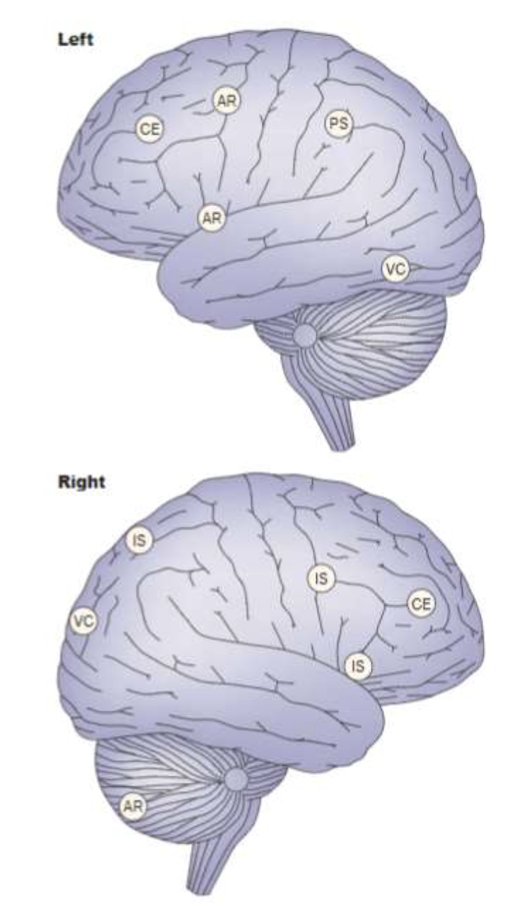

How do different brain regions interact to support working memory, as illustrated in lecture slide 23?
Working memory involves interactions between frontal and posterior brain regions, supporting attentional control, temporary maintenance and coordinated behaviour.
Main brain regions and functions:
Prefrontal areas — Central executive
Represent higher-level cognitive goals and contribute to cognitive control.
Broca's area — Phonological loop
Supports speech-related processing and verbal rehearsal.
Frontal eye fields — Visuospatial sketchpad
Involved in eye-movement control and spatial attention.
Parietal cortex — Visuospatial sketchpad
Supports spatial attention and visuospatial information processing.
Inferior parietal cortex — Attentional priorities
Contributes to selecting and prioritising relevant information.
Posterior visual and motor coordination regions
Contribute to integrating visual information with movement.
Key principle: Working-memory components do not correspond to isolated brain regions; they rely on interconnected networks.
What is the difference between working-memory duration and capacity?
Working memory has limitations in both duration and capacity.
Duration: How long information can remain available.
Unrehearsed information in the phonological system lasts approximately 2 seconds.
Rehearsal can help maintain information for longer.
Capacity: How many items or chunks can be maintained simultaneously.
Capacity is limited and depends partly on how information is organised.
Key distinction: Duration = how long; capacity = how much.
What did Miller (1956) propose about working-memory capacity, and why was this estimate later questioned?
Miller proposed a capacity of approximately 7 ± 2 items (around 5–9).
Evidence:
Digit span: People could typically recall approximately 5–9 digits.
Magnitude ratings: People could reliably distinguish approximately seven alternatives.
Limitation:
Participants could organise information into meaningful groups (chunks).
This may have inflated estimates of how many independent items could be maintained.
Key idea: Miller's estimate may partly reflect the benefits of organising information rather than pure storage capacity.
What is chunking, and how can it increase the amount of information remembered without increasing working-memory capacity?
Chunking is grouping strongly associated items into meaningful units that are treated as single chunks.
Example:
Individual letters:
F B I A B C I B M→ 9 items.Meaningful chunks:
FBI – ABC – IBM→ 3 chunks.
Instead of remembering nine separate letters, a person familiar with the abbreviations can remember three meaningful units.
Key principle: Chunking increases the amount of information represented per chunk, not necessarily the number of chunks working memory can hold.
What did Cowan (2000) propose about working-memory capacity, and how does it differ from Miller's estimate?
Cowan proposed that working-memory capacity is closer to approximately four items or chunks, rather than Miller's 7 ± 2.
Supporting evidence:
Unattended speech: Around four digits could be recalled unexpectedly.
Enumeration: Small sets of approximately 3–4 objects could be counted particularly efficiently.
Multiple-object tracking: People could successfully track approximately 3–4 moving objects.
Key distinction: Miller = approximately 7 ± 2; Cowan = approximately 4.
Capacity estimates can vary depending on chunking, task demands and information complexity.
How did Cowan et al. (1999) investigate working-memory capacity using unattended speech, and what did they find?
Method:
Participants performed a primary picture-matching task.
Spoken digits were presented simultaneously as secondary, unattended information.
On occasional unexpected trials, participants were asked to recall the digits.
Finding:
- Participants recalled approximately four items from the unattended speech.
Conclusion:
- This supported Cowan's smaller capacity estimate, without relying on intentional memorisation of long digit sequences.

How do enumeration and multiple-object tracking provide evidence for a working-memory capacity of approximately 3–4 items?
1. Enumeration task
Participants report how many objects are briefly displayed.
1–3/4 objects: Reaction times increase very little.
More objects: Reaction times increase more steeply.
Suggests a small number of objects can be processed particularly efficiently.
2. Multiple-object tracking
Participants track several moving objects simultaneously.
3–4 objects: Tracking is relatively manageable.
More objects: Performance declines.
Conclusion: Both tasks support a limited ability to maintain or track multiple object representations.

How did Luck & Vogel (1997) investigate visual working-memory capacity using a change-detection task?
Research question: How many visual objects can be temporarily maintained in memory?
Procedure:
Memory array: Several coloured objects are briefly presented.
Retention interval: The display disappears, requiring participants to maintain the colours and locations.
Test array: A new display appears.
Response: Participants indicate whether an object's colour has changed.
Key principle: Accurate performance requires maintaining visual object representations across the delay.
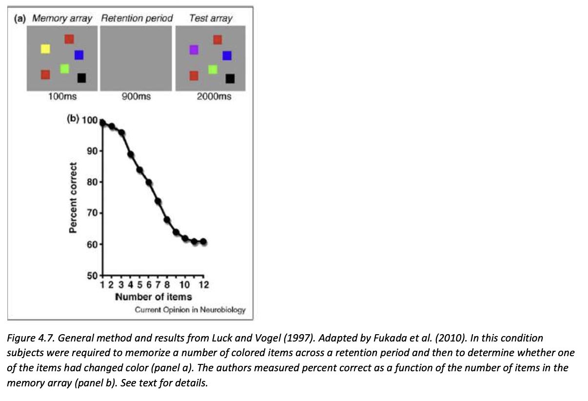
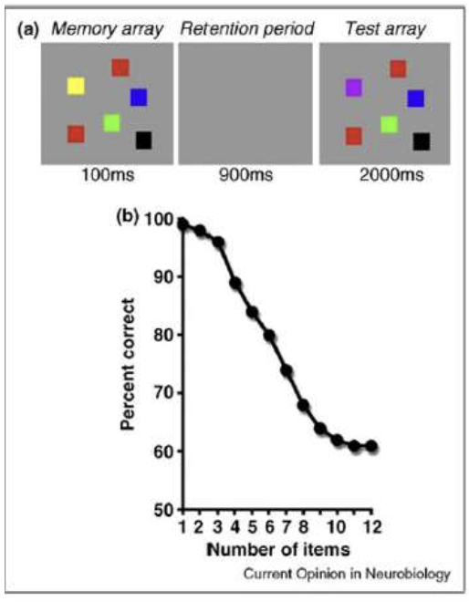
What did Luck & Vogel (1997) find about visual working-memory capacity, and how does this support Cowan's estimate?
Results:
1–3 objects: Change-detection accuracy remained very high.
4 or more objects: Accuracy declined as the number of objects increased.
Estimated capacity: Approximately 3–5 objects, commonly summarised as around four.
Conclusion:
Visual working memory can maintain only a limited number of object representations.
The findings support Cowan's estimate of approximately four items rather than a fixed capacity of seven.
Important: Capacity is approximate and can vary with the complexity of the information and task demands.
How did Xu & Chun (2006) investigate where visual working memory is stored and whether capacity depends on object complexity?
Research questions:
Which brain regions maintain visual objects in working memory?
Does object complexity influence working-memory capacity?
Method: Visual change-detection task using fMRI.
Procedure:
Sample display: Participants briefly viewed several objects and memorised their identities and locations.
Retention interval: Objects disappeared, requiring participants to maintain the information.
Test display: One object appeared at a previously occupied location.
Response: Participants judged whether that object had changed.
Two conditions:
Simple: Detect a change in whether an object contained a gap.
Complex: Detect a change in the object's shape.
Measurements: Behavioural performance and fMRI activity while varying the number and complexity of objects.

Which three brain regions did Xu & Chun (2006) investigate, and what are their proposed roles in visual working memory?
1. Superior intraparietal sulcus (superior IPS)
Associated with maintaining object identities.
Storage capacity is sensitive to object complexity.
2. Inferior intraparietal sulcus (inferior IPS)
Associated with maintaining spatial locations.
Storage capacity is relatively unaffected by object complexity.
3. Lateral occipital complex (LOC)
Associated with maintaining object identities.
Storage capacity is sensitive to object complexity.
Key distinction:
Superior IPS + LOC: What the objects are.
Inferior IPS: Where the objects are.

How did Xu & Chun (2006) use changes in fMRI activity to estimate the storage capacity of different brain regions?
The researchers assumed that activity in a storage-related region should reflect the amount of information maintained.
Expected relationship:
Few objects: Relatively little information must be maintained → lower activity.
Increasing objects: More representations must be maintained → activity increases.
Capacity limit reached: Additional objects cannot all be stored → activity levels off (plateaus).
Interpretation:
The point where activity plateaus provides an estimate of the region's storage-capacity limit.
Comparing plateau points across conditions reveals whether capacity depends on object complexity.
Key idea: An activity plateau may indicate that the storage system has reached its capacity.
-This is the study's interpretation of the activity pattern, not proof that fMRI activity directly measures the exact number of stored objects.
What did Xu & Chun (2006) find about the effect of object complexity on visual working-memory capacity?
Behavioural findings:
Simple feature changes:
Capacity ≈ 4 objects.
Complex feature changes:
Capacity ≈ 2 objects.
Interpretation:
Complex objects require more detailed information to be maintained.
Consequently, fewer complex objects can be remembered successfully.
Conclusion: Visual working-memory capacity depends not only on the number of objects but also on their complexity.
Memory cue: Simple = 4; Complex = 2.
What did Xu & Chun (2006) find about activity in the superior IPS and LOC, and what does this suggest about their functions?
fMRI findings — Superior IPS and LOC:
Simple objects:
Activity increased until approximately 4 objects, then plateaued.
Complex objects:
Activity increased until approximately 2 objects, then plateaued.
Interpretation:
Their estimated capacity decreases when object complexity increases.
This suggests involvement in maintaining object identities and their visual characteristics, rather than merely recording how many spatial locations are occupied.
Key conclusion: Superior IPS and LOC show complexity-sensitive storage capacity.

What did Xu & Chun (2006) find about inferior IPS activity, and why does this suggest a role in spatial-location storage?
fMRI findings — Inferior IPS:
Simple objects:
Activity increased until approximately 4 objects, then plateaued.
Complex objects:
Activity also increased until approximately 4 objects, then plateaued.
Interpretation:
Activity depended on the number of objects/locations rather than object complexity.
This suggests the inferior IPS primarily maintains spatial-location representations, rather than detailed visual identities.
Key conclusion: Inferior IPS capacity is relatively complexity-insensitive.
Memory cue: Inferior IPS = where, not detailed what.

How did Xu & Chun (2006) provide additional evidence that the inferior IPS maintains spatial locations rather than object identities?
Method:
Multiple objects were presented sequentially at the same spatial location.
Researchers measured inferior IPS activity as the number of presented objects increased.
Finding:
- Inferior IPS activity did not increase with the number of objects.
Interpretation:
Although several objects were presented, only one spatial location needed to be represented.
If inferior IPS primarily maintained separate object identities, increasing the number of objects might have increased its activity.
Conclusion: The findings further support the role of inferior IPS in maintaining spatial locations rather than detailed object identities.
What are the main conclusions of Xu & Chun (2006), and how can the fMRI results distinguish object-identity storage from spatial-location storage?
Main conclusions:
1. Object complexity affects WM capacity:
Simple objects ≈ 4; complex objects ≈ 2.
2. Different posterior regions have specialised roles:
Superior IPS + LOC: Object identities; activity plateaus earlier for complex objects.
Inferior IPS: Spatial locations; activity plateaus around 4 regardless of complexity.
3. Visual WM relies on distributed processing:
Multiple posterior cortical regions cooperate to maintain objects and their locations.
These regions also contribute to visual perception.
Graph interpretation:
Different plateau points across conditions → Complexity-sensitive storage (superior IPS/LOC).
Similar plateau points across conditions → Complexity-insensitive storage (inferior IPS).
Key principle: Visual working memory involves specialised, interacting brain regions rather than one general-purpose storage area.

What are the fusiform face area (FFA) and parahippocampal place area (PPA), and what are their visual specializations?
Certain posterior cortical regions are specialised for processing particular categories of visual information.
Fusiform face area (FFA):
Particularly involved in processing faces.
Parahippocampal place area (PPA):
Particularly involved in processing places, buildings and scenes with spatial layouts.
Key idea: Visual processing is partly organised into specialised cortical regions, rather than all visual information being processed identically.

How did O'Craven & Kanwisher (2000) investigate whether visual perception and mental imagery involve the same specialised brain regions?
Research question: Are the FFA and PPA activated during both actual perception and mental imagery of their preferred stimulus categories?
Method: fMRI measurements of FFA and PPA activity across two conditions.
Perception condition:
Participants viewed familiar faces or scenes.
Imagery condition:
Participants heard the names of familiar faces or scenes.
They closed their eyes and mentally imagined the named stimuli.
Key comparison: Brain activity during seeing a face or scene versus imagining it.
What did O'Craven & Kanwisher (2000) find about FFA and PPA activity during visual perception and mental imagery?
Results:
- Fusiform face area (FFA):
Showed greater activity for faces than scenes.
Activated during both face perception and face imagery.
- Parahippocampal place area (PPA):
Showed greater activity for scenes than faces.
Activated during both scene perception and scene imagery.
- Perception versus imagery:
Activity was generally stronger during perception than imagery.
Key finding: Both regions retained their stimulus-category preferences even when the stimuli were only mentally imagined.

What do O'Craven & Kanwisher's (2000) findings reveal about the relationship between visual perception, mental imagery and top-down processing?
Main conclusions:
1. Shared neural involvement:
The same category-selective cortical regions can be activated during perception and mental imagery.
FFA responds to faces; PPA responds to scenes.
2. Top-down processing:
Mental imagery can activate posterior perceptual regions even without corresponding visual input.
This demonstrates that these regions can be influenced by internally generated information.
3. Perception versus imagery:
They involve overlapping neural mechanisms, but imagery generally produces weaker activity.
Connection to Lecture 2: Consistent with Ganis et al. (2004), which also found overlapping brain involvement in visual perception and imagery.
Key principle: Imagining an object can recruit some of the same specialised brain regions involved in actually seeing it.
What role does the dorsolateral prefrontal cortex (dlPFC) play in protecting working memory from distraction?
The dlPFC helps protect task-relevant working-memory representations when distracting information is present.
Posterior cortical regions: Maintain representations of information.
dlPFC: Exerts top-down control over these representations.
Becomes particularly active when distraction increases the need for cognitive control.
Two possible control mechanisms:
Target enhancement: Strengthening representations of relevant information.
Distractor suppression: Reducing representations of irrelevant information.
Key question: Does the dlPFC protect working memory by strengthening targets or suppressing distractors?
How did Feredoes et al. (2011) investigate the dlPFC's role in protecting working memory from distraction?
Research question: Does the dlPFC protect working memory through target enhancement or distractor suppression?
Method: Working-memory task combining TMS and fMRI.
Procedure:
Targets: Participants memorised a sequence of faces or houses.
Retention interval: Targets had to be maintained temporarily.
On some trials, distractors from the opposite category appeared.
TMS: Applied to the dlPFC on half the trials.
Memory probe: Participants judged whether a subsequent stimulus was one of the original targets.
Brain regions monitored:
FFA: Face representations.
PPA: House representations.

What were the four experimental conditions in Feredoes et al. (2011), and why were faces and houses used?
Condition | Targets | Distractors |
|---|---|---|
A | Faces | None |
B | Faces | Houses |
C | Houses | None |
D | Houses | Faces |
Why faces and houses?
They are processed preferentially in different posterior cortical regions:
FFA: Faces.
PPA: Houses/scenes.
This allowed researchers to distinguish neural activity associated with targets from activity associated with distractors.
Key comparison: Conditions with versus without distraction, combined with TMS versus no TMS.
Why did Feredoes et al. (2011) combine TMS and fMRI, and how did this reveal dlPFC interactions with posterior regions?
The two methods served different purposes:
- TMS — Influence dlPFC activity
Applied to the dlPFC to amplify its ongoing modulatory influence on connected posterior regions.
Was not simply intended to disrupt dlPFC functioning.
- fMRI — Measure posterior activity
Recorded activity in the FFA and PPA during the memory task.
Experimental logic:
If the dlPFC was modulating a posterior region, TMS should change activity in that region.
If the dlPFC was not significantly modulating that region, TMS should have little or no detectable effect there.
Key idea: Combining TMS and fMRI allowed researchers to investigate which posterior representations were influenced by prefrontal control.
What were the predictions of the target-enhancement and distractor-suppression hypotheses in Feredoes et al. (2011)?
1. Target-enhancement hypothesis
The dlPFC strengthens relevant working-memory representations.
Face targets + house distractors: TMS should increase FFA activity.
House targets + face distractors: TMS should increase PPA activity.
2. Distractor-suppression hypothesis
The dlPFC suppresses irrelevant representations.
House distractors: TMS should influence PPA activity.
Face distractors: TMS should influence FFA activity.
Key distinction:
Enhancement: Effects in the region representing the targets.
Suppression: Effects in the region representing the distractors.
The prediction concerns which region is affected; the direction of a TMS effect depends on how stimulation changes the ongoing interaction.
What did Feredoes et al. (2011) find about the effects of dlPFC stimulation on FFA and PPA activity?
Main findings:
1. TMS effects depended on distraction:
Significant changes in posterior activity occurred only when distractors were present.
2. Face targets + house distractors:
TMS increased activity in the FFA (target-processing region).
3. House targets + face distractors:
TMS increased activity in the PPA (target-processing region).
4. Distractor-processing regions:
No corresponding significant TMS effects were found in regions representing the distractors.
Key result: TMS strengthened activity associated with relevant targets, not irrelevant distractors.

What did Feredoes et al. (2011) conclude about how the dlPFC protects working memory during distraction?
Main conclusion: The dlPFC protects working-memory contents primarily through target enhancement, rather than distractor suppression.
Supporting evidence:
TMS increased activity in posterior regions representing relevant targets.
No significant corresponding effects occurred in regions representing distractors.
Effects were observed specifically when distracting information was present.
Broader implications:
PFC: Provides goal-directed, top-down control.
Posterior cortex: Maintains task-relevant representations.
Their interaction: Helps preserve relevant information when distraction increases cognitive demands.
Key principle: Cognitive control can protect working memory by strengthening relevant information, not merely by inhibiting irrelevant information.
Qualification: The findings favour target enhancement in this task; they do not establish that the PFC never suppresses distractors.
What is the difference between bottom-up and top-down processing in terms of their driving influences, information flow and behavioural effects?
1. Bottom-up processing — Stimulus-driven
Driven by: External sensory information.
Information flow: Sensory regions → higher-level/response-related regions (feedforward).
Function: Enables environmental stimuli to automatically attract attention or trigger responses.
Example: Automatically looking towards a suddenly appearing object.
2. Top-down processing — Goal-directed
Driven by: Goals, expectations and cognitive control.
Information flow: Higher-level control regions → lower-level processing regions (feedback).
Function: Modulates processing and enables responses that may conflict with automatic tendencies.
Example: Ignoring a distracting stimulus to complete a task.
Key distinction: Bottom-up = environment drives processing; top-down = goals guide processing.
Important: Both processes interact and can conflict, requiring executive control.
How does the Stroop task demonstrate conflict between bottom-up and top-down processing?
Task: Participants must name the ink colour of a written word rather than read the word itself.
Two conditions:
1. Congruent trials:
Word meaning and ink colour match.
Example: The word "RED" printed in red.
No conflict between reading and colour naming → faster responses.
2. Incongruent trials:
Word meaning and ink colour differ.
Example: The word "RED" printed in blue.
Reading produces a competing response → slower responses.
Processing conflict:
Bottom-up: Automatic tendency to read the word.
Top-down: Goal-directed control to name the ink colour instead.
Key principle: Incongruent trials require cognitive control to overcome the competing reading response.


Why are responses slower during incongruent Stroop trials, and what role does the prefrontal cortex play?
Stroop interference occurs when the automatic reading response conflicts with the required ink-colour response.
During incongruent trials:
The written word automatically activates a reading response.
This response competes with the task goal of naming the ink colour.
Executive control, involving the PFC, helps inhibit the competing response and select the correct one.
Result:
Incongruent trials typically produce slower response times than congruent trials.
The additional control required makes the task more demanding.
Key conclusion: The PFC contributes to overriding automatic responses when they conflict with behavioural goals.
How do prosaccades and anti-saccades differ, and how do they illustrate bottom-up versus top-down control?
A saccade is a rapid eye movement between points of fixation.
Task: Participants initially fixate on a central cross. A stimulus then appears to the left or right.
1. Prosaccade — Bottom-up
Look towards the appearing stimulus.
Follows the natural, stimulus-driven tendency to orient towards new visual information.
Requires relatively little executive control.
2. Anti-saccade — Top-down
Look away from the stimulus, in the opposite direction.
Requires inhibition of the automatic prosaccade.
Requires programming an alternative, goal-directed eye movement.
Example: Stimulus appears on the right → prosaccade = look right; anti-saccade = look left.
Memory cue: Pro = towards; Anti = opposite.

Why are anti-saccades slower and more error-prone than prosaccades, and what does this reveal about cognitive control?
Main findings:
Prosaccades: Faster and generally more accurate.
Anti-saccades: Slower and produce more errors.
Common error: Looking towards the stimulus instead of away from it.
Explanation:
A suddenly appearing stimulus automatically creates a tendency to look towards it.
To perform an anti-saccade, frontal control regions must:
Inhibit the automatic eye movement towards the stimulus.
Program a new eye movement in the opposite direction.
Execute the goal-directed response.
Conclusion: Anti-saccades require additional top-down executive control to overcome a stimulus-driven response.
Key idea: Greater cognitive-control demands lead to slower responses and increased opportunities for errors.
How do feedforward and feedback processing contribute differently to prosaccades and anti-saccades?
1. Feedforward processing — Bottom-up
Direction: Sensory regions → higher-level/response-related regions.
Function: Transmits incoming sensory information towards systems that can generate a response.
Prosaccade: A visual stimulus can rapidly trigger an eye movement towards its location.
2. Feedback processing — Top-down
Direction: Higher-level frontal control regions → earlier processing/response systems.
Function: Modulates sensory processing and response selection according to task goals.
Anti-saccade: Frontal control helps suppress the automatic response and guide an eye movement in the opposite direction.
Key distinction: Feedforward carries stimulus-driven information; feedback allows higher-level control to influence ongoing processing.
Important: Both directions operate within interacting networks rather than entirely separate processing systems.
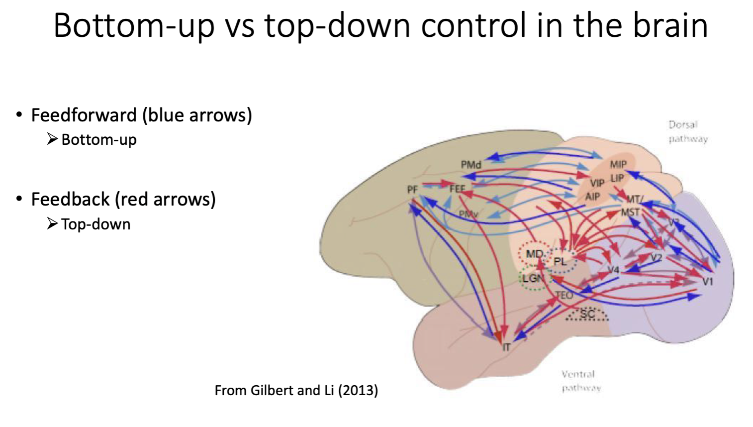
How does top-down feedback processing contribute to response inhibition, working memory, attention and predictive coding?
Top-down processing allows higher-level brain regions to influence lower-level processing according to goals and expectations.
Applications:
1. Response inhibition
Overcomes automatic, stimulus-driven responses.
Examples: Stroop task and anti-saccades.
2. Working memory
Helps maintain or enhance task-relevant representations in posterior regions.
Example: dlPFC target enhancement during distraction (Feredoes et al., 2011).
3. Attention
Modulates processing to prioritise information relevant to current goals.
4. Predictive coding
Higher-level regions send predictions to earlier processing areas.
These predictions help determine prediction errors.
Overall conclusion: Cognition depends on continuous interactions between bottom-up sensory input and top-down feedback, rather than a one-way flow of information.