Ch 8 Membrane Transport
 Call Kai
 Learn
 Practice Test
 Spaced Repetition
 Match
 Flashcards
 Knowt Play
1/49

There's no tags or description
Looks like no tags are added yet.
50 Terms
Passive mediated transport
something helping across membrane
no E required, moving down a gradient
Active transport
requires E, moving against concentration gradient
such an endergonic process must be coupled to a sufficiently exergonic process to make it favorable
non-mediated transport
simple diffusion
O2 from Hb → Mb
Mediated transport
needs carrier
can be passive or active
transport of acetate (b/c charged)
_________ and ___________ dictate exergonicity
concentrations and membrane potential
chemical potential

chemical potential difference
if concentration is greater outside, things flow in spontaneously
if concentration is greater inside, active mediation via ATP or another gradient is required
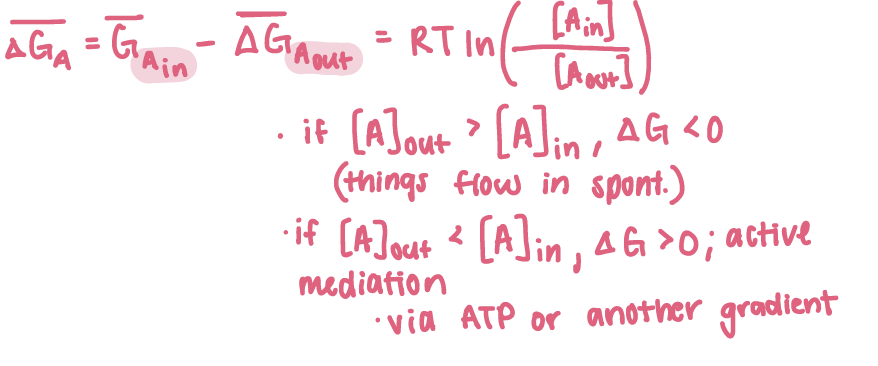
electrochemical potential
for charged species: Na+, Ca2+, K+, Cl-, CO32-, R-COO-, R-NH3+

passive-mediated transport
aka facilitated diffusion, molecules flow high→low
ionophores
porins
ion channels
aquaporins
transport proteins
ionophores
increase increase permeability of membranes to ions
there effect can only be to equilibrate concentrations on either side of membrane
carrier ionophores - actually moves in membrane
channel-forming ionophores - allows flow
channel-forming ionophores
form transmembrane channels or pores through which select ions can diffuse

Carrier ionophores
increase permeability of membranes to their selected ions by binding it, diffusing through membrane, and releasing ion on other side
their ionic complexes are soluble in nonpolar solvents

porins
H2O, ions, small, molecules
beta-barrels (antiparallel beta-strands)
select solutes using diameter and a.a. residues
found in outer membrane of gram (+) mitochondria and chloroplast
ion channels
highly selective and may be gated
aquaporins
mediate transmembrane passage of H2O but not protons
transport proteins
mediate uniport, symport (coupling), or antiport transport
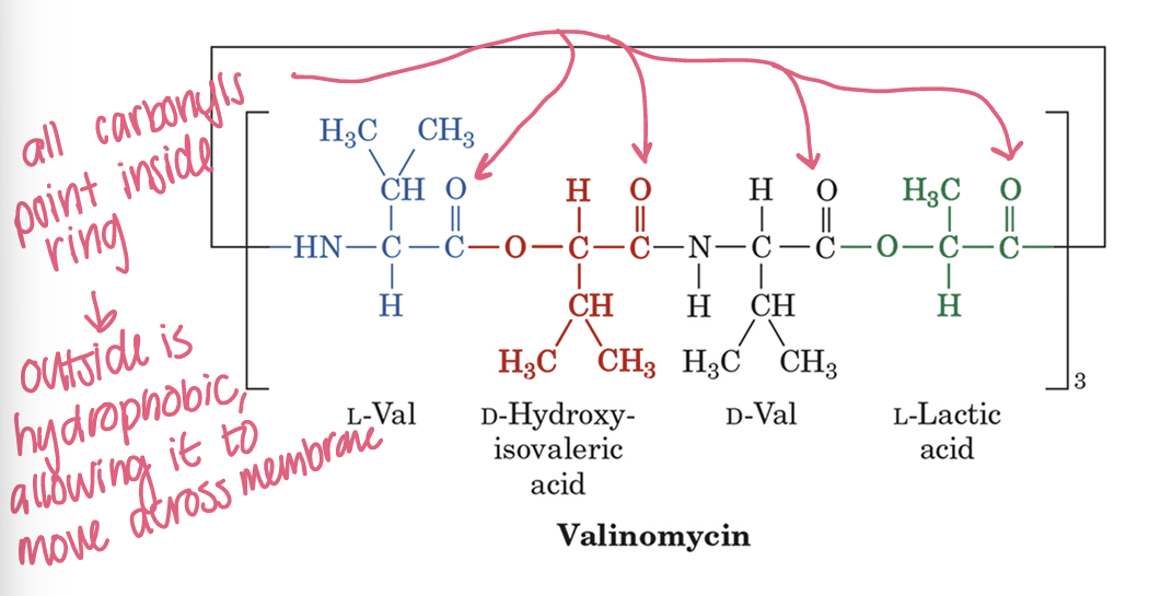
Valinomycin
carrier ionophore
cyclic molecule of alternating D- and L-amino acid residues with hydroxy acids
repeating trimer of L-Val, D-Hydroxyisovaleric acid, D-Val, and L-Lactic acid
peptide bonds and ester linkages
all carbonyls point inward, coordinating a complex bound to K+
nonpolar side chains creates a hydrophobic exterior that allows it to diffuse freely across lipid bilayer membrane
the pre-organized ring size provides strict specificity for K+ ions
its cyclic structure makes it specific, generating a size-selective cavity
Maltoporin
channel-forming ionophore, large B-barrel structure spanning outer membrane
selectively permits transport of specific solutes based on diameter and amino acid side-chain properties lining interior
greasy slide mechanism: interior is full of aromatic a.a. side chains creating a greasy slide → this interact specifically with the flucose rings of maltodextrins
Low Energy Barrier: smooth surface provided by the hydrophobic aromatic residues ("greasy slide") minimizes friction
Multiple polar groups located at the channel constriction allow for a continuous exchange of hydrogen bonds as the maltodextrin molecule glides smoothly through the constriction zone
K+ Channel KcsA
ion channel
potassium ions passively diffuse from the cytoplasm to extracellular space through transmembrane proteins known as K+ channels
diffusion of K+ is due to selectivity filter
backbone carbonyls
3 angstrom diameter → remove solvation shell
entrance = 4 anionic residues attract cations and repel anions
Gated Passive-Mediated Transport
Mechanosensitive
ligand-gated
signal-gated
voltage-gated
Mechanosensitive
open in response to local deformations in lipid bilayer
physical stimuli - touch, sound changes in osmotic pressure
Ligand-gated
open in response to an extracellular chemical stimulus like a NT or in response to building an intracellular signal such as Ca2+ ion
Signal-gated
intracellular signals
Voltage-gated
open in response to changes in membrane potential
nerve impulses
Action potential
initiation & depolarization: a ligand/stimulus causes voltage-gated Na+ channels ot open, driving an influx of sodium into the cell
spiking the membrane potential toward the Na+ equilibrium potential (+60mV)
repolarization: peak depolarization triggers voltage-gated K+ channels to open, driving repolarization and an overshoot into hyperpolarization near K+ equilibrium potential (-75mV)
resting recovery: ion gradients are eventually reset using ion pumps to restore
Nerve impulses - constant amplitude and coding stimulus intensity
constant amplitude: AP propagates along a nerve cell, continuously renewing itself so that the strength remains constant
coding stimulus intensity: because all AP share the same amplitude, the magnitude (strength) of stimulus is controlled by firing rate
Voltage-gating in Kv Channels
voltage-gated K+ channels = Kv channels
voltage gating is triggered by motion of a positively charged protein helix
S4 segment is packed with positive charges
Gate opens:
Normally, the inside of a resting cell is negative, which keeps this gate firmly shut
When a nerve impulse arrives, the inside of the cell suddenly becomes positive (this is called depolarization)
Because like charges repel, the positive electrical charge inside pushes against the positive charges on the S4 part, forcing it to slide outward
That outward physical movement pulls the door open, letting potassium rush out of the cell.
Gate stops:
channel has built in stopper, attached to the inside of the channel is a small piece (inactivation ball on flexible chain)
right after the channel opens, this ball swings up and physically plugs the hole, stopping flow of ions
Cl- Channels are an example of anion channels
basic a.a. line the entrance, selectivity filter made of N-termini and hydroxyl groups
homodimer with each subunit forming an anion-selective pore
Aquaporins mediate…
AQP1 = homotetrameric glycoprotein
each of its subunits consists of 6 transmembrane alpha helixes plus 2 shorter helices in the bilayer
transmembrane water movement
found largely @ kidneys, salivary glands, sweat glands, and lacrimal glands which are tissues that rapidly transport of H2O
How do aquaporins select for JUST H2O
lined with hydrophobic groups that lack strong interactions with H2O
for a H2O molecule to transmit the constriction region, it must be separate from other H2O molecules to which it is H-bonded
this is facilitated by Arg and His
aquaporin forms hydrogen bonds from the side chain NH2 groups of 2 Asn residues in pore
Gap junctions
important intracellular communication channels
small molecules (not macromolecules) are allowed to pass through
GLUT-1 in RBC
glucose binds to the protein which is specific to glucose
conformational change closes the 1st binding site on other side of membrane
glucose dissociated from protein
transport cycle is completed by reversion of GLUT-1 to its initial conformation in absence of bound glucose

all known transport proteins appear to be ______ situated transmembrane proteins that alternated between __ conformational states…
asymmetrically situated transmembrane proteins that alternate between 2 conformational states in which ligand binding sites are exposed to opposite sides of membrane
mediated vs non-mediated
speed and specificity
saturation
mediated = hyperbolic
nonmediated = linear rate
mediated transport is competitive
protein transport can be inactivated
uniport translocation system
involves the movement of a single molecule at a time
ex: GLUT-1

Symport translocation system
simultaneously transports 2 different molecules in the same direction

Antiport translocation system
simultaneously transports 2 different molecules in opposite directions

5 general ATP-driven families for Active Transport
P-type ATPases
F-type ATPases
V-type ATPases
A-type ATPases
ABC transporters
P-type ATPases
undergo phosphorylation of a specific Asp residue within the P domain
as they transport cations like Ca2+, K+, Na+ across membrane
transport ions in opposite directions
Na+/K+-ATPase
E1 and E2 possess different structures, catalytic activities, and different ligand specificities…
In E1 state, ions to be exported and ATP bind from cytosolic side
ATP phosphorylates aspartate
complex undergoes conformational E1→E2 change
At E2, ADP is released and channel opens to exterior and ion binding affinity decreases, leading it to release the ions
exterior ions bind to E2 and aspartyl phosphate = hydrolyzed
Pi is released
Protein undergoes a conformational change back to E1
release of counterions completes cycle

Na+/K+-ATPase Transports Ions in Opposite Directions
maintains low [Na+] and high [K+] inside cells
3 Na+ out and 2K+ in
Ca2+ ATPase Pumps Ca2+ Out of Cytosol
Ca2+ pump actively pumps 2 Ca2+ ions out of cytosol at expense of ATP hydrolysis, while counter transporting 2-3 protons
after muscle contraction: pumps Ca2+ ions back to SR
F-type ATPases
found in mitochondrial and bacterial membranes
proton transport complexes
proton-gradient driven ATP synthesis
V-type ATPases
found in acidic vesicles and plant vacoules
similar to f-type
A-type ATPases
anion transport across membranes
ABC transporters
ATP Binding Cassette
bind ATP and hydrolyze ATP to move amphipathic substances across membrane
P-Glycoprotein
conformational change at transmembrane alpha-helices
ABCG2
found in human cells
ABC transporter
pumps out chemotherapeutics and maintains physiological barriers (blood-brain and maternal-fetal)
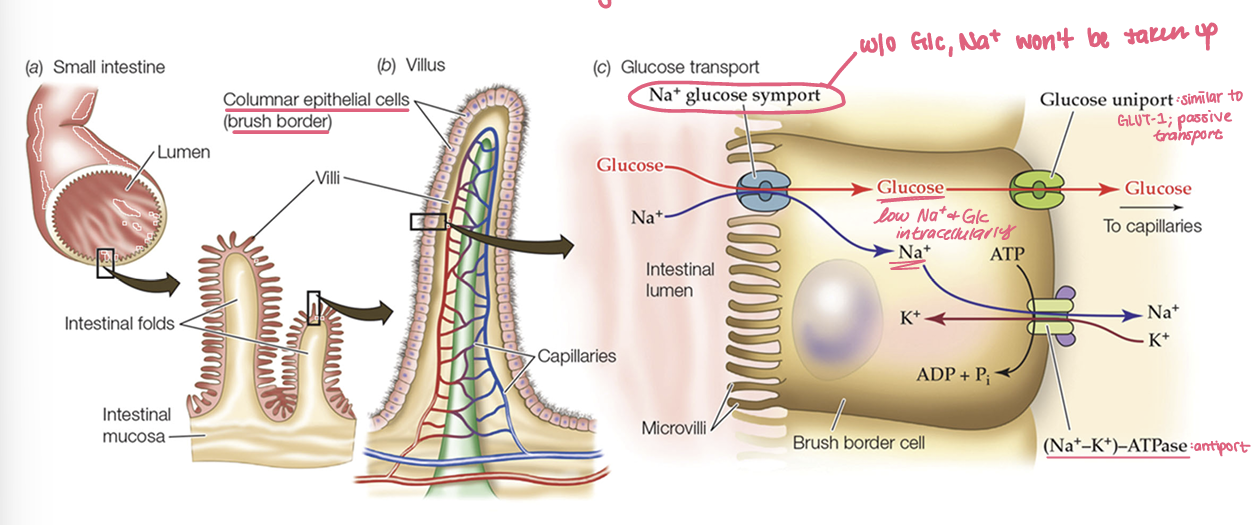
Glucose Transport in Intestinal Epithelium
oral rehydration solution: Na+ (lesser extent K+), glucose
Lactose Permease in E. coli
symport transporter: uses energy stored in H+ gradient to drive lactose across the bacterial cell membrane against its concentration gradient
2 main conformational states:
E-1 (Inward-facing): The binding site faces the cytoplasm and has low affinity for lactose
large internal hydrophilic cavity is open to the cytoplasmic side
E-2 (Outward-facing): The binding site faces the periplasm (outside) and has high affinity for lactose.
Step-by-step cycle for Lactose Permease in E. coli
Outside binding
proton binds to the outward-facing transporter (E2) followed by a molecule of lactose
because it is facing outside, it has a high affinity for lactose
Transport - crossing the membrane
The transporter changes shape, moving the bound H+ and lactose across the membrane toward the inside (cytoplasm)
This movement is powered by the proton gradient (higher H+ concentration outside / low pH, moving toward higher pH/ lower H+ inside).
Release (inside)
now facing the cytoplasm, the transporter shifts to the E1 state, dropping its affinity for lactose
lactose and proton are released into cytoplasm
Recovery (reset pump)
empty transporter is rest
cell oxidative metabolism uses a proton pump to continuously pump H+ back out of the cell, maintaining the proton gradient needed to drive future transport