Mosby's ECG: Interpreting the 12-Lead ECG
 Call Kai
 Learn
 Practice Test
 Spaced Repetition
 Match
 Flashcards
 Knowt Play
1/33

There's no tags or description
Looks like no tags are added yet.
34 Terms
Rhythm
Measuring atrial and ventricular rhythms
- Regular?
- Irregular?
If variations are less than 0.04 seconds (1 small box), consider the rhythm regular.
If the difference between the longest and shortest P-P intervals is greater than 0.12 seconds (3 small boxes), consider sinus arrhythmia.
Rate
Measuring atrial and ventricular rates
- Tachy?
- Brady?
Count 6 second tracing and multiply by 10.
Count large or small boxes.
Use the sequencing method.
P Waves
Present?
- Location: should precede every QRS; if after QRS, consider junctional or ventricular origin
- Amplitude: should be no more than 2.5 mm high
- Duration: should be no more than 0.10 seconds
- Configuration: should be smooth, rounded, uniform
- Positive in lead II
Absent? Probably afib.
PR Intervals
Duration: should be 0.12 - 0.20 seconds in adults
- if longer, 3rd degree AV block
- if shorter, ectopic origin or accessory pathway
Constant
- if variable, consider AV block
QRS Complexes
Duration: should be 0.06 - 0.10 seconds
- if longer, aberrant conduction, block, or ventricular
Configuration
- leads I, II, and III should be positive
Q-wave duration should be less than 0.04 seconds.
Q-wave amplitude should be less than 25% of R wave.
ST Segments
Amplitude: 0.04 seconds, less than 1 mm
T Waves
Location: follow ST segment
Amplitude: should be 5 mm or less
Configuration: rounded, smooth, asymmetrical, upright in leads I, II, V2-V6
- if tented or peaked, consider myocardial injury
- if symmetrical, consider ischemia, electrolyte imbalance, nervous system disorder
- if inverted, consider myocardial ischemia
- if notched or pointed, consider pericarditis
QT Intervals
Duration: varies; usually 0.36 - 0.40 seconds
U Wave
Not always there.
If there, consider electrolyte imbalance.
If negative, consider ischemic heart disease.
Flutter or Fibrillation
Flutter = sawtooth, regular
Fibrillation = craziness, irregular
Ischemia
Heart's demand for oxygen exceeds supply.
Causes
- atherosclerosis
- vasospasm
- thrombosis
- embolism
- tachycardia
- severe hypotension
- aortic valve disease
S/S
- angina
- ST depression
- T wave inversion
Injury
If ischemia is not resolved, it will lead to injury.
Infarction
Death of myocardial cells.
Serious business.
Most commonly due to thrombus blocking the coronary artery.
Q Wave
Small, septal Q waves typically appear in leads I, aVL, V5, and V6.
Pathologic Q Wave
- indicates necrosis
- more than 0.04 seconds
- more than 25% of the following R wave
Key Leads for Infarction
Right Coronary Artery
- lead II
- lead III
- aVF
Left Coronary Artery
- lead I
- aVL
- V1-V6
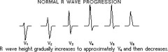
R-Wave Progression
As the precordial electrodes move left, the R wave becomes taller and more positively deflected.
Poor R-wave progression means:
- LBBB
- left ventricular hypertrophy
- severe COPD (emphysema)
- old anteroseptal or anterior infarction
Anterior Wall Infarction
Leads
- V3
- V4
ECG Changes
- ST elevation
- tall T waves
- pathologic Q wave
Complications
- left ventricular dysfunction
- heart failure
- cardiogenic shock
Lateral Wall Infarction
Leads
- I
- aVL
- V5
- V6
ECG Changes
- ST elevation
- reciprocal changes in leads II, III, and aVF
- AV block
Inferior Wall Infarction
Leads
- II
- III
- aVF
ECG Changes
- ST elevation
- reciprocal changes in leads I and aVL
Septal Infarction
Leads
- V1
- V2
ECG Changes
- absence of normal septal R waves
- absence of normal septal Q waves in leads I, II, III, aVF, and V4-V6
- ST elevation and tall T waves in V1 and V2
- R and LBBB
- 2nd Degree AV block type II
- complete AV block
Posterior Wall Infarction
Leads
- no leads directly view this wall
- reciprocal changes may be viewed in anterior view
ECG Changes
- ST depression in V1-V3
- tall R waves in V1-V3
Right Ventricle Infarction
Leads
- II
- III
- aVF
ECG Changes
- when ECG changes suggest acute inferior wall MI
Symptoms
- JVD
- liver tenderness
- pulmonary edema
- hypotension
Bundle Branch Fascicles
Anterior Fascicle
- anterior and lateral walls of LV
- thin and vulnerable to disruptions in impulse transmission
Posterior Fascicle
- posterior and inferior walls of LV
- short, thick, rarely disrupted, dual blood supply
Septal Fascicle
- midseptum
ECG Characteristics
View Lead V1
- LBBB may have a notch
Complete RBBB or LBBB
- QRS complex is greater than 0.12 seconds
Incomplete RBBB or LBBB
- QRS complex is 0.10 - 0.12 seconds
Wide QRS complex or delay
- QRS complex is wide but no BBB pattern is present
Right Bundle Branch Block
Septum is activates by the left posterior fascicle and is depolarized from left to right.
Then the LV is stimulated and depolarizes from right to left.
Then the RV is depolarized, causing a second R wave (R') in lead V1
Left Bundle Branch Block
Alters septal activation.
Causes RV to depolarize before the LV.
Atrial Englargement
Right Atrium
- P wave is abnormally tall at the upslope
- tall, peaked, normal duration
- 2.5 mm or more in leads II, II, and aVF
- possibly biphasic in lead V1
Left Atrium
- P wave may be notched
- 0.10 seconds or more
- may be biphasic in V1
Ventricular Enlargement
Right Ventricle
- reversed R-wave progression in precordial leads
Left Ventricle
- deeper-than-normal S waves and small R waves in V1 and V2
- taller-than-normal R waves and small S waves in V5 and V6
Hypercalcemia
- prolonged PR interval
- prolonged QRS complex
- shortened QT interval
Hypocalcemia
- long, flattened ST segments
- prolonged QT intervals
Hypermagnesemia
- prolonged PR interval
- prolonged QRS complex
- elevated T wave
Hypomagnesemia
- diminished voltage of P waves and QRS complexes
- flattened T wave
- widened QRS
- prolonged QT
- prominent U wave
Hyperkalemia
- peaked T wave
- widened QRS
- prolonged PR
- flattened ST
- flattened or absent P
Hypokalemia
- depressed ST
- flattened T wave
- prominent U wave