MCB 244 exam 3
 Call Kai
 Learn
 Practice Test
 Spaced Repetition
 Match
 Flashcards
 Knowt Play
1/238

There's no tags or description
Looks like no tags are added yet.
239 Terms
Functions of Muscles
Movement, Stability, Control of body openings and passages, Heat production (thermogenesis), Hormone secretion, Glycemic control
Endomysium
thin sleeve of loose connective tissue around each fiber, allows room for capillaries and nerve fibers; provides chemical environment for muscle fiber and nerve ending
Perimysium
thicker layer of connective tissue that wraps fascicles (bundles of muscle fibers wrapped together), Carries nerves, blood vessels, and stretch receptors
Epimysium
fibrous sheath surrounding entire muscle; blends with fascia and deeper connective tissues
Fascia
sheet of connective tissue between muscles or muscle groups
Fusiform muscles
thick in the middle with fascicles converging on each tapered end
Parallel muscles
uniform width and parallel fascicles
Triangular (convergent) muscles
broad at one end with fascicles converging on the other, narrower end
Pennate muscles
feather-shaped; fascicles attach obliquely on a tendon that runs full length of muscle
Circular muscles (sphincters)
fascicles form rings around body openings and passages
Intrinsic muscle
entirely contained within a region
Extrinsic muscle
acts on a designated region but arises from another region
Excitability
responsiveness to chemical signals, stretch, and electrical changes across the plasma membrane
Conductivity
local electrical excitation sets off a wave of excitation that travels along the muscle fiber
Contractility
shortens when stimulated
Extensibility
capable of being stretched between contractions
Elasticity
returns to its original rest length after being stretched
Skeletal muscle
voluntary, striated muscle usually attached to bones
Sarcolemma
plasma membrane of a muscle fiber
Sarcoplasm
cytoplasm of a muscle fiber
Myofibrils
long protein cords occupying most of sarcoplasm
Glycogen
carbohydrate stored to provide energy for exercise
Myoglobin
red pigment; provides some oxygen needed for muscle activity
Sarcoplasmic reticulum (SR)
smooth ER that forms a network around each myofibril
Terminal cisterns
dilated end-sacs of SR which cross the muscle fiber from one side to the other
Transverse (T) tubules
tubular infoldings of the sarcolemma which penetrate through the cell and emerge on the other side
Myoblasts
stem cells that fused to form each muscle fiber early in development, each contributing one nucleus
Satellite cells
unspecialized myoblasts remaining between the muscle fiber and endomysium
Structure of a Skeletal Muscle Fiber

Thick filaments
made of several hundred myosin molecules (myosin is a motor protein)
Fibrous (F) actin
thin filament; two intertwined strands of globular (G) actin subunits, each with an active site that can bind to head of myosin molecule
Tropomyosin
thin filament; each blocks six or seven active sites on G actin subunits
Troponin
thin filament; small, calcium-binding protein on each tropomyosin molecule
Elastic filaments
made of a huge, springy protein called titin
regulatory proteins
Tropomyosin and troponin
contractile proteins
myosin and actin
Dystrophin
links actin in outermost myofilaments to membrane proteins that link to endomysium
Striations and Sarcomeres

Sarcomere
segment from Z disc to Z disc; functional contractile unit of muscle fiber
Motor unit
one nerve fiber and all the muscle fibers innervated by it; they behave as a functional unit
Synapse
point where a nerve fiber meets its target cell
neuromuscular junction (NMJ) or motor end plate
Synapse when target cell is a muscle fiber
Axon terminal
swollen end of nerve fiber; Contains synaptic vesicles with neurotransmitter called acetylcholine (ACh), Nerve impulse causes synaptic vesicles to undergo exocytosis releasing ACh into synaptic cleft
Synaptic cleft
narrow gap between axon terminal and sarcolemma
Postsynaptic membrane folds (junctional folds) of sarcolemma
increase surface area, maximize number of ACh receptors
basal lamina
thin layer of collagen and glycoprotein separating muscle fiber and nerve ending from surrounding connective tissue
acetylcholinesterase (AChE)
enzyme that breaks down ACh, allowing for relaxation
electrical potential, or voltage
Any difference in charge between two points
resting membrane potential (RMP)
The plasma membrane is electrically polarized with a negative charge
Spastic paralysis
a state of continual contraction of the muscles; possible suffocation
Tetanus (lockjaw)
a form of spastic paralysis caused by toxin Clostridium tetani
Botulism
type of food poisoning caused by a neuromuscular toxin secreted by the bacterium Clostridium botulinum
Excitation
action potentials in motor nerve fiber lead to action potentials in the muscle fiber
Excitation–contraction coupling
events that link the action potentials on the sarcolemma to activation of the myofilaments, thereby preparing them to contract
Relaxation
when stimulation ends, a muscle fiber relaxes and returns to its resting length
Length–tension relationship
the amount of tension generated by a muscle depends on how stretched or shortened it was before it was stimulated
rigor mortis
hardening of muscles and stiffening of body beginning 3–4 hours after death
Myogram of a Muscle Twitch

Threshold
minimum voltage that causes a muscle twitch
Recruitment or multiple motor unit (MMU) summation
the process of bringing more motor units into play with stronger stimuli
temporal (wave) summation
Higher frequency stimuli (for example, 20 stimuli/s) produce these
incomplete tetanus
Only partial relaxation between stimuli resulting in fluttering

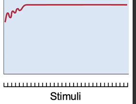
complete (fused) tetanus
Unnaturally high stimulus frequencies (in lab experiments) cause a steady, contraction called
Isometric contraction
contraction without a change in length
Isotonic contraction
contraction with a change in length but no change in tension
Isotonic contraction
contraction with a change in length but no change in tension
Concentric contraction
muscle shortens as it maintains tension (example: lifting weight)
Eccentric contraction
muscle lengthens as it maintains tension (example: slowly lowering weight)
Isometric and Isotonic Phases of Contraction

Anaerobic fermentation
enables cells to produce ATP in the absence of oxygen; yields little ATP and lactate, which needs to be disposed of by the liver
Aerobic respiration
produces far more ATP; does not generate lactate; requires a continual supply of oxygen
Myokinase
transfers Pi from one ADP to another, converting the latter to ATP
Creatine kinase
obtains Pi from a phosphate-storage molecule creatine phosphate (CP) and gives it to ADP
Anaerobic threshold (lactate threshold)
point at which lactate becomes detectable in the blood
Potassium accumulation in the T tubules
reduces excitability
ADP and Pi accumulation
slows cross-bridge movements, inhibit calcium release and decrease force production in myofibrils
Maximum oxygen uptake (VO2 max)
the point at which the rate of oxygen consumption plateaus and does not increase further with added workload
Excess post-exercise oxygen consumption (EPOC), or oxygen debt
elevated rate of oxygen consumption following exercise
Slow-twitch, slow oxidative (SO), red fibers, or type I fibers
Thin cells with abundant mitochondria, capillaries, myoglobin (deep red color) and contain a form of myosin with slow ATPase, and a SR that releases calcium slowly
Fast-twitch, fast glycolytic (FG), white fibers, or type II fibers
Contain a form of myosin with fast ATPase and a large SR that releases calcium quickly, Fibers are thick and strong; low myoglobin gives them pale color
Factors that affect muscular strength
Muscle size, Fascicle arrangement, Size of active motor units, Multiple motor unit summation, Temporal summation, The length–tension relationship, Fatigue
Cardiac Muscle
Striated, relatively short and thick, and surrounded by endomysium (but no peri or epimysium), Cardiomyocytes branch and join to other cells at connections called intercalated discs
Multiunit smooth muscle
individual myocytes respond independently of all the others
Unitary (single-unit) smooth muscle
myocytes coupled by gap junctions, so contract as a unit
Muscular dystrophy (MD)
group of hereditary diseases in which skeletal muscles degenerate and weaken, and are replaced with fat and fibrous scar tissue
Myasthenia gravis
autoimmune disease in which antibodies attack neuromuscular junctions and bind ACh receptors together in clusters
Central nervous system (CNS)
brain and spinal cord
Peripheral nervous system (PNS)
nerves and ganglia
Nerve
a bundle of nerve fibers (axons) wrapped in fibrous connective tissue
Ganglion
a knot-like swelling in a nerve where neuron cell bodies of PNS are concentrated
Sensory (afferent) division
carries signals from receptors (sense organs) to CNS
Somatic sensory division
carries signals from receptors in the skin, muscles, bones, and joints
Visceral sensory division
carries signals from the viscera (heart, lungs, stomach, and urinary bladder)
Motor (efferent) division
carries signals from CNS to effectors (glands and muscles that carry out the body’s response)
Somatic motor division
carries signals to skeletal muscles; causes voluntary muscle contraction and automatic reflexes
Visceral motor division (autonomic nervous system, ANS)
carries signals to glands, cardiac and smooth muscle; no voluntary control; responses called visceral reflexes
Sympathetic division of ANS
stimulates and prepares the body for action
Parasympathetic division of ANS
has a calming effect on the body
An enteric plexus within digestive tract wall
enables coordination and communication within digestive tract
Secretion
when signal reaches end of axon, the neuron secretes a neurotransmitter that stimulates the next cell