cross bridge cycle
 Call Kai
 Learn
 Practice Test
 Spaced Repetition
 Match
 Flashcards
 Knowt Play
1/14

There's no tags or description
Looks like no tags are added yet.
15 Terms

step 1
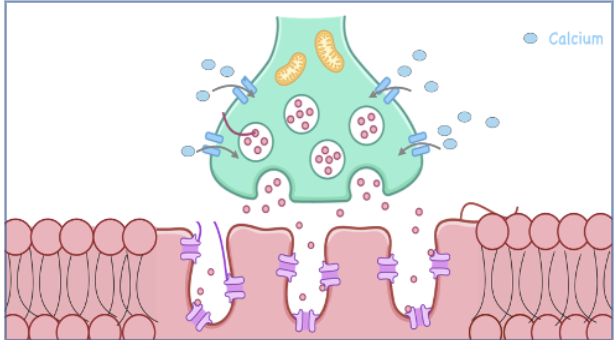
an action potential arrives at the axon terminal of the motor neuron
step 2
the calcium channels open, allowing calcium from the extracellular fluid to enter the synaptic terminal

step 3
the vesicles within the synaptic terminal fuse with the pre-synaptic membrane, allowing acetylcholine to be released into the synaptic cleft

step 4
the released acetylcholine attaches to the acetylcholine receptors on the motor end plate

step 5
sodium from the extracellular fluid can enter the sarcolemma (i.e. depolarization)

step 6
the action potential continues traveling through the transverse tubule

step 7
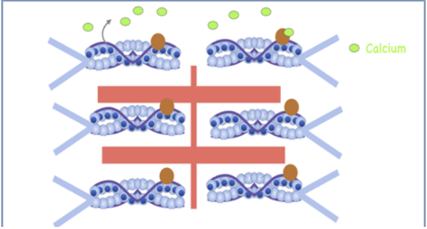
the action potential’s travel triggers the release of calcium from the sarcoplasmic reticulum into the sarcoplasm
step 8
calcium binds to troponin; tropomyosin shifts, exposing actin’s active site
step 9
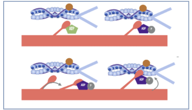
ATP binds to myosin and is hydrolyzed, while ADP and phosphate remain. The myosin head is cocked in a high energy position. Myosin binds to actin’s active site, forming a cross-bridge

step 10
the release of phosphate, followed by the release of ADP from the myosin head triggers a power stroke. This causes the myosin heads to pull actin towards the center of the sarcomere

step 11
a new ATP molecule attaches to the myosin head, causing the cross bridge to detach from actin

step 12
the myosin head hydrolyzes ATP into ADP and phosphate. Myosin returns to its cocked position

step 13
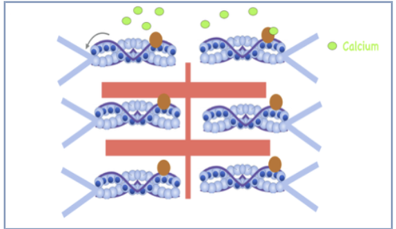
calcium is actively pumped back into the sarcoplasmic reticulum, which requires the use of ATP to make this possible
step 14
calcium detaches from the troponin; tropomyosin shifts, covering actin’s active sites-stopping the cross-bridge-cycling and allowing the muscle fiber to relax

step 15
acetylcholine is broken down by acetylcholinesterase, terminating the signal at the neuromuscular junction, preventing further depolarization of the muscle fiber