Quiz 7 (Rest of L21-L23)
 Call Kai
 Learn
 Practice Test
 Spaced Repetition
 Match
 Flashcards
 Knowt Play
1/22

There's no tags or description
Looks like no tags are added yet.
23 Terms
Given two monosaccharides, draw the disaccharide that results from formation of a specified O-glycosidic bond (e.g., 1 -> 3, etc.); identify the glycosidic bond and the anomeric C involved in the bond.
Anomeric Carbon: 1st carbon(right of the O in pyranose)
Alpha vs Beta: the location of the -OH group
For Furanose, Alpha OH is down; Beta OH is up
For Pyruanose, Alpha OH is trans to CHOH; Beta OH is cis to CHOH

Distinguish between O- and N-glycosidic bonds and recognize examples of each
O-glycosidic bonds take anomeric carbon of sugar and links to another glucose
N-glycosidic bonds take anomeric carbon of sugar and links to a nucleotide that contains nitrogen
Both invovles removal of H2O

Recognize abbreviations for glucose, galactose and ribose.
Glucose: Glc
Galactose: Gal
Ribose: Rib

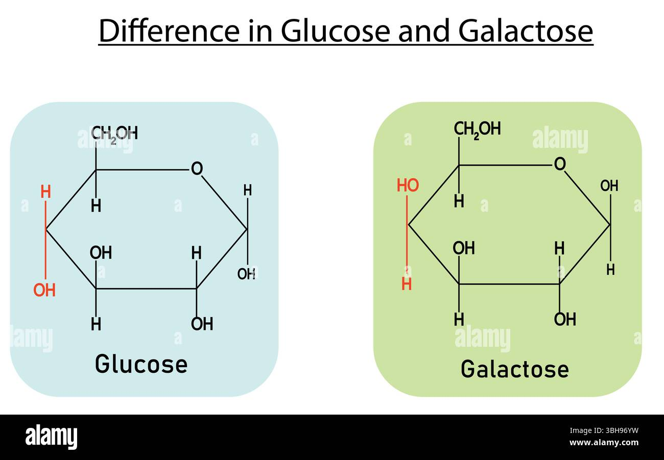
Recognize the structural difference between Glc and Gal
The hydroxyl group on the C4 carbon is on the bottom for glucose and on the top for galactose.
Define the terms monosaccharide, disaccharide, oligosaccharide and polysaccharide.
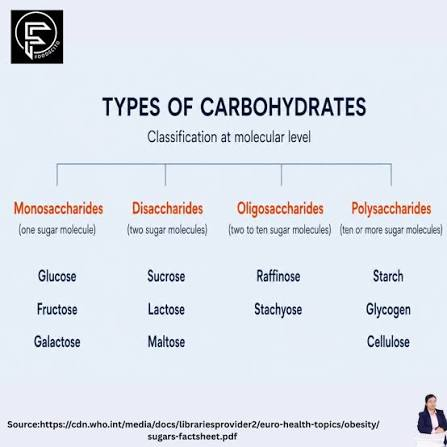
Monosaccharide: one sugar subunit
Disaccharide: two sugar subunits linked by glycosidic linkage
Oligosaccharide: 3-10 sugar subunits
Polysaccharide: more than 10 sugar subunits
Explain why all linkages between two saccharides involve the anomeric C of one saccharide that is attacked by an OH of the other.
Anomeric carbon is subject to nucleophilic attack by the hydroxyl group from the other saccharide. This causes disaccharide to form.
Explain why linkages between pyranose forms of saccharides can be (1 -> 1), (1 -> 2), (1 -> 3), (1 -> 4), or (1 -> 6), but NOT (1 -> 5).
The 5th carbon OH is a member of the ring for glucose so it is not available for glycosidic linkage between pyranose forms of saccharides

Describe how Tamiflu works.
Tamiflu binds to the active site of the virus neuraminidase enzyme, which prevents the release of new virions. Therefore, it slows down the spread of the virus between cells.
Spread of virus requires hydrolysis of sialic acid(using neuraminidase enzyme) that binds virion to the cell surface.

Describe amylose, amylopectin, glycogen, cellulose, chitin, and glycosaminoglycans, and proteoglycans.
Found in what types of organisms (e.g., animals, plants, arthropods/mollusks)
What saccharides are used in them
Type(s) of linkages, whether linear or branched polymers
Function/purpose (energy storage, structural, recognition)
Amylose | Amylopectin | Glycogen | Cellulose | Chitin | |
Organism type | Plants | Plants | Animals | Plant Cell Walls(50%of all organic matter on Earth | Arthropod and mollusk eaxoskeleton |
Reactants | Glucose | Glucose | Glucose | Glucose | Modified glucose |
Linkages | Alpha (1,4) Not branched (linear) | Alpha (1,4) and Alpha (1,6) Somewhat branched | Alpha (1,4) and Alpha (1,6) Highly branched | Beta (1,4) Linear | Beta (1,4) Linear |
Function | Energy storage | Energy storage | Energy storage | Structural | Structural |
Glycosaminoglycan: Found in ECM, a long chain of carbohydrate(many negative charge)
proteoglycan: Found in ECM, a core protein + GAG group(many negative charge); highly slippery therefore lubrication

Explain the basis for blood types O, A, B, and AB; apply these principles to donor and acceptor roles of each blood type (e.g., why someone with Type O is a “universal donor” while someone with Type AB is a “universal acceptor” but can only donate blood to someone with Type AB.
Type O (“universal donor”): Doesn’t have A/B antigens so the antibody cannot attack to destroy the antigen, so can be given to anybody. Only accept O.
Type A: Donates to A or AB, accepts A or O
Type B: Donates to B or AB, accepts B or O
Type AB (“universal acceptor”): Ability to recognize both A and B antigens on the surface. Only donate AB.

What is the History of ATP & Basic Propoerty of ATP
Brief History of ATP
ATP is an ancient biomolecule, predating (coming before) proteins
Many proteins evolved to bind to ATP
Basic Properties of ATP
Inside cell, ATP always complexed to metal ion (Mg2+)
The 7 non bridging oxygens are nearly equivalent in electronegativity value (~ -0.7)
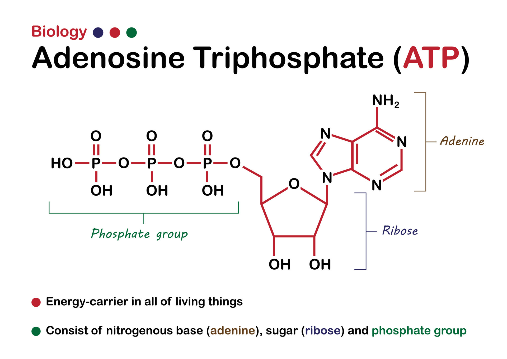
Between each phosphate, the bond is “phosphoanhydride” (i.e. water is removed from 2 Pi when bond is formed)
For “high energy molecules”, draw hydrolysis products, give reasons why hydrolysis rxn is high energy
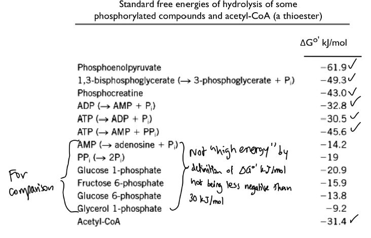
Big Picture: “High energy molecules” are those with a ΔG°’ (delta G standard prime) equal or higher in magnitude than ATP at -30.5 kJ/mol (< - 30.5 kJ/mol)
Examples of “high energy molecules” with their hydrolysis products + rxn reasoning
ATP
→ More resonance stabilization of hydrolysis products
→ More favorable water-binding of products, including H+ (more water solvation)
→ Decreased electrostatic repulsion in products
→ Favorable entropy to have more product molecules than reactant molecules
→ Favorable H+ release

Examples of “high energy molecules” with their hydrolysis products + rxn reasoning
1,3 BPG
→ Phosphate resonance
→ Carboxylate resonance
→ More favorable water-binding of products, including H+ (more water solvation)
→ Favorable entropy to have more product molecules than reactant molecules
→ Favorable H+ release

Examples of “high energy molecules” with their hydrolysis products + rxn reasoning
Phosphocreatine
→ Phosphate resonance
→ Guanidino resonance
→ Better solvation of products
→ Favorable entropy to have more products than reactants

Examples of “high energy molecules” with their hydrolysis products + rxn reasoning
Phosphoenolpyruvate (PEP)
→ Phosphate resonance
→ Keto is very stable (more than enol)
→ Better solvation of products
→ Favorable entropy to have more products than reactants

Examples of “high energy molecules” with their hydrolysis products + rxn reasoning
Thioester
→ Lack of thioester resonance stabilization (size of sulfur → little orbital overlap with C=O pi-orbital)
→ Carboxylate resonance
→ More favorable water-binding of products, including H+ (more water solvation)
→ Favorable entropy to have more product molecules than reactant molecules
→ Favorable H+ release

Calculate ΔG (and know equation!) for these cases:
The concentration cell (diffusion across a permeable barrier)
Concentration of Cell:
Equation: G = G°' + RT ln(products/reactants) , where G° is the standard state at biological conditions (pH = 7)
There is a “driving force” to produce product or reactant solely based on concentration when moving same molecule
G°' = 0 if reactant and product contain same molecule
G = 0 if at equilibrium
Standard State for H+ is 10-7 M otherwise are 1M
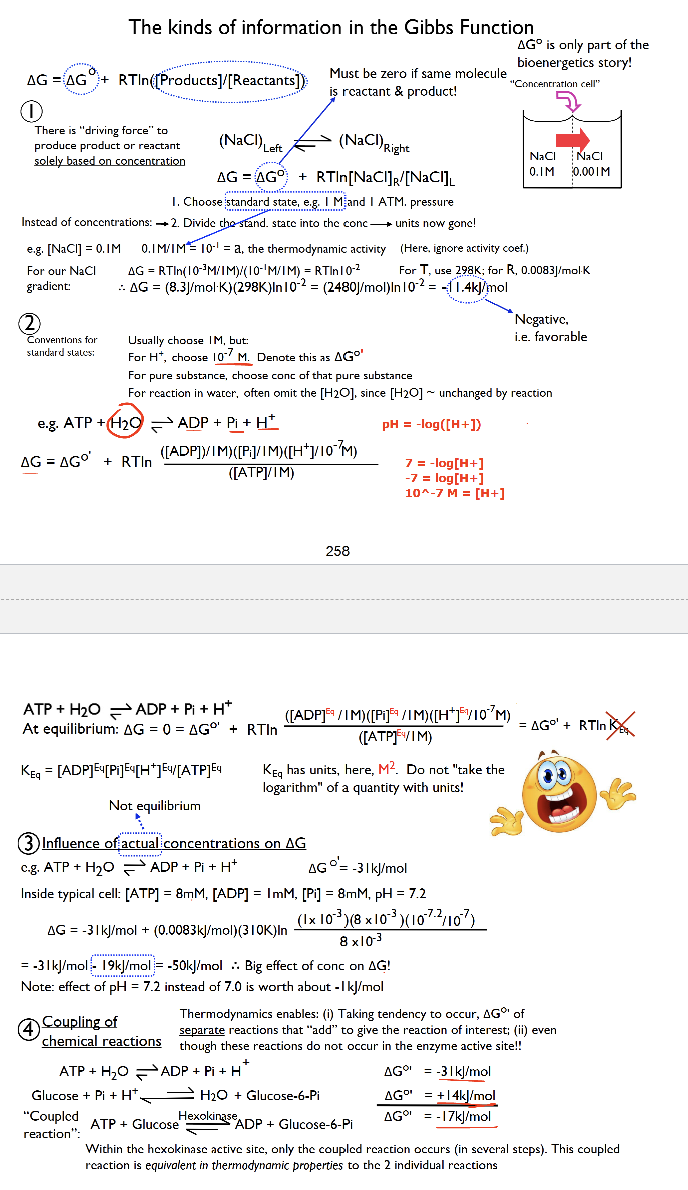
Calculate ΔG (and know equation!) for the following case:
Transport of ions across a cell membrane
Equation: GTransport = RT ln( going to / coming from)+zFΨ
→ z = charge on ion, F = Faraday’s constant, ΔΨ = change in voltage
Inside the cell is negative while outside the cell is positive for Ψ sign consideration

For given redox half reactions and concentrations, be able to calculate ΔG for a given overall reaction.
Overview: All redox reactions behave like “coupled” reactions → termed “redox half reactions” (involves flow of electrons from one molecule to another) + cells use NADH/NAD+ and FADH2/FAD for redox reactions
ΔG°' = -nFE°'
Gibbs Function: ΔG = -nF(E°' of half-reaction #1 + E°' of half-reaction #2) + RT ln( productsreactants)
n = number of electrons transferred from the reaction,
F = Faraday’s constant,
E°' = difference in the standard reduction potential (E°' of half-reaction #1 + E°' of half-reaction #2)
The better the e- acceptor (oxidizing agent in the reactant), the more positive the E°'(the more negative G°' will be)
Negative half reaction → product is a better reducer (electron donor)
Positive half reaction → product is a better oxidizer (electron acceptor)
→ Opposite rxn = Flip voltage sign
e- transfer depends on chemical properties of the molecule and e- transfer depends separately (in an additive way) on the chemical properties of each molecule with each step involving e- lost from one molecule and e- gained by another molecule

Recognize oxidation levels (see eBook Figure 13-22)
C-C = 1 electron for C
C-H = 2 electron for C
C-O = 0 electron for C

Compare and contrast the light-driven reduction of CO2 by photoautotrophs (plants,algae, cyanobacteria) and the energy-yielding oxidation of glucose by chemoheterotrophs (all animals, most fungi, protists, bacteria).
Phototrophs - Use Carbon Dioxide + Water →(input of light) Glucose + Oxygen
light driven reduction
Chemotrophs - Use Oxygen + Glucose → ATP + Carbon Dioxide + Water
energy yielding oxidation of glucose

Explain why photoautotrophs and chemoheterotrophs are "locked in a vital ecological loop".
They are locked in a vital loop because the products of photoautotrophs (glucose and oxygen) serve as the essential reactants for chemoheterotrophs, while the metabolic end products of chemoheterotrophs (Carbon dioxide + H2O) supply the carbon and electron sources required for photoautotrophy