Cardiovascular System
 Call Kai
 Learn
 Practice Test
 Spaced Repetition
 Match
 Flashcards
 Knowt Play
1/127

There's no tags or description
Looks like no tags are added yet.
128 Terms
Arteries
AWAY from the heart
Veins
To the heart
Capillaries
Sites of exchange
Pulmonary circulation
Deoxygenated blood from right side of heart to lungs then through left side of heart and out
Systemic circulation
Oxygenated blood from left side of heart to systemic capillaries and back to right side of heart
Anastomosis
When 2 or more arteries provide blood to same body region
End arteries
Only artery to supply a body region
Functional end artery
Anastomoses so small they might as well be an end artery
Venous anastomosis
2 or more veins supplying blood to one region, more common than in arteries
Companion vessels
Veins and artery pair supplying blood to same region

Mediastinium
Cavity in medial thoracic cavity where heart and other organs are located
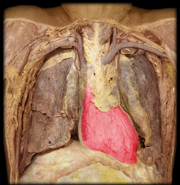
Fibrous pericardium
Outer, dense connective tissue layer of the heart

Parietal layer of serous pericardium
Protective sac that encloses heart
Pericardial cavity
Space between parietal pericardium and visceral pericardium containing lubricating fluid

Visceral layer of serous pericardium
Aka epicardium
Outer covering of heart
Myocardium
Muscle layer of heart, causes contractions
Endocardium
Innermost lining of heart

Interatrial septum
Partitions two atria (membrane

Interventricular septum (membranous here)
Myocardial tissue separating two ventricles
(Muscular here)


Apex of the heart

Superior vena cava
Drains to right atrium
Recieves blood from head and upper body
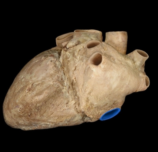
Inferior vena cava
Receives from legs and trunk
Drains into right atrium
Coronary veins
Return deoxygenated blood from myocardium to coronary sinus

Coronary sinus
Recieves from coronary veins
Drains into right atrium

Right atrium
Recieves from vena cava and coronary sinus


Right auricle
Anterior right atrium

Pectinate
Muscular ridges between both auricles and anterior walls of atria

Right atrioventricular valve
Prevents backflow into right atrium
Aka tricuspid valve

Right ventricle
Sends blood to pulmonary trunk

Trabeculae carnae
Ridges of muscle in endocardium of ventricles

Papillary muscle
Fasten chordae tendinae to ventricular wall

Chordae tendinae
Connective tissue holding valves in position during contractions

Pulmonary valve
Prevents backflow from pulmonary trunk to right ventricle

Pulmonary trunk
Recieves from right ventricle
Sends to lungs via pulmonary arteries

Pulmonary arteries
Direct deoxygenated blood to lungs

Pulmonary veins
Return oxygenated blood to left atrium

Left atrium
Recieves from pulmonary veins

Left auricle
Anterior left atrium


Left atrioventricular valve
Prevents backflow into left atrium
Aka bicuspid and mitral valve

Left ventricle
Thick myocardial wall for contractions against high pressure

Aortic valve
Prevent backflow from aorta to left ventricle

Ascending aorta
Recieves from left ventricle

Right coronary artery
Send to myocardium

Arch of aorta
Descending aorta
Descends from aortic arch continuing inferiorly until it splits into common iliac arteries


Umbilical cord
Large vein and 2 arteries
Umbilical vein
Oxygen and nutrients from mother to fetus
Turns into round ligament of liver
Umbilical artery
Deoxygenated blood from fetus to placenta
Turns into medial umbilical ligaments
Ducts venosus
Branch of umbilical vein enters vena cava, bypasses portal system of liver
Turns into ligamentum venosum
Foramen ovale
Opening in interatrial septum to bypass pulmonary circulation
Turns into fossa ovalis
Ducts arteriosus
Shunt between pulmonary trunk and aorta, bypasses pulmonary circulation
Turns into ligamentum arteriosum
Branches of aortic arch
Brachiocephalic trunk
Left common carotid artery
Left subclavian artery

Brachiocephalic trunk
First branch of aortic arch
Supplies to right arm and head through right subclavian and right common carotid arteries

Left common carotid artery
Middle branch of aortic arch
Supplies left side of neck and head through external and internal carotids

Left subclavian artery
Last branch of aortic arch
Supplies left arm through axillary artery

Common carotid artery
Right artery supplies to right side of head and neck

External carotid artery
Divides into branches that supply all parts of head except for brain

Internal carotid artery
Enters skull via carotid canal, supplys to cerebrum and orbit
Vertebral artery
Passes through transverse foramen, forms basilar artery

Right subclavian artery

Internal thoracic artery

Axillary artery

Brachial artery


Radial artery
Branch of brachial artery

Ulnar artery
Branch of brachial artery

Intercostal arteries

Celiac artery
Celiac trunk Divides into splenic, left gastric and common hepatic arteries


Splenic artery
Supplies spleen

Left gastro-omental artery
Supplies greater curve of stomach

Left gastric artery
Supplies lesser curve of stomach, forms loop with right gastric artery

Common hepatic artery
Supplies liver, gallbladder, stomach, duodenum, pancreas

Proper hepatic artery
Supplies liver

Right gastric artery
Lesser curve of stomach

Hepatic arteries
Left and right sides of liver

Gastroduodenal artery
Suppliespyloric region and duodenum, splits into branches that supply pancreas and greater curvature

Right gastro-omental artery
Greater curve of stomach

Superior mesenteric artery
Majority of small intestine and part of large intestine

Renal arteries
Supply kidneys

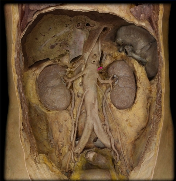
Suprarenal artery
Adrenal glands

Gonadal artery

Inferior mesenteric artery
Supplies part of large intestine and rectum

Lumbar artery
Lumbar muscles and spinal cord

Median sacral artery
Supplies region of coccyx and sacrum

Common iliac arteries


Internal iliac artery
Genital organs and gluteal muscles

External iliac artery
Continues as femoral artery

Femoral artery
Muscles of thigh
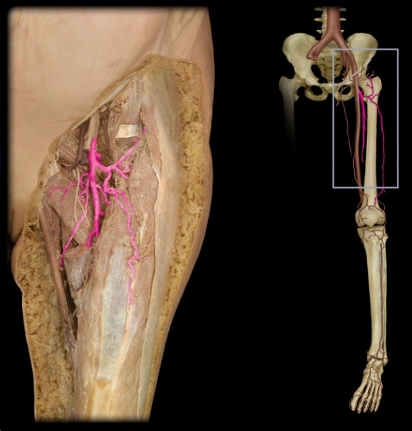
Deep femoral artery
Posterior thigh muscles

Popliteal artery
Posterior knee

Anterior tibial artery
Extensor muscles of lower leg, ankle and dorsal side of foot

Posterior tibial artery
Flexor and peroneal muscles of lower leg, sole, toe

External jugular vein
Drains into subclavian vein

Internal jugular vein

Brachiocephalic vein
Enters superior vena cava

Axillary vein
Merges with cephalic vein to become subclavian vein

Subclavian vein
Becomes brachiocephalic vein

Brachial veins


Basilic vein
Merges with brachial to become axillary vein

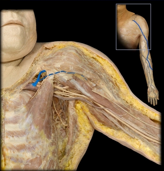
Cephalic vein
Merges with axillary
Drains superficial portion of hands
