Lesson 79: Structure and Function of the Small Intestine in Domestic Animals
 Call Kai
 Learn
 Practice Test
 Spaced Repetition
 Match
 Flashcards
 Knowt Play
1/32

There's no tags or description
Looks like no tags are added yet.
33 Terms
The average length of the small intestine in various animals
➢ Dog – 4 m (13 feet)
➢ Cat – 1.72 m (5.6 feet)
➢ Horse – 22 m (73 feet)
➢ Pig – 18 m. (59 feet)
➢ Bovine – 46 m. (150 feet)
➢ Small ruminant – 26 m (85 feet)
Function of the SI: Digestion of Food
Enzymatic Breakdown: Digestive enzymes from the pancreas and intestinal mucosa break down carbohydrates, proteins, and fats.
In ruminants, the small intestine continues the digestion of microbial protein from the rumen
Function of the SI: absorption of carbs
Broken down into monosaccharides (glucose, fructose) and absorbed into the bloodstream
Function of the SI: absorption of proteins and lipids
Digested into amino acids and peptides for absorption.
Lipids: Emulsified by bile, broken down by lipases, and absorbed as fatty acids and monoglycerides
Function of the SI: absorption of vitamins and minerals
Fat-soluble vitamins (A, D, E, K) require bile for absorption.
Water-soluble vitamins (B-complex, C) are absorbed directly.
Minerals (Ca, P, Na, K) are absorbed through active and passive transport.
Function of the SI: Movement of Chyme
Peristalsis: Coordinated contractions move food along the intestine.
Segmentation: Mixing contractions help enhance digestion and absorption.
Function of the SI: Immune function
Gut-associated lymphoid tissue (GALT):
Peyer’s patches (especially in the ileum) help detect and respond to pathogens.
Secretory IgA and mucosal immunity prevent infections
Function of the SI: water and electrolyte balance
absorbs a large portion of water from digestive secretions.
Electrolyte Transport: Sodium, chloride, and potassium balance is maintained for cellular function.
Function of the SI: species specific adaptations
Ruminants: The small intestine absorbs microbial protein and volatile fatty acids.
Horses: The small intestine is crucial for protein and fat absorption, as the hindgut
ferments fiber.
Carnivores (Dogs & Cats): Highly adapted for protein and fat digestion.
Pigs & Omnivores: Efficient digestion of both plant and animal matter
The duodenum
The proximal part of the small intestine → Fairly fixed and attached to body wall
Connecting peritoneum is called mesoduodenum
Three segments: Descending duodenum, Transverse duodenum, Ascending duodenum
Anatomy of the duodenum

Descending duodenum
Proximal and the most fixed part of the duodenum and small intestine.
On the right of the median plane/mesentery
Contacts right abdominal wall
Proximal ‘S’ shaped OR Cranial duodenal flexure
Major and Minor duodenal papillae
Descending duodenum: Major and Minor duodenal papillae
Major duodenal papilla – Opening of the common bile duct and major/main pancreatic duct
Minor Duodenal papilla – Opening of the lesser/accessory pancreatic duct
The Minor Duodenal papilla is?
Inconstant in cats(Present only in 20% cats)
Transverse Duodenum
➢ Also called caudal flexure of the duodenum
➢ Crosses the median plane
➢ Mostly along the roof
Ascending Duodenum
On the left side, Mostly along the roof
Duodenojejunal flexure: A sharp bend where it becomes the jejunum
➢ Duodeno-colic fold
Small intestine - The Jejunum
From ascending duodenum to the Ileum
Long, coiled loops with a thin, flexible wall
Very flexuous portion, remains free in the abdominal cavity and forms bulk of the small intestine
Connecting peritoneum of the jejunum
Mesojejunum /Jejunal mesentery/ mesentery is very long
Occupies most of the abdominal space in the dog and cat
Cranioventral and caudoventral
The Mesentery/Great mesentery” and “Root of the mesentery”

Small intestine - The Ileum
Shortest portion of the small intestine, located in the caudal abdominal cavity.
Slightly thicker wall than the jejunum.
Suspended by the mesentery and connected to the cecum via the ileocecocolic junction
The antimesenteric artery can be used to?
demarcate the ileum from the jejunum
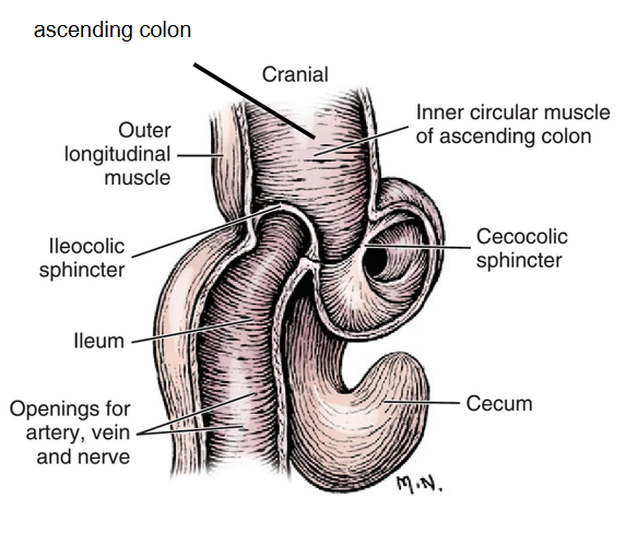
The ileocolic junction (Ileocecocolic junction) → Ileum opens into the ascending colon
The ileal (ileocolic) orifice, The ileal papilla, The Ileocolic sphincter
The duodenum - blood supply
Cranial part: Hepatic a. → Gastroduodenal a. → Cranial pancreaticoduodenal a.
Caudal Part: Cranial Mesenteric Artery → caudal pancreaticoduodenal a.
Cranial mesenteric artery (#2) supplies almost entire small intestine and major part of the large intestine
Jejunal aa. #9 (Multiple)
Common trunk aka Ileocolic a. (#3)
Mesenteric ileal a. (#7)
Antimesenteric ileal a. (#8)

SI: venous return
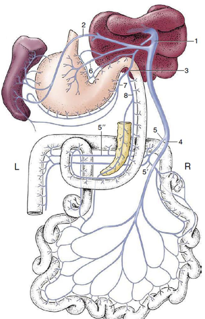
Hepatic Portal vein: Gastroduodenal vein (#3), Cranial pancreaticoduodenal vein (#8)
Hepatic Portal vein: The Cranial mesenteric vein (#4)
Semischematic dorsal view of the formation of the portal vein (dog).
1, Portal vein;
2, splenic vein;
3, gastroduodenal vein;
4, cranial mesenteric vein;
5, caudal mesenteric vein;
5′, ileocolic vein;
5″, middle colic vein;
6, left gastric vein;
7, right gastroepiploic vein;
8, cranial pancreaticoduodenal vein
SI: sympathetic innervation
originates from the thoracic spinal cord (T5–T12)
Travel via the greater splanchnic nerve (T5–T9) and lesser splanchnic nerve (T10–T12)
Motor and sensory
SI: pre and postganglionic fibers
The preganglionic fibers synapse in the Prevertebral ganglia
the celiac ganglia, and cranial mesenteric ganglion
The postganglionic fibers travel along the branches of hepatic artery (for duodenum) and cranial mesenteric artery (jejunum and ileum) to reach the organs.
SI: Parasympathetic innervation
Originates from the dorsal motor nucleus of the Vagus n. in the medulla oblongata of the brainstem
Dorsal and Ventral Vagal Trunks – Plexuses
Ventral Vagal trunk – Duodenum and pancreas
Dorsal Vagal trunk - Jejunoileum
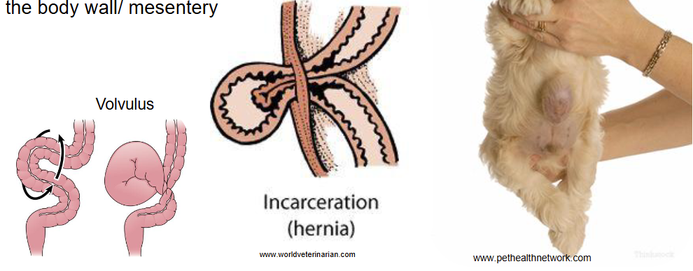
Hernia/Incarceration: Through natural/pathological openings in the body wall/ mesentery
Intussusception – telescoping(one part of intestine sliding into another)
➢ Intussusceptum (inner)
➢ Intussuscipiens (Outer)

The Small intestine – Common procedures: Intestinal anastomosis (End to end anastomosis)
