NBEO Part 3 - PEPS
 Call Kai
 Learn
 Practice Test
 Spaced Repetition
 Match
 Flashcards
 Knowt Play
1/347

There's no tags or description
Looks like no tags are added yet.
348 Terms
Corneal foreign body: signs and symptoms
Sx: Sudden onset pain, photophobia, watering, possible FB sensation.
S: Conjunctival injection, FB embedded in cornea, may need fluorescein to see
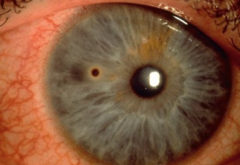
Corneal Foreign Body - Testing
NaFl, Lid eversion, Retroillumination
Corneal Foreign Body (metal and not) - Treatment in office and after
Remove foreign body with cotton-tipped applicator, or instill Proparacaine and remove with instrument, remove rust ring with Alger brush, place BCL if large epithelial defect, Moxifloxacin 0.5% qid x1 wk, prn: oral NSAID and frequent PFATs
Corneal Foreign Body - FU times
1 day if BCL, otherwise 1 wk
Metal FB pt. education
You have a piece of metal in your eye that we have removed. We placed a bandage lens on your eye to help your cornea heal, so you need to return tomorrow so that we can remove it. We've given you an antibiotic to use 4x per day, and we also rcmnd using PFATs 6-8x throughout the next several days. In the future, it is important to remember to wear safety glasses when working with metal.
Recurrent Epithelial Erosion (RCE): Symptoms and signs
Sx: Pain upon opening eyes, ripping or tearing sensation, tearing, blurred vision, redness, photophobia
S: subtle loose epithelium

Suspected RCE/EBMD - Testing
NaFl, Lid eversion
RCE - Treatment for subtle loose epithelium
Erythromycin ointment QID, NaCl 5% QID until healed. Then NaCl QHS and Genteal gel QHS x3-6 months
RCE - Treatment in office and after for heaping epithelium
Propraracaine and debride 1-2mm beyond lesion, Place BCL if not a previous CL wearer, Moxifloxacin 0.5% qid x1 wk, Nacl 5% soln qid x2 wk then taper to qhs for 3 months, frequent PFATs
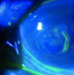
RCE -Recalcitrant treatment
Do in office anterior stromal puncture
RCE - FU times
1 day for BCL, Otherwise 3-4 weeks
RCE from previous trauma or EBMD - Education
You have a condition called EBMD, in which the top layer of your cornea does not adhere well to the bottom layers. Sometimes, this can lead to the top layer of the cornea being ripped off, leaving a large abrasion. (OR - Because of a past trauma to your eye, the top layer of your cornea has difficulty staying adhered to the bottom layers). We removed some of the top layer of your cornea so that it can heal properly, and placed a bandage lens to speed up the healing process. We also gave you an antibiotic to prevent any infection while this heals, and a drop that helps to dehydrate the cornea. This drop can help to prevent this from happening again, but may need to be continued for the next few months. If this recurs, you should return to our office.
Chemical Burn - workup and testing
Irrigation for 15 mins, lid eversion and irrigation/swabbing of fornices, Testing PH with litmus paper after 5 minutes (normal is 7-7.4), NaFl
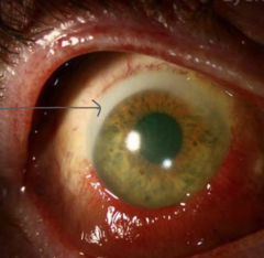
Chemical Burn - Treatment
Debridement of any loose epithelium, Moxifloxacin 0.5% qid x1 week, Prednisolone Acetate 1% qid x1 week then taper, frequent PFATs
Chemical Burn - FU
1 day
Chemical Burn - Education
You have gotten a chemical into your eye which has burned a layer of your cornea. I've removed all of the loose tissue to help the corneal heal better. I want you to use an antibiotic to prevent infection and an anti-inflammatory to help with swelling and inflammation. You should use PFATs 6-8x per day while this heals. I'd like to see you tomorrow to make sure your eye is healing well. In the future, it is important to use safety glasses while using chemicals.
IIH: Symptoms and signs
Sx: HA, transient vision loss, back pain, pulsatile tinnitus, nausea
S: Bilateral ON edema

Suspected IIH - Testing
RNFL OCT (elevated nerves), VF (enlarged blind spot), Fluorescein AutoFluorescence (FAF) (r/o ONH drusen), Red cap (decreased), BP, Order urgent MRI/MRV and LP afterwards with CSF analysis
Confirmed IIH - Treatment
Acetazolamide 250mg po BID. Refer to PCP/OBGYN to discuss weight loss and changing birth control drug/method.
IIH - FU from initial dx
4 weeks
IIH - Education
You most likely have Idiopathic Intracranial Hypertension or IIH, a condition in which there is increased pressure inside the brain. The optic nerve is being affected by this increased pressure, causing it to swell and leading to visual changes, headaches and nausea. Two potential causes of this increased pressure may be due to being overweight, or the type of birth control you're using. It's important that we rule out any other causes of increased pressure in the brain, so I am referring you for an urgent MRI and lumbar puncture. I would also like you to see your PCP who can offer different treatments to help bring down the pressure and may recommend you d/c your birth control. I'd like to see you back in a month to check the health of your optic nerve.
Non-arteritic Ischemic optic neuropathy (NAION) -symptoms and signs
Sx: sudden, painless, unilateral vision loss
S: Unilateral ON edema (diffuse or segmental) that is hyperemic NOT pale, flame hemes, vessel obscuration
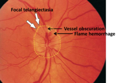
NAION - Testing and orders
In office: RNFL OCT (elevation), VF (altitudinal or central), Pupils (+RAPD), Red cap (reduced), BP (high), EOMs (r/o neuritis)
Order: ESR/CRP/platelet count (r/o GCA)
NAION - Treatment
Refer to PCP for management of BP/BS/Cholesterol
NAION - FU
1-2 months
NAION - Education
You have a condition called Non-Arteritic ischemic Optic Neuropathy or NAION, in which blood supply to your optic nerve has been cut off. This is often due to systemic issues like uncontrolled high BP, BS, or cholesterol. First, I want to refer you urgently for blood work to rule out dangerous inflammation in the body. I also want to refer you to your PCP to help get your BP/BS/cholesterol under control as there is a risk of this happening to the other eye as well. Up to 40% of patients show mild improvement in vision over 3 to 6 months in some studies. I'd like to see you back one month after you've seen your PCP.
Arteritic Anterior Ischemic Optic Neuropathy (AAION) -Symptoms and signs
Sx: HA, scalp tingling, jaw claudication, temporal artery pain, sudden acute unilateral vision loss
S: Chalky white edematous ON, vessels are not obscured
Suspected AAION - Testing and orders
RNFL OCT, VF (altitudinal or central), Pupils (+RAPD), Red cap (reduced), Palpation of temporal artery, TA biopsy, ESR/CRP/CBC with diff -platelets (elevated. nl: ESR (age+10)/2 (F), CRP < 10)

AAION - Treatment
Refer to ER for blood work and IV methylprednisolone, Switch to oral steroids if (+) TA biopsy and cont for 6-12 months
AAION - FU
3-4 wks
AAION - Education
You have Arteritic Anterior Ischemic Optic Neuropathy or AAION/GCA. This condition leads to inflammation of the medium and large blood vessels in the body, leading to your fever, headaches, and jaw pain. This inflammation is also affecting the blood supply to your optic nerve, leading to vision loss. Unfortunately, there is only a small chance that the vision you have lost will return. The primary goal now is to prevent this from happening in the other eye. I am referring you for urgent blood work, as well as steroids at the hospital. They will likely keep you on steroids for 6-12 months after this to keep inflammation in your body low. I'd like to see you back in about a month.
Optic Neuritis -Symptoms and Signs
Sx: Gradual vision loss over 10-14 days, periocular pain worse with eye movement, photopsia or phosphenes (flashes of light), Uhtoff's phenomenon (vision dims after exercise or going from A/C to outside)
S: based on testing results. Rarely has ON edema, usually retrobulbar.
Optic Neuritis - Testing in office and orders
Color vision (decreased), contrast sensitivity (decreased), VF (central or arcuate), pupils (+RAPD), red cap (reduced), EOMs (pain), OCT ONH/RNFL (normal if acute), Urgent MRI and LP
Optic Neuritis - Treatment
Refer for imaging and oral steroids if within the first 1 -2 weeks of onset. Refer to neurology for MS management.

Optic Neuritis - FU
1 month. After that, every 3 months
Optic Neuritis - Education
You have a condition called optic neuritis. This means that the optic nerve connecting your eye to your brain is swollen, leading to your blurry vision. Your vision will return after the swelling has gone down. However, this kind of swelling is often associated with multiple sclerosis. MS is an autoimmune disease in which your body's own immune system attacks the tissues in your nervous system. It's important that we refer you for an MRI and to a neurologist to help make that diagnosis. I'd like to see you back in 1 month, and we'll check your vision every 3 or 6 months from here on out.
ONH Drusen -Symptoms and signs
Sx: none
S: ON that is not round, could look edematous, but it is not.
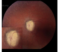
ONH Drusen - Testing
FAF (appear bright), B-scan (hyper-reflective), VF (generalized depression), RNFL OCT (shows waterfall defect, bumps)

ONH Drusen - Treatment
Observation
ONH Drusen - FU
q 6 or 12 months
ONH Drusen - Education
You have ONH drusen, which are calcified deposits within the optic nerve. Usually these are benign and will have no effect on your vision. Rarely, they can start to compress the nerve tissue and lead to vision loss. I'd like to see you every 6-12 months to make sure that they are not impacting your vision. There is no treatment for this condition.
Horner Syndrome -Symptoms and signs
Sx:
S: Same sided pupil meiosis and ptosis, possibly reverse ptosis as well, possible anhydrosis

Horner Syndrome - Testing and orders
Pupil sizing (one pupil is smaller, more obvious in the dark) and testing (light and near testing are normal), MRD 1 (2mm ptosis), old photos (will show if long-standing), 1% apraclonidine testing (dilates Horner pupil), RTC 1 day for 1% hydroxyamphetamine testing (dilates if pre-ganglionic)
If acute: order chest CT (Pancoast tumor), MRI/MRA of brain/neck (carotid artery dissection [esp if neck pain]), CBC w/ diff
Horner Syndrome - Treatment
Refer for neuroimaging if acute
Horner Syndrome - FU
FU after imaging
Horner Syndrome - Education
You have a condition called Horner syndrome which is the cause of your lid droop and change in pupil size. This can be caused by several things, but there is likely inflammation or compression of the nerve that connects parts of your eye to the brain. It may be due to headaches or migraines, or something more serious like a mass. I need to refer you for imaging to rule out that possibility. I will follow up with you after the imaging results.
Adie's Tonic Pupil -Symptoms and signs
Symptoms: May have unilateral photophobia and blur at near, unilateral accommodative paresis (trouble focusing at D after N)
Signs: Dilated pupil in bright light
Adie Tonic Pupil - Testing
Pupil sizes (affected pupil dilated in bright light, poor reaction to light, sector paralysis under SLE), Near testing (pupil constricts at near), old photos show normal pupils, 0.125% Pilocarpine (constriction, but have to wait 60 minutes)

Adie Tonic Pupil - Treatment
Colored CL for cosmesis, Vuity or Vizz if near vision is bothering patient, sunglasses for light sensitivity
Adie Tonic Pupil - FU
1 year
Adie Tonic Pupil - Education
Your pupils are different sizes because of something called Adie's tonic pupil. This is a temporary condition that should resolve on its own. It could get better in a couple weeks or it could take up to six months. I recommend wearing sunglasses when outside to help with light sensitivity. If you do not like the way your eyes look, I can prescribe you colored contacts that would hide the size of the larger pupil, or a drop that helps to constrict the pupil. If you're having trouble with near work, the drop would help with that. Because this could happen in your other eye, I recommend coming in every year.
Myasthenia Gravis -Symptoms and signs
Sx: By end of day, eyelids get droopy and sometimes gets diplopia
S: ptosis
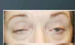
Myasthenia Gravis - Testing and orders
MRD1, MRD1 after 2 min sustained upgaze, Ice pack test over ptotic eye (improves ptosis), pupils (normal),
Order: Bloodwork (T3, T4, TSH) and Neck CT (r/o thymoma), edrophonium testing
Myasthenia Gravis - Treatment
Prism for diplopia. Upneeq for ptosis. Refer to neurology for treatment (steroids may help if early in the disease course, pyridostigmine if trouble breathing),
Myasthenia Gravis - FU
4 months
Myasthenia Gravis - Education
You have a condition called MG. This is an autoimmune condition in which your body's own immune system attacks your tissues, leading to a droopy eyelid and double vision towards the end of the day. It may also lead to difficulty breathing. I am referring you to a neurologist who can treat you. I am also are ordering blood work, as this condition is often associated with thyroid and thymus issues. If the double vision is consistent and bothersome to you, we can also prescribe prism in your glasses to help. I'd like to see you back in 4 months.
Bell Palsy -Symptoms and signs
Sx: One-sided cannot close eye, cannot smile, cannot raise eyebrow
S: Acute one-sided facial paralysis

Bell palsy - Testing
NaFL, TBUT, ask patient to raise their eyebrows. If can raise both eyebrows more likely a stroke and need to refer immediately to ER
Bell palsy - Treatment
R/O stroke, Refer to neurology, PFATs, Ointment at bed time, Lid taping or eye mask at night, Turn off fans while sleeping
Bell palsy - FU
1 month
Bell palsy - Education
You have a condition called Bell palsy. This is a condition where inflammation or a previous viral infection has caused drooping on the whole side of your face. As this can often mimic a stroke, I want to refer you to the ER for a stroke workup. I am also referring you to neurology for further care. This condition is usually temporary and resolves in a few months. Since your eye can not close properly, it's important to make sure your eyes do not dry out. I recommend frequent PFATs throughout the day, an ointment at bed time, and wearing a sleeping mask at night. Turning off fans while sleeping can also help. I'd like to see you back in 1 month.
Ocular Hypertension -symptoms and signs
Sx: none
S: IOP is consistently greater than 21
OHTN - Testing
RNFL/GCC OCT, 24-2 VF, Pachymetry, IOP, VEP, Rabin cone color contrast testing
OHTN - Treatment
Monitor if
OHTN - FU
RTC 4 weeks after starting prostaglandin to repeat IOP. Follow up again in 3 months
OHTN - Education (for IOP lower than 28)
You have OHTN. This means that the pressure inside your eye is higher than normal. This can be a risk factor for glaucoma, so I would like to see you again in 3 months to recheck your eye pressure. If the pressure is still high, we may choose to give you a drop that helps reduce the eye pressure and prevent glaucoma from developing.
Primary Open Angle Glaucoma -symptoms and signs
Sx: none
S: Enlarged C/D ratio (especially vertical) or asymmetric, NRR thinning (esp ST or IT), vascular changes (bayonetting, nasalization, bean potting), Drance heme

POAG - Testing
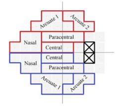
RNFL/GCC OCT (shows loss), 24-2 HVF (nasal step, arcuate) Pachymetry (thin CCT), IOP (> 21mmHg), Gonio (shows open)
POAG - Treatment
1st line - Latanoprost 0.05% qhs (CI in pregnancy; pigment changes, lash growth, hyperemia), Timolol 0.25-0.5% qAM (CI in CHF, bradycardia, COPD, asthma, MG), Brimonidine 0.2% bid (CI with MAOI and kids
POAG - FU
4 weeks for IOP check. Monitor 6 months
POAG - Education
You have a condition called POAG. There are many risk factors for glaucoma, including (+) FOHx, eye pressure >24, C/D size, African/Hispanic, and PDS/PXS. Increased eye pressure pushes on the optic nerve, leading to nerve tissue loss and progressive peripheral vision loss. The only known way to tx glaucoma is through lowering the eye pressure. I am going to start you on an eye drop to use once per day before bed. This drop can lead to irritation, so use PFATs throughout the day. You may also notice a darkening of your iris color or growth of your eye lashes. I want to see you back in 1 month to re-check the eye pressure, then we can monitor you every 6 months with scans.
Normal Tension Glaucoma -Symptoms and signs
Sx: none
S: IOP is WNL, but enlarged C/D ratio (especially vertical) or asymmetric, Drance heme, NRR thinning (esp ST or IT), vascular changes (bayonetting, nasalization, bean potting), nocturnal hypotension
NTG - Testing
RNFL/GCC OCT (shows loss), 24-2 HVF (nasal step, arcuate) Pachymetry (thin CCT), IOP (< 21mmHg), Gonio (shows open)
NTG - Treatment
1st line - Drops; Neuroprotective drops are preferred (Brimonidine 0.2% bid or Betaxolol 0.25-0.5% bid). 2nd line - Latanoprostene bunod 0.024% qhs (most effective drop in NTG; expensive)
NTG - FU
FU in 1 month for IOP check. Monitor q 6 mo.
NTG - Education
You have a condition called NTG. When we look at the nerve in the back of your eye we can see nerve tissue loss, indicating you have glaucoma. Often, glaucoma is associated with high eye pressure. Even though your eye pressures today were normal, the only known way to treat glaucoma is to lower the eye pressure. I'm going to start you on a drop that you'll use twice a day and I want to see you back in 1 month to re-check your pressure. After that, I want to monitor you every 6 or 8 months.
Acute Angle Closure Glaucoma -symptoms and signs
Sx: Ipsilateral HA, eye pain, photophobia, blur, halos around lights, nausea/vomiting
S: fixed, mid-dilated pupil, glaucomflecken (anterior lens crystalline opacities), hyperemia, corneal edema

AACG - Testing
IOP (>40 mmHg), Gonio (180+ of iridotrabecular contact [ITC], or if IOP is not hight, significant peripheral anterior synechiae [PAS]) Ant-seg OCT (plateau iris or double hump sign)

AACG - Treatment
3 drops, 1 drop every 15 min (Apraclonidine 0.5%, Timolol 0.5%, Dorzolamide 2%) - &/OR - Acetazolamide PO 250mg x2. Recheck IOP in 1 hour and repeat drops if still high. Once the cornea is clear, perform LPI. Send home on Acetazolamide 250mg bid and Timolol 0.5% bid
AACG - FU
1 day; RTC 2-7 days for LPI in fellow eye if narrow angles
AACG - Education
You are experiencing AACG. The drainage system in the eye that allows fluid to filter out is blocked, leading to increased eye pressure. This leads to the headache, eye pain, nausea and distorted vision you are experiencing. We are going to try to lower your eye pressure in-office using drops/pills, and may perform/refer out for an LPI surgery. This surgery prevents the drainage system from getting blocked again so that the eye pressure won't spike. If it goes well, we may choose to perform an LPI preventatively in the other eye as well. I'm also going to send you home with medicine to keep your eye pressure low. I'd like to see you tomorrow to re-check your eye pressure.
Diabetic Retinopathy -symptoms and signs
Sx: asymptomatic or may have large shift in prescription
S: macular aneurysms at all stages, dot blot hemes, CWS, exudates, IRMA, venous beading, neovascularization
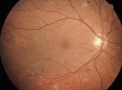
Diabetic Ret - Testing and orders
Fundus photo (more easily see MAs, hemes, etc), Mac OCT (SRF), IVFA (neo or perfusion abnormalities) or FAF, Gonio (check for NVI)
Order A1C, BP
Diabetic Ret - Grading
All stages may have CME. Mild - hemes. Moderate - more hemes, CWS, or exudate. Severe - 4 quadrants of hemes, 2 of beading, 1 of IRMA. Proliferative - NVD, NVE, NVI, or vitreous heme. High Risk Proliferative - NVD >1/4 size of the nerve, or any NVD/NVE associated with pre-retinal or vitreous heme

Diabetic Ret - Treatment
Refer for AntiVEGF for significant CME or NV, PRP for high risk NV, Vitrectomy for persistent vitreous heme. Otherwise, Monitor.
Diabetic Ret - FU
None - 12 mo. Mild - 6 mo. Moderate - 4 mo. Severe - 3 mo. Proliferative - 2 mo.
Diabetic Ret - Education
You have diabetic retinopathy. When your blood sugar is high or fluctuating, it makes the small blood vessels in the back of the eye fragile and leak. Blood and protein start to leak into the retina. It can also leak into the macula and cause swelling, leading to blurry vision. I'm going to refer you to a retinal specialist - OR - monitor. I'd like to see you in ___ months.
Hypertensive retinopathy -symptoms and signs of early
Sx: asymptomatic, or if acute then vision loss, HA, eye pain
S: generalized and focal arterial narrowing, AV nicking, copper wiring (for early stage)
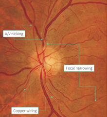
Hypertensive Ret - Testing
Fundus photo, BP (elevated), OCT-A, IVFA
Hypertensive Ret - Staging
1 - Attenuation. 2 - AV crossing. 3 - CWS, hemes, exudate, micro/macroaneurysms, venous or arterial obstruction, neo, Elschnig spots. 4 - ONH edema, macular star, venous engorgement.
Hypertensive Ret - Treatment
Refer to PCP for better BP control. Refer to ER if malignant HTN (BP is > 180/120). Focal laser if leaking macroaneurysm
Hypertensive Ret - FU
2 or 3 months
Hypertensive Ret - Education
You have hypertensive retinopathy. Uncontrolled high BP can lead to changes in the back of the eye such as bleeding, and can ultimately cause vision loss if this continues long term. I'm going to refer you back to your PCP to better control your BP.
CRAO -symptoms and signs
Sx: Sudden, painless, unilateral vision loss
Acute sign: cherry red spot surrounded by retinal whitening (only lasts 1-2 days)
Chronic signs: diffuse ischemic whitening of posterior pole, could have disc splinter heme , ON edema, +RAPD
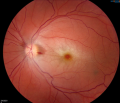
RAO - Testing and orders
OCT, fundus photo, IVFA (shows lack of blood flow), VF, BP, gonio (if longer than 2 days, may show NVI)
Order ESR/CRP/CBC, Carotid doppler, ECG

RAO - Treatment
Refer for stroke workup, Refer to PCP for systemic management, Monitor monthly for neovascularization. If presents within 24 hours, attempt gonio massage, breathing into a paper bag, Acetazolamide 2 250mg tablets, Timolol 0.5% q15 minutes.
RAO - FU
once a month for next 6 months to monitor for neovascularization if CRAO, 3-6 mo for BRAO
RAO - Education
You have a blockage in the main artery in the back of your eye this is preventing blood flow and oxygen from reaching the retinal tissue. Without blood flow, that tissue dyes off, leading to permanent vision loss. This may have been caused by a blockage in your carotid artery or from heart valve disease. I want to send you for urgent blood work and a carotid doppler to check for other blockages; if there is an artery blockage in the back of the eye, there could also be one in the brain that could lead to stroke. I also want you to FU with your PCP. I want to see you back in 1 month to make sure there are no complications developing from this, such as glaucoma.
CRVO -symptoms and signs
Sx: Sudden, painless, unilateral vision loss
S: Blood and Thunder (widespread heme, CWS), +RAPD, edema, VA worse than 20/200

RVO - Testing and orders
Mac OCT (macular edema), IVFA (confirm if ischemic or non-ischemic), ERG (reduced), Gonio (if been about a month), BP (high)
Order Lipid panel