VTMD 206 - Neuro 3
 Call Kai
 Learn
 Practice Test
 Spaced Repetition
 Match
 Flashcards
 Knowt Play
1/97

There's no tags or description
Looks like no tags are added yet.
98 Terms
Brain
Major Area Responsible for Motor Control
Integrates sensory information and initiates movement
Integrates sensory information
Initiates and ensures proper goal-directed movement
Brainstem
Major Area Responsible for Motor Control
Refinement area
Unconscious motor control
Relay and augmentation of locomotion/voluntary movement, posture, respiration, cardiovascular output
Spinal Cord
Major Area Responsible for Motor Control
Dorsal=sensory
Ventral=motor output
Simple reflexes (fully contained within the spinal cord, independent from the brain)
Sensory input (dorsal horn) and motor output (ventral horn)
Where rhythmic movements reside and are generated
Muscle
Major Area Responsible for Motor Control
Contraction and sense through proprioceptors
Motor end plate onto muscle fibre cells and proprioceptors (sensory afferents and can sense muscle tone and send information to the spinal cord)
Brain → motor neurons in the spinal cord ventral horn → motor end plates → neuromuscular junction → ACh release → muscle contraction → feedback on relative muscle tone provided by sensory neuron innervation
How are Muscle Cells Engaged?
Reflexive
Main Type of Motor Control
Spinal cord loops => quick responses
Voluntary
Main Type of Motor Control
Goal-directed movement
Rhythmic
Main Type of Motor Control
Maintenance of repetitive movements (locomotion)
Musculoskeletal proprioceptors
__________ ____________ sense stretch, position, and force
Present in muscles, at the junction of tendons, and joints
Also often needs input from your vestibular system and eyes
Sensory (afferent) neurons carry information from muscle to spinal cord
Aα fibre
Afferents that carry info from proprioceptors to ventral horn are Aα fibres
Very fast myelinated fibers
Synapse into dorsal horn
Projections up to brain to refine movements
Golgi tendon organs
Type of Muscle Proprioceptor
Muscle tension
Present in skeletal muscle
Present at connections between muscles and tendons
Sense tension of muscle
Stops muscle from receiving too much tension, preventing damage to the muscle
_____ _____ reflex prevents this when a high amount of tension on the muscle is sensed
Between muscle fibre cells and tendon
Significant activation leads to reflexive loss of tension
Muscle spindles
Type of Muscle Proprioceptor
Muscle length via stretch
Present in skeletal muscle
Sits in the center of every muscle
Lie parallel to muscle fibre cells, contained within intrafusal fibres
Intrafusal fibers help sense and give feedback on the amount of stretch a muscle is experiencing
When there is significant stretch, a message is sent from spindle to spinal cord
Innervated by motor neurons for feedback!
Gamma motor endings are important to gauge overall stretch of muscle, as opposed to relative/changes in stretch
Autonomic, Somatic, Upper, Lower, Alpha, Gamma
______ motor neurons – activate smooth and cardiac muscle
______ motor neurons – activate skeletal muscle
Upper vs. lower motor neurons
_____ motor neurons = cell bodies in the brain
_____ motor neurons = cell bodies in the spinal cord/horn, receive info from upper motor neurons
_____ motor neurons – direct projections onto muscle fibres (motor end plate)
Majority of motor neurons
_____ motor neurons – smaller diameter projections, projections onto intrafusal muscle fibres
Only regulate sensitivity of muscle spindle
Spinal cord
Dorsal horn = sensory
Ventral horn = motor control
White matter vs. grey matter
White matter is more highly myelinated
Upper motor neurons project onto lower motor neurons (which then produce their effects on muscle)
Excitatory MN - Glutamate
Inhibitory MN - GABA
Modulates responses
Direct spinal loops for immediate sensory activation from dorsal horn to motor effect in ventral horn
Circuits for generation of rhythmic behaviours
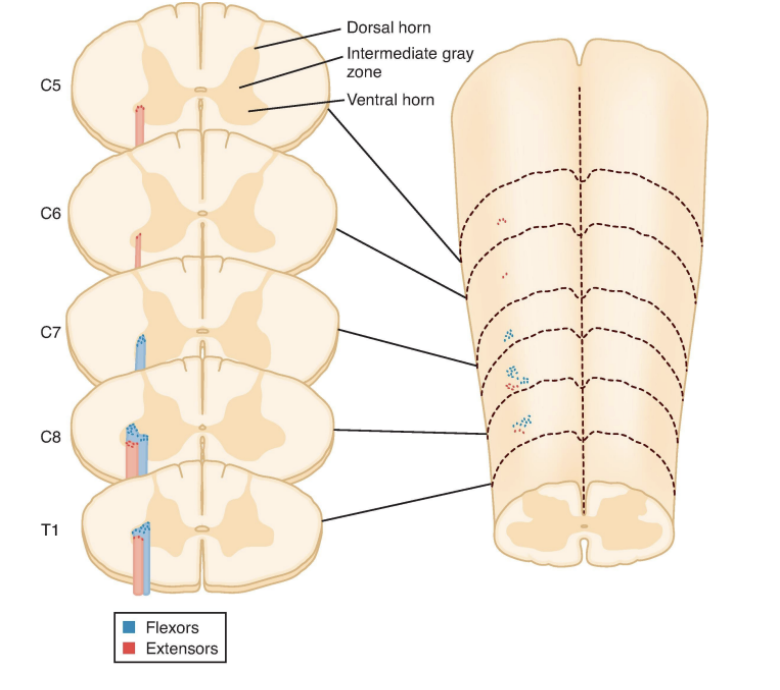
Somatotopically arranged
As you move down SC, innervating different areas of the body
Ex. thoracic is forelimbs
Proximal muscles closer to the center
Patellar (Stretch) Reflex
Triggered by stretch of quadriceps muscles (caused by hammer knock)
This activates muscle spindles and their Ia sensory afferents
Direct synapse onto alpha motor neuron (motor efferent fibers)
Contraction of quadriceps muscle
Also referred to as myotatic reflex
This reflex modulates the sensitivity of the muscle spindle
Reflex is there to maintain posture
Primary Motor Cortex (M1)
Part of the Cerebral Cortex
Sits anterior to the somatosensory cortex in all animals
Postcruciate/precruciate gyrus and rostral suprasylvian gyrus (in dogs)
Plans and initiates voluntary movements
Upper motor neurons here project to lower motor neurons in spinal cord
Neurons here are somatotopically organized and have different representation for different species
Left brain = right limb (decussation)
Premotor and supplementary motor areas (~M2)
~precruciate gyrus/ frontal gyrus
Helps to plan and sequence complex voluntary movements, along with prefrontal cortex
Set the stage/outline steps for movement before actual movement has even occurred
Most projections are to M1
Basal Ganglia
Causes action selection and sends signals to motor cortex to initiate movement
Collection of several subcortical nuclei
Modulate motor output via projections to thalamus (and onto M1) and brainstem motor areas
Tonic inhibition of motor system => release of tonic inhibition by basal ganglia initiates a given movement
Regulation of motor and premotor regions to pick the right movement and facilitate smooth movement
Influenced heavily by the prefrontal cortex (involved in decision making)
Role = paroxymal dyskinesia
Paroxysmal = sudden, uncontrolled
Dyskinesia = uncontrollable, involuntary movements
Pyramidal tracts
Motor and Descending (Efferent) Pathways
Includes:
Lateral corticospinal tract
Anterior corticospinal tracts
Spinocerebellar Tracts
Tract that provides proprioceptive feedback to the brain
Spinal cord → Cerebellum
More unconscious perception
Helps to fine tune unconscious movements
Ex. maintain posture/balance
Includes:
Posterior __________ tract
Anterior ___________ tract
Dorsal Column Medial Lemniscus
Tract that provides proprioceptive feedback to the brain
More conscious perception
Includes:
Gracile fasciculus
Cuneate fasciculus
Cerebellum
Fine-tunes and ensures movements are correct, maintains posture and balance
Receives input from proprioceptors (spinocerebellar tract), motor cortex, and vestibular system
Can be divided into 3 sections
No direct projections to lower motor neurons
Done through other mechanisms
Executes commands to the spinal cord via the brainstem and motor cortex
Vestibulocerebellum
Section of the Cerebellum
Balance and eye movements
Spinocerebellum
Section of the Cerebellum
Posture and limb coordination
Cerebrocerebellum
Section of the Cerebellum
Planning and timing of complex movements
Pain
An aversive sensory and emotional experience typically caused by, or resembling that caused by, actual or potential tissue injury
Refers to both the sensory and psychological perception of nociceptive stimuli
Subjective, it varies between individuals and varies day to day for an individual
Usually adaptive, but times when it becomes chronic and is less adaptive
Nociception
Refers to the physiological detection and processing of painful stimuli. Does not consider emotional/psychological aspects of the pain response
Acute Pain
Protective
Warns of existing or imminent tissue damage
Normal peripheral and central nervous system
Well defined temporal onset
Can clearly define the moment when you felt that pain
Variety of effective treatment options
Chronic Pain
No known defensive or beneficial function
Neuropathic or inflammatory
Aberrant peripheral and central nervous system
Poorly defined temporal onset
Hard to tell when you started/in general felt pain
Limited treatment options
Nociceptor
A sensory neuron that is activated by stimuli capable of causing tissue damage
Free nerve endings
Three types of nociceptors
Mechanical (Mechanical trauma)
Thermal (noxious heat and cold)
Chemical
Also classified according to the afferent fibre (A-delta or C-fibre)
Aδ
Axon/Fibre of Nociceptor
Fast Pain, temperature
Myelination, faster
Second smallest in diameter
Fire first – sharp, jabbing pain
C
Axon/Fibre of Nociceptor
Slow Pain, Temperature, Itch
No myelination, slower
Smallest in diameter
Fire after – low, throbbing pain
Lateral spinothalamic tract, spinireticular tract, spinomesencephalic tract
3 Ascending Pain Pathways (Anterolateral System)
Along the lateral side of the spinal cord
Carry info up spinal cord to the brain
Lateral Spinothalamic Tract
Ascending Pain Pathway
Projects to thalamus, then to sensory cortex
Intensity & location
Spinoreticular Tract
Ascending Pain Pathway
Projects to reticular formation and then to diverse brain areas (thalamus, hypothalamus)
Autonomic, motor, and sensory responses to pain
Motivational, affective and aversive aspects of pain
Emotional aspects of pain
Spinomesencephalic Tract
Ascending Pain Pathway
Projects to midbrain
Activates periaqueductal gray (PAG) = produces endogenous analgesia
Affective & aversive behaviours of pain (i.e fear)
Gate Control Theory of Pain
Proposed by Melzack and Wall (1965)
Explains how sensory information entering the spinal cord can either increase of decrease the transmission of pain signals to the brain
Concept: non-painful sensory input (touch or pressure) can inhibit transmission of pain signals at the spinal cord level
Ex.
Hit thumb with hammer – gates are open
When you put pressure on thumb – gates close more
Depends on pain fibers and inhibitory interneurons
Spinal cord receive signals from the periphery and from descending axons from the brain
Aβ fibers: Large, myelinated sensory fibers that transmit non-painful touch and pressure
Aδ and C fibers: Nociceptive fibers that transmit information about potentially damaging stimuli.
Spinal inhibitory interneurons: Neurons that can reduce activity of nociceptive projection neurons
Connect between different neurons
A lot of interneurons in the SC contain GABA and Glycine
Inhibitory NT

Periaqueductal Grey Matter, Locus Coeruleus, Rostral Ventromedial Medulla
3 Descending Inhibitory Pain Pathways
Transmit signals from the brain to the SC to dampen pain
Periaqueductal Grey Matter (PAG)
Descending Inhibitory Pain Pathway
Release enkephalins
Mediated by opioid receptors
Locus Coeruleus (LC)
Descending Inhibitory Pain Pathway
Release norepinephrine
Released into SC and acts on A1 and A2 receptors
2-adrenergic receptors inhibit pre- and post-synaptic pain transmission
1-adrenergic receptors release GABA from interneurons
Rostral Ventromedial Medulla (RVM)
Descending Inhibitory Pain Pathway
Release serotonin
5-HT (serotonin receptor) – multiple subtypes and different coupling; not well understood
Allodynia
Type of Pain Hypersensitivity — Characteristic of Chronic Pain
Painful response to something that is usually non-painful
Lowered pain threshold
Innocuous stimulus elicits pain
Innocuous touch (mechanical allodynia)
Cooling (cold allodynia)
Hyperalgesia
Type of Pain Hypersensitivity — Characteristic of Chronic Pain
Increased/exaggerated response to pain to something that is normally painful
Enhanced pain response to noxious stimuli
Typically evoked by heat stimulation
Spontaneous Pain
Type of Pain Hypersensitivity — Characteristic of Chronic Pain
Pain that just occurs, no clear stimulus
Pain in the absence of an overt stimulus
Recurring pain
100% of chronic pain patients affected
Peripheral Sensitization
Ex. cut finger, whole bunch of responses that cause ________ _________, fires much more strongly/robustly
Occurs bcs when you injure tissue, there is an inflammatory response and molecules come in, cytokines released, inflammation sensitizes neurons so they fire more readily
Greater output for a stimulus bcs fibers are sensitized
Tissue injury leads to activation of immune cells, which release signaling molecules often referred to as the inflammatory soup
Signaling molecules activate ion channels (ASICs, purinergic receptors) and other receptors (TrkB) • Signaling molecules bind to and change gating of other channels (TRPV1, etc.)
Net result is an amplification of nociceptor responses = more output for the same input
Requires input to be sustained
Central sensitization
Within the CNS
Pain pathways in brain and spinal cord respond way more strongly to a painful or non-painful stimulus due to changes in pathways, makes pathways function way more robustly in terms of sending pain signals
Over the top response
Composite measure pain scales
Facial expression (“grimace”) scales
List 2 validated pain assessment scales
Central Pattern Generators
a network of thoracolumbar spinal circuits that generate rhythmic motor output, do 2 things:
Alternating flexor-extensor activity
Opposing activity on contralateral side
Coordinate movement between all 4 limbs as one
Activated by excitatory drive from the brainstem
Brain input to the spinal cord can modulate the circuit, but is not required to maintain
Present in all vertebrates at birth
Ex.
Breathing
Mastication
Heart pumping
Flying
Swimming
Blinking
Swallowing
Half Center Circuit
The ___________ Hypothesis
Each side of the spinal cord contains one “__________”
Bcs it drives one half of the stride (stance and swing)
Each circuit produces alternating activity in flexor and extensor muscles in a single limb
In ventral horn of the spinal cord, lower motor neurons, one sends response to flexor muscle and one sends response to extensor muscle
Also have interneurons that can send signals between extensor and flexor motor neurons
Inhibitory interneurons connect flexor and extensor interneurons, so that when one muscle is extending (extensor signals being sent), muscle cannot flex (no flexor signals being sent), and vv
Flexor afferent activates the excitatory flexor interneuron
This activates the flexor motor neuron, and contracts the flexor muscle
Activation of the excitatory flexor interneuron also activates an inhibitory (GABA) neuron
This inhibits the extensor muscle

Mesencephalic locomotor region
Part of the brainstem (midbrain)
Receives input from cortex motor areas and the basal ganglia
Projects to the reticular nuclei, which then project onto lower motor neurons in the ventral horn
Activation of the ___________ is sufficient to drive central pattern generators (intensity drives change from walk to trot to gallop)
Cortex Motor Areas and Basal Ganglia → MLR → Reticular Nuclei → Lower Motor Neurons
Process of initiating and maintaining gait
Descending supraspinal input
Responsible for modulation of Central Pattern Generators
Input, initiates, modulates, and stops locomotion action via CPGs
Once it starts, keeps going until it receives further input from the brain
Can be a voluntary modulation (faster or slower)
Can be a modulation in response to sensory input (proprioceptive, nociceptive, vestibular, visual, auditory)
Some modulation can occur through direct spinal reflexive circuits involving sensory input (e.g. proprioceptive)
Reticular nuclei
Red nucleus
Vestibular nuclei
The cerebellum executes commands to the spinal cord via the brainstem and motor cortex
3 Important Areas:
Reticular nuclei
Part of cerebellum responsible for initiation and modulation CPGs
Also referred to as the reticular formation
Communicates bidirectionally with many motor areas as an integration network
Also receives some direct input from proprioceptors
Direct projection to ventral horn (mostly to interneurons) to drive lower motor neurons (reticulospinal tract)
Reticulospinal tract is very important for gait!
The MLR projects here to initiate, maintain, modulate, and terminate CPGs in the spinal cord
Red nucleus
Part of cerebellum responsible for coordination and modification of gait
Much more active in non-primate mammals (especially in cats)
Receives input from cerebellum and motor cortex
Direct projection to ventral horn (mostly to interneurons) to influence lower motor neurons (rubrospinal tract)
Helps coordinate limb movements, including swing-phase movements by the flexor muscle and skilled forelimb actions (cat dodging obstacle)
Projecting directly to lower motor neurons and receives input from cerebellum
Vestibular nuclei
Part of cerebellum responsible for control of balance and equilibrium
Receive info from vestibular system, control balance while walking
Direct projection to LMN
Communicates bidirectionally with cerebellum to take in vestibular information from world and process it
Receives input from proprioceptors, vestibular organs in ears, visual system, skin receptors, etc.
Direct projection to ventral horn (mostly to interneurons) to influence lower motor neurons (vestibulospinal tract)
Corrects balance and muscle tone, stabilizes eye movements
Directs many postural reflexes
Rubrospinal tract
Reticulospinal tract
Vestibulospinal tract
Extrapyramidal tracts
Controlled by parts of the cerebellum
Cervical and lumbar intumescence
Spinal cord is thicker where the brachial plexus and the lumbar plexus are located, associated with spinal nerves that are going to innervate the appendicular skeleton
Cauda equina
Collection of segmental nerves that are collected into the spinal column before they emerge
Lumbosacral interarcuate space
Area where CSF can be collected just above the sacrum
More caudal epidural space can also be used for epidural injections
Filum terminale
Connective tissue coming off the surface of the spinal cord and coming back caudally where it will fuse with the dura in that space, terminates on the caudal vertebra
Dorsal Funiculus
Funiculus of the White Matter of the Spinal Cord
Mostly ascending tracts
On top
Funiculi are composed of ascending and descending nerve fibers that are grouped within bundles to form tracts
Lateral Funiculus
Funiculus of the White Matter of the Spinal Cord
Space between ventral and dorsal root
Ascending tracts laterally
Ascending and descending tracts located medially
Funiculi are composed of ascending and descending nerve fibers that are grouped within bundles to form tracts
Ventral Funiculus
Funiculus of the White Matter of the Spinal Cord
Contain ascending and descending tracts
Located on the bottom
Funiculi are composed of ascending and descending nerve fibers that are grouped within bundles to form tracts
Intervertebral foramen
Where spinal nerves exit
Suprascapular nerve
Nerve of the Brachial Plexus
from spinal nerves C6-C7, enters between the supraspinatus and subscapularis muscles near the neck of the scapula, crosses over to the lateral side via the scapular notch
Innervates supraspinatus and infraspinatus muscles
Subscapular nerve
Nerve of the Brachial Plexus
From spinal nerves C6-C7, very short, enters the subscapularis muscle, sometimes two nerves
Innervates subscapularis muscle
Musculocutaneous nerve
Nerve of the Brachial Plexus
from spinal nerves C6-C8, travels down the brachium between the biceps muscle cranially and brachial vessels caudally, provides a communicating branch to the median nerve in the dog, then it courses deep to the insertion of the biceps
Innervates coracobrachialis, biceps and brachialis muscles, also provides a branch that innervates the skin of medial antebrachium
Axillary nerve
Nerve of the Brachial Plexus
from spinal nerves C7-C8, enters the leg between the subscapularis and the teres major level with the neck of the scapula
Innervates teres major, teres minor, deltoideus and part of subscapularis muscles
Radial nerve
Nerve of the Brachial Plexus
from spinal nerves C7-C8, T1-T2, enters the triceps distal to the teres major, spirals around the humerus on the caudal aspect
Innervates triceps, tensor fasciae antebrachii, anconeus, extensor carpi radialis, supinator, common digital extensor, lateral digital extensor, ulnaris lateralis muscles
Median nerve
Nerve of the Brachial Plexus
from spinal nerves C8, T1-T2, runs with the ulnar nerve, this common trunk lies on the medial head of the triceps, median is the cranial division and passes just cranial to the epicondyle of the humerus to enter the forearm
Innervates pronator teres, flexor carpi radialis, pronator quadratus, superficial digital flexor, deep digital flexor muscles
Ulnar nerve
Nerve of the Brachial Plexus
from spinal nerves C8, T1-T2, runs with the median nerve, this common trunk lies on the medial head of the triceps, ulnar is the caudal division and crosses the elbow caudal to the medial epicondyle of the humerus
Innervates flexor carpi ulnaris, deep digital flexor, interossei muscles, as well as skin of caudal antebrachium/palmar paw/fifth digit
Horse
In the _____, it is the median and musculocutaneous nerves that run together down the forelimb
Cattle
The musculocutaneous nerve in ______ loops under the axillary artery to join the roots of the median and ulnar nerves. All three nerves run a short distance down the forelimb before the ulnar branches off caudally and then smaller musculocutaneous branches leave variously as the nerve travels down the limb. Also, the axillary and radial nerves run together proximally before becoming separated to innervate their respective muscles.
Obturator nerve
Nerve of the Lumbosacral Plexus
from spinal nerves L4-L6, it is formed within the caudomedial portion of the iliopsoas muscle and leaves the muscle dorsomedially, runs along the body of the ilium and leaves the pelvis by passing through the obturator foramen
Innervates adductor, external obturator, pectineus and gracilis muscles
Femoral nerve
Nerve of the Lumbosacral Plexus
from spinal nerves L4- L6, pursues a course through the psoas muscles to reach the dorsocaudal corner of the flank and the iliopsoas muscle, on entering the thigh it runs between the sartorius and pectineus muscles
Innervates iliopsoas and quadriceps group (rectus femoris, rectus lateralis, rectus medialis, rectus intermedius) muscles
Saphenous nerve
Nerve of the Lumbosacral Plexus
branch of the femoral nerve that dives between the rectus femoris and vastus medialis to branch within
Innervates the sartorius muscles group
Caudal gluteal nerve
Nerve of the Lumbosacral Plexus
variably from spinal nerves L7 and/or S1-2, passes over the ischiatic notch medial to the middle gluteal muscle and enter the medial surface of the superficial gluteal
Innervates superficial gluteal muscle
Cranial gluteal nerve
Nerve of the Lumbosacral Plexus
from spinal nerves L7 and S1, passes over the greater ischiatic notch, crosses the lateral surface of the ilium at the origin of the deep gluteal muscle
Innervates middle and deep gluteal muscles
Sciatic nerve
Nerve of the Lumbosacral Plexus
from spinal nerves L6-L7 and S1-S2, passes over the greater ischiatic notch, caudally over the hip medial to the greater trochanter and travels distally, caudal to the femur on the lateral side of the adductor muscle (most you exposed this when you lifted the biceps femoris muscle up), sciatic nerve terminates in the thigh as the tibial and common fibular nerve
Innervates gemelli, internal obturator, quadratus femoris, biceps femoris, semimembranosus and semitendinosus muscles
Tibial nerve
Nerve of the Lumbosacral Plexus
Larger of the two sciatic branches, heads caudally and at the stifle it passes between the two heads of the gastrocnemius muscles
Innervates gastrocnemius, superficial digital flexors, popliteus, deep digital flexor muscles
Common (peroneal) fibular nerve
Nerve of the Lumbosacral Plexus
smaller of the two sciatic branches, heads craniolaterally deep to the thin part of the biceps femoris muscle, it crosses the lateral head of the gastrocnemius muscle and then passes between the lateral digital flexor and fibularis longus, divides into smaller branches
Innervates cranial tibial, long digital extensor muscles, lateral digital extensor, fibularis longus muscles
Lumbosacral trunk
Name for Cranial gluteal nerve and Caudal gluteal nerve together
Horse and Cow (horse earlier than cow)
In the _____, the sciatic splits into its tibial and peroneal branches much earlier than in the dog.
Brain or brainstem
Neuroanatomical Localization
Abnormal mentation = lesion in the __________
Brainstem (multiple) or Peripheral Cranial Nerve (single)
Neuroanatomical Localization
Cranial Nerve Deficits = lesion in the __________
Cerebellum, Vestibular, Proprioceptive
Neuroanatomical Localization
Ataxia = lesion in the __________
Spinal cord — Lower motor neuron
Neuroanatomical Localization
Reduced/Absent Reflexes = lesion in the __________
Spinal Cord — Upper Motor neuron
Neuroanatomical Localization
Normal/Increased Reflexes = lesion in the __________
Mentation
Refers to a patient’s mental activity
Originates mainly at the level of the Cerebrum and brainstem (to a lesser extent)
Alterations leads to various levels of alertness
I.e. Dull, stuporous, comatose
Dogs with altered ______ do strange things
Head pressing, stargazing, wandering, ignoring owners etc
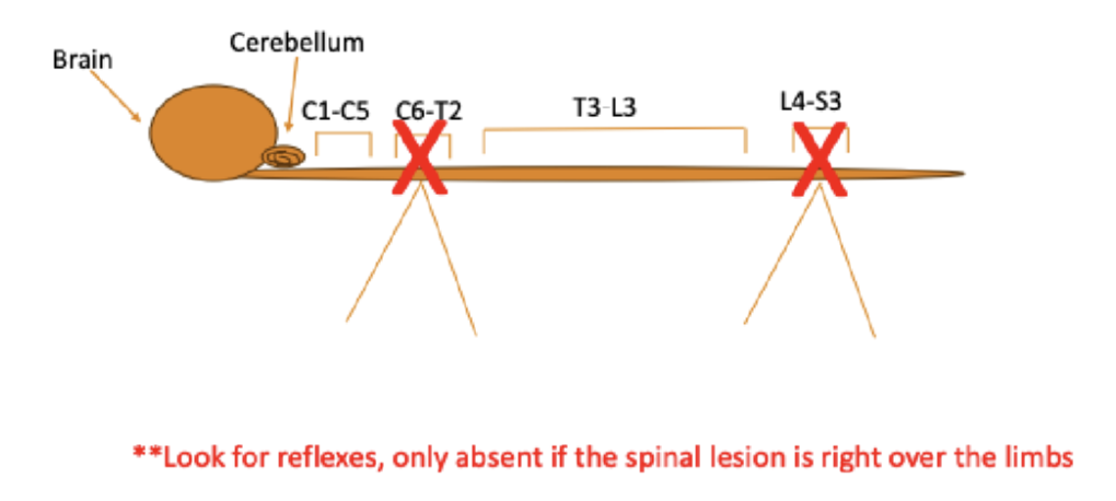
C6-T2 or L4-S3
If reduced reflexes (LMN issue), the spinal lesion is either between _____ or _____
Sedation
CNS depression with drowsiness and some degree of centrally induced relaxation
Still rousable, will respond to noxious stimuli but generally unaware of surroundings
Very relaxed, but rousable and respond to noxious stimuli
Tends to preserve autonomic reflexes
Can have tranquilizing effects
Anxialistic – reduces anxiety
Are part of anesthesia protocol
“Twilight Anesthesia” in humans
Misnomer, actually ______
Premedication
Given prior to induction of anesthesia
Get things started before putting them under anesthesia
Usually 2 or more agents
Goals
Relieve anxiety, apprehension, fear
Counteract unwanted side effects of anesthetic agents
Shorten procedure overall (amount of time they are under anesthesia)
Anesthetic sparing (use less of it)
Increased safety
Perioperative analgesia (pain control)
General anesthesia
From Greek meaning ‘insensibility’ or ‘without feeling’
Reversible unconsciousness that is characterized by controlled depression of the CNS
Not rousable by noxious stimuli
Reflexes attenuated (sensory, motor, autonomic)
Amnesia
General vs dissociative
Levels of consciousness
Dissociative state = do not respond, but look awake
More helpful for short procedures
Administered through various routes
Inhalational, IV, IM
Analgesia
Absence of pain in response to stimulation that would normally be painful
Varying degrees of efficacy
Agent
Timing of administration matters
Hypoalgesia - lowering of pain
Part of anesthesia and sedation protocols
Can be sedative themselves
Not always related to drug administration, other routes
Electroacupuncture
Laser therapy
Can be combined
Local anesthesia
Reversible block of the generation and propagation of electrical impulse through nerves
Only way to achieve 100% analgesia, if they work
Non-specific
Affect on sensory and motor
Successful block results in
Decreased anesthetic requirements
Sustained analgesia
Reduced mortality
Neuroleptanalgesia
Gave sedation and a potent pain medication/analgesic with it
Multimodal Analgesia
Doing more than one thing to keep your patient comfortable
Using multiple ways to block many parts of the pathways to prevent pain in patient
Balanced anesthesia
Induces:
Unconsciousness
Analgesia
Reflex suppression
Muscle relaxation
Amnesia
Produced by combination of 2-3+ drugs
Use less of each drug = increased safety