Clinical Chem Quiz 1
 Call Kai
 Learn
Learn Practice Test
 Spaced Repetition
 Match
Match Flashcards
 Knowt Play
1/78

There's no tags or description
Looks like no tags are added yet.
Name | Mastery | Learn | Test | Matching | Spaced | Call with Kai | Chat |
|---|
No analytics yet
Send a link to your students to track their progress
79 Terms
Essential amino acids
Arginine
Histidine
Isoleucine
Leucine
Lysine
Methionine
Phenylalanine
Threonine
Tryptophan
Valine
Gold standard for IEM screening
HPLC Tandem Mass Spec
Plasma
Cell free portion of blood that includes coagulants
Serum
Portion of blood that has no coagulants (especially fibrinogen)
>2 peptide link protein measurement
Proteins added to reagent with copper sulfide and alkalines
Copper breaks down links and creates color change
Color change is measured at 540nm
Not sensitive to protein <0.1mg/dL
Major serum proteins
Prealbumin (transthyretin)
Albumin
Alpha 1 antitrypsin
Alpha 2 macroglobulin
Beta 2 transferrin
Gamma immunoglobulins (G, A, M, CRP)

Nephrotic syndrome SPEP profile
Kidneys passing too much protein, increase in alpha 2 molecules to make up and keep up oncotoic balance

Polycloncal gammapothy
Increase in gamma globulins, many types

Monocolonal gammopathy
increase in one type of monoclonal antibody

Hypogammaglobinemia
Lack of gamma globulins
Acute phase inflammation response INCREASES IN…
C-reative protein
Procalcitonin
Haptoglobin
Fibrinogen
ALpha-1-trypsin
Ceruloplasmin
Ferritin
Serum amyloid A
Acute phase inflammation response DECREASE IN…
Albumin
Transferrin
Prealbumin
Retinol binding protein
Alpha-fetoprotein
Albumin
Contributes to oncotic pressure and fluid balance
3.5 to 5.2 g/dl
Marker for nutrition status and liver capacity
Heavy and negative charged
Measured through Bromocresol green dye binding
Microalbumin (urine albumin)
Albumin in urine (should be low)
>300mg/day is considered positive (BAD)
Measured via turbidity or nephelometry
24 hour urine collection is preferred
Prealbumin (transthyretin)
Good marker for nutrition status
Transports thyroxine and retinol
Normal range 18-39 mg/dl
Measured via turbidity of nephelometry
Anti-tryspin 1
Inhibits proteases from breaking down self proteins (protects liver and lungs from neutrophil breakdown)
Can lead to emphysema (damage to alveoli) or liver disease
Haptoglobin
Found in serum
Marker for hemolysis (acute phase reactant)
Ceruloplasmin
Copper transport protein
Can leads to Wilson disease (defect in ceruloplasmin binding copper, leading to excess)
Manifests as Fleischer rings in eyes
High liver copper amounts, low serum levels and high urine levels
Can leads to Menkes‘ disease (defect in copper transport)
Manifests as neurodegenerative disorders
Low liver copper
Transferrin
Binds to ferric iron (fe3+)
Measures via turbidity
Presents as high transferrin in Iron Deficiency Anemia
Ferritin
Stores iron
Measures via sandwich immunoassay
Presents as low ferritin in Iron Deficiency Anemia
Presents as high ferritin in Iron Overload
B2 transferrin
Marker for CSF
Detection via immunofixation
B2 microglobulin
Marker for kidneys
Measures renal tubular disorders (low serum levels, high urine levels), kidney heavy metal damage, multiple myeloma)
C-reatice protein
<10 mg/L indicates low probability of bacterial infection
Phenylketonuria
Missing Phenylalanine hydroxylase (PAH)
Body cannot break down phenylalanine, leading to toxic build up
Treated via diet change
Alkaptonuria
Build up of homogentisic acid (by product of alanine to tyrosine), leading to black urine
Treated via diet change
Maple syrup urine disease
Body cannot breakd down branched amino acids leading to maple-syrup-smelling urine
Treat with low protein diet, consider liver transplant
Cofactor
Inorganic non protein that is required for enzyme activity
Coenzyme
Organic protein or molecule that is required for enzyme activity
Enzyme function
Biomolecule that catalyzes (speeds up) a reaction
Rate of catalyzed reaction

Vmax
The max rate of a reaction
kM (Michelis constant)
Substrate concentration at ½ the Vmax
Zero and first order
First order - exponential portion of reaction, not dependent on substrate concentration
Zero order - plateauing portion of reaction where enzymes are occupied
Enzyme present assay
We put excess substrate to get to V max and measure
or
We put enzyme in excess so that V is proportional to concentration of substrate
or
We add a ton of substrate and let it run to completion
Competitive inhibitors
Binds to active site (decreases the amount of enzyme available)
kM increases, Vmax stays the same
Non competitive inhibitor
Binds to somewhere on the active site to (slows it down)
kM stays the same, Vmax decreases
Uncompetitive inhibitor
Binds to the substrate enzyme complex
Enzyme activity constant
End point method (stop the reaction at a fix time, measure absorbance of initial vs final)
Kinetic method (where we have multiple measurements at specific time intervals)
Enzyme activity measurement
IU
or
Umol/min
or
Umol/min/L
Creatine Kinase/Phosphatase (CK) source, function and diagnosis
Source: Highest around the skeletal muscle, heart and brain
Function: Creatine-P to Creatine
Diagnosis: Increase = source tissue disorder
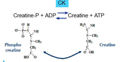
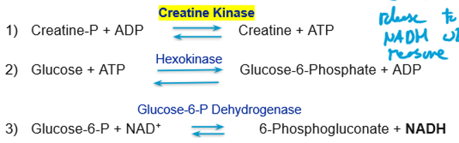
Creatine Kinase/Phosphatase (CK) measurement and error source
Measurement: Creatine P to creatine + ATP, leads to NADH with 2 enzymes
Error: Hemolysis
CK isoenzymes
CK-MM (Skeletal muscle)
CK-BB (Brain) (>500U/L is severe, >205U/L) is death
CK-MB (Heart)
Lactate dehydrogenase (LDH) Source and diagnosis and function
Source: Heart, liver, skeletal muscles
Elevations: Tissue cell and inflammation
Function: Lactate to pyruvate
GREAT marker for reinforcing diagnosis
LDH measurement and error
Measurement: LActate to pyruvate creates NADH which is measurable
Error: Hemolysis
Aspartate aminotransferase (AST) source, elevation and measurement
Source: heart>liver>skeletal muscle>RBCs>serum
Elevation: liver damage (moderate = cirrhosis, large = toxic hepatitis)
Measurement: AST, then use MD to create NAD which is measurable

AST Error
Source: hemolysis (very much so), only stable for 3 to 4 days when refrigerated
Alanine aminotransferase (ALT) Source and diagnosis
Source: Many tissues but high concentration in the liver
Elevation: high ALT and AST? liver disease, normal ALT, high AST? non-liver source, AST/ALT ratio >3? alcoholic hepatitis or cirrhosis
ALT error and measurement
Error: Stable for 3-4 days, unaffected by hemolysis
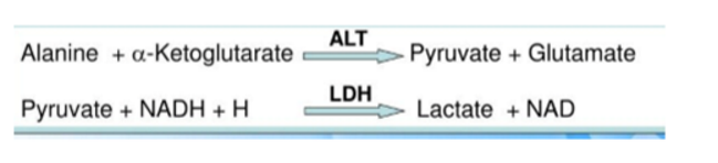
Measurement: Reaction coupled with LDH to generate NAD
Alkaline phosphate ALP source, diagnosis and function
Source: Liver, bone, spleen, placeneta
Diagnosis: Hepatobiliary disorder, bone disorder
Function: Catalyzes phosphate monoester cleavage
ALP error and measurement
Measurment: removal of phosphate from P-nitrophenylphosphate, produces P-nitrophenolate which is yellow at 405NM
Error: hemolysis
y-Glutamyltransferase GGT source, diagnosis
Source: Liver, bile ducts, kidney, brain, pancreas
Diagnosis: hepatobiliary disoder, alcoholism, pancreatitis, diabetes mellitus
Measurement: Production of P-nitroanaline which can be measured at 405NM
If AST, ALT greater than ALP, GGT then…
Primary liver disease, acute hepatitis or jaundice
If ALP, GGT greater than ASP or ALT then…
Obstructive liver disease or jaundice, post hepatic jaundice or hepatobiliary disorder
Glucose -6-phosphate dehydrogenase G6PD source, function and diagnosis
Source: adrenal cortex, spleen, thymus, lymph nodes, mammary glands
Function: G6P to 6Phosphogluconate with release of NADPH
Diagnosis: Toxic drug reactions and heinz bodies
G6PD measurement
Measurement: Hemolysate
Macroenzymes
High molecular mass forms of enzymes in serums (bound to Ig)
High activity detected as they are too big to be passed through kidney
Purpose of hexose-monophosphate shunt
Generation of NADPH and precursors to nucleotides

Glucose reference ranges
UW 62-125mg/dL
Hypoglycemia
Low blood glucose (less than 62mg/dL)
Manifests as hunger, sweating, nausea
Hyperglycemia
Increase in plasma glucose caused by imbalance of hormones (>125mg/dL)
Symptoms exhibited at >200mg/dL
Polyuria (lots of pee)
Fasting plasma glucose
When you have no foods or drinks 8-12 hours before a test
Normal fasting: 70-99mg/dL
Impaired fasting 100-125mg/dL
Provisional diabetes >126mg/dL
Type 1 diabetes mellitus effect and risks
Hyperglycemia caused by damaged pancreas makes little to no insulin (moves sugar to blood stream into cells)
Autoantibodies attack spleen Beta-cells that make insulin
Leads to excess glucagon
Risk factors: family history, infection, virus
Type 2 diabetes mellitus effect and risks
Greater tendency to get into coma
Family history, obesity, unhealthy diet
Insulin resistance: often obese, high blood pressure, dark velvety skin patches
Fasting glucose >100mg/dL
Gestational diabetes mellitus
Gestational
Placental estrogen and cortisol block the mother's insulin leading to insulin resistance and hyperglycemia
Treat by changing diet
Done at 24 to 48 weeks
Gestational diabetes mellitus tests
Glucose challenge
Oral glucose
Glucose challenge test
Administer 50g of glucose, measure serum glucose after 1 hour
Normal <140mg/dL, abnormal >140mg/dL
Oral glucose test
Pregnant patient fasts for 8-14 hours
Take baseline plasma glucose then administer 75g in 250-300mL
Normal < 140mg/dL, impaired glucose tolerance 140-199mg/dL, diabetes >200mg/dL
Criteria of diabetic diagnosis
HBA1C > 6.5%
Random glucose level >200mg/dL
Fasting plasma glucose >125mg/dL
Oral glucose tolerance test >200mg/dL after 2 hours after taking 75g glucose load
Metabolic syndrome
Hypertension
Increased serum glucose
Abnormal lipids
Increased body fat
Risk factor, often those with it have diabetes
Specimen tubes
Lime green (Plasma separation tube)
Orange (Rapid serum tube)
Gold (Serum separation tube)
Grey sodium fluoride/potassium oxalate (glycolysis prevention)
Red top (no preservatives)
Hemoglobin A1C test
reflects 2-3 months of glycemic control
Normal: 4-5.6%, prediabetic: 5.7-6.4%
Requires lavender top
Hemoglobin A1C test methods
HPLC and boronate affinity chromatography
Seperates different hemoglobin species
A1c immunoassay
Monoclonal antibody to glucose attaches to glycated hemoglobin beta chain
Enzyme assay
Cleaves N terinal valine, oxidation and colormetric detection
Glucose oxidase reaction

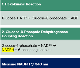
Hexokinase reaction
Lactate measurement

Type 1 von gierke disorder
Glucose 6 phosphatase deficiency
Can't make glycogen
type 3 Cori disease
Deficiency of gylcogen debrancher
Type 4 Amylopectinosis
Deficiency of branching enzyme, abnormal glycogen build up
Galactosemia
Inability to metabolism galactose
Can't metabolism galactose to lactose leading to build up of galactose in blood
Leads to brain damage, cataracts, jaundice, kidney damage