physio e3l5: cardiac function/electrical activity
 Call Kai
 Learn
Learn Practice Test
 Spaced Repetition
 Match
Match Flashcards
 Knowt Play
1/28

There's no tags or description
Looks like no tags are added yet.
Name | Mastery | Learn | Test | Matching | Spaced | Call with Kai | Chat |
|---|
No analytics yet
Send a link to your students to track their progress
29 Terms
3 parts cardiovascular system
what is its function?
heart ,blood vessels, blood
transport of substances (O2, wastes, hormones, immune cells, clotting proteins)
heart structure:
4 chambers
4 valves
2 septums
base and apex?
draw it
r atrium, l atrium, r ventricle, l ventricle
valves
atrioventricular (AV) valves = r tricuspid, l bicuspid (mitral valve)
middle level
semilunar (SL) valves = r pulmonary and l aortic
bottom level
base = inferior point of left ventricle, apex = broad superior/posterior surface

blood vessels:
closed system?
5 vasculature levels in order/ what they are
why do arterioles have high resistance?
aorta = ?
starts and ends in heart
5 blood vessels:
heart → arteries = large branching vessels
arterioles = small branching vessels
high resistance
capillaries = site of exchange
venules = small converging vessels
veins → heart = large converging vessels
high SA, low velocity
largest artery/first one out of heart
blood components:
erythrocytes RBCs
Leukocytes WBCs
thrombocytes Platelets
Plasma
which 3 are cellular components
transport
immunity
cell fragments that clot/hemostasis
fluid + solutes
first 3
path of blood through heart/vasculature:
2 circuits = where start/end
function?
where 2 main capillary beds
what kind of flow
circuits
pulmonary = BVs from heart, through lungs, back to heart
oxygenation of blood
systemic = BVs from heart, through systemic tissues, back to heart + coronary circulation
supply tissues w/ O2 and nutrients
lungs and tissue
series flow through cardiovascular system:
systemic circuit = start and end points? 5 steps?
pulmonary circuit = start and end points? 5 steps?
draw it
systemic = LV, aorta, tissue, vena cava, RA
pulmonary = RV, PA, alveoli, Pulmonary veins, LA

Oxygen exchange:
pulmonary capillaries = where?
blood entering v. blood leaving
what happened?
systemic capillaries
deoxygenated blood = ?
pulmonary capillary beds
blood entering lungs = deoxygenated
O2 diffuses alveoli → blood
blood leaving lungs = oxygenated
systemic capillaries = pick up CO2 and release O2 in tissue
Blood entering tissues = oxy
O2 diffuses blood → tissues
blood leaving tissues = deoxy
deoxygenated = less O2 not none
parallel flow in cardiovascular system:
what kind of flow/why important
which oxygenated/deoxygenated
arteries
arterioles
capillaries
venules
veins
parallel flow allows independent regulation of blood flow to organs via dilation/constriction
which oxygenated/deoxygenated
arteries = oxy
arterioles = oxy
capillaries = both
venules = deoxy
veins = deoxy
parallel flow in cardiovascular system:
portal circulation: what is it/3 locations?
how unique?
coronary circulation
coronary arteries = where
blood path
myocardia ischemia?
myocardial infarction?
portal circulation = if 2 capillary beds
hypothalamus/ant pit
intestines/liver
nephrons of kidney
portal circulation = series flow instead of just parallel
coronary circulation = heart’s independent blood supply
coronary artery → arteriole → cap → venue → vein
l and r coronary arteries arise from aorta
doesn’t use what it pumps on inside
myocardia ischemia = less blood in coronary art due to blockage… leads to
myocardial infarction/heart attack = heart low O2 supply
pericardium:
what is it
2 layers
pericardial cavity
pericardial fluid
pericarditis?
sac around heart
2 layers w/ cavity in b/t
visceral pericardium/epipericardium = on heart
pericardial cavity = contains pericardial fluid
decrease friction of heart movement in tight space
parietal = away from heart
pericarditis = inflammation/infection of pericardium

3 layers of the heart wall:
epicardium
myocardium
left v right
endothelium
epicardium= external membrane
myocardium = middle, cardiac muscle, action
left is thicker/stronger
endothelium = inner layer of endothelial cells
heart valves:
what do they do
what determines if they open
2 things heart chamber pressure dependent on
normal direction of flow
directional, prevent blood back flow
open/close based on pressure gradient: high → low
amount of blood in chamber/ if muscle contracting
clockwise = atria to ventricle, ventricle to arteries
atrioventrical AV valve:
chord tendinae/papillary muscle = what do they do and how
why unidirectional
SL valves:
why unidirectional?
keep AV valves from everting
Contraction of papillary muscles tightens chord tendinae and prevent valve cusps from being pushed into atria
ventricle relaxed v contracted
ventricle relaxed = AV valve open = blood flows
ventricle contracted = pap muscle contracted = AV valve closed
1, unidirectional bc of valve curvature
electrical activity of the heart leads to
mechanical activity
conduction system of heart:
autorhythmic? how?
autorhytmic cells
myogenic
2 parts conduction system
heart is autorhythmic = generates own rhythm
pacemaker cells
spread excitation throughout heart
myogenic = heart muscle myocardia contracts w/o NS input
conduction system = pacemaker cells and conduction fibers
conduction system of the heart:
pace maker cells/pacemaker potentials
what are they why important
2 places they are located/what
pacemaker cells have pacemaker potentials that
spontaneous APs
provide heart rhythm
myocardium
sinoatrial AV node = myocardium pacemaker cells = pacemaker of the heart, the source
AV node = myocardium pacemaker = secondary, picks up what’s coming from AV node
conduction system of heart:
why is it important
3 step process of conducting APs
auto rhythmic cells
2 types
conduction fibers
speed compared to other muscle fibers
contractile cells/myocardium
2 types
electrical activity of the heart is what leads to mechanical activity
autorhythmic cells generate AP → conduction fibers pass on APs → contractile cells/myocardium
autorhythmic cells = have pacemaker cells/generate pacemaker potentials, pacemakers of the heart
SA node (SAN) = source, main pacemaker
AV node (AVN) = secondary
conduction fibers = conduct APs to contractile cells/myocardium
much faster 4m/s v .4m/s
contractile cells/myocardium
Ventricle
atrium
conduction system of heart:
which auto rhythmic cells Firing rate at rest is fastest:
why is that important?
cardiac cells linked by
SA node 70-80 AP/min = pacemaker bc faster depolarizing than all the other auto rhythmic cells
SA node = main node, reason why resting heart rate is 70-80 bpm**
gap junctions
conduction system of the heart:
how does excitation b/T cells spread
how is that coordinated
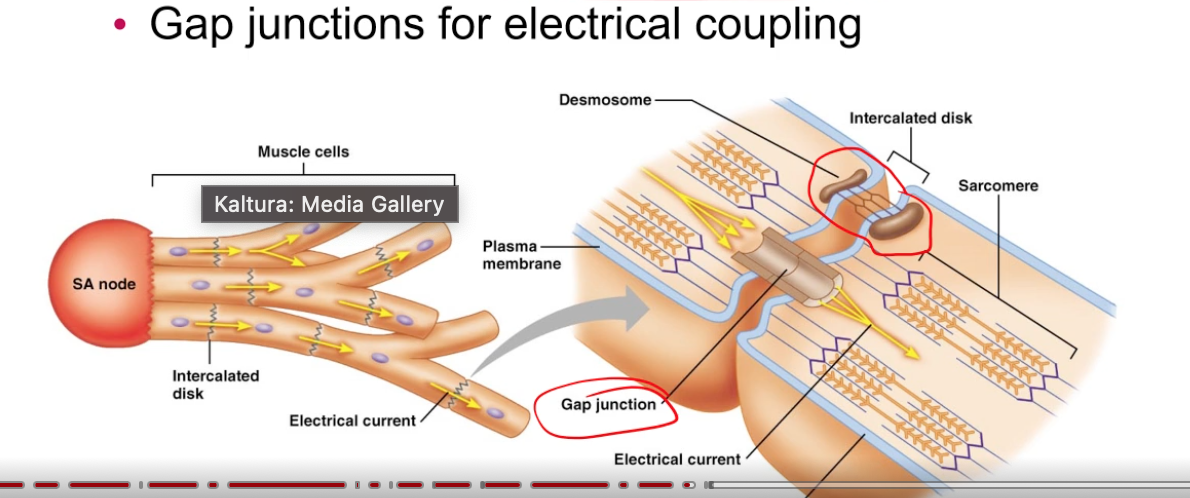
intercalated disks?
2 parts
atria contract → ventricle contract
contracting order coordinated by gap junctions and conduction pathways
intercalated disks = junctions that connect cardiac muscle
desmosomes = resist mechanical stress breaking electrical connection
gap junctions for electrical coupling
Cardiac System Flow 8 steps:
Blue
deox blood from tissue → RA
superior vena cava = blood from waist up
inferior vena cava = waist down
RA pumps deox blood → RV
through tricuspid valve
RV pumps deox blood → pulmonary artery
through pulmonary valve
pulmonary artery (deoxy) → Lungs → pulmonary vein (oxy)
Red
oxy blood pulmonary vein → LA
LA pumps oxy blood → LV
through bicuspid/mitral valve
LV pumps oxy blood → tissue
through aortic valve into aorta
which side of heart stronger
pulmonary vein and pulmonary artery
atrium or ventricle stronger
function of the septum
ventricle and atrium opposite functions
passive filling vs active filling
left side = left bc pumping to tissue
opposite to normal. pulmonary artery = deox
ventricle bc pumping out
separate oxy from deox blood
when one contracts the other relaxes
passive filling = atrium → ventricle just bc valve opened, most blood
active filling = atrium → ventricle contraction gets remaining out
AP plot for auto rhythmic vs contractile cells of heart:
auto rhythmic cell
what are they, resting membrane potential
4 phases of the plot/ why does it look like that?
contractile cells 6 phases
draw it
auto rhythmic cell = ASAN and AVN
Phase 4 =RMP -60 not static, depol start Na+ enters in ‘funny channels’
funny bc always open even at low voltage
Phase 0 = Ca+ enters L-Ca (lung) channels open
AP climbs
apex of AP = L-Ca+ close
phase 3 = K+ leaves via K+ channels
phase 4 = back at RMP

AP plot for contractile cells:
6 phases
contractile cell = ventricles and atriums
Phase 4 = RMP -90
Phase 0 = Na+ influx
Phase 1 = apex, Na+ influx closes, K+ leaves
phase 2/plateu phase = K+ leaves, but Ca+ enters
plateau bc Ca+ enters
end of phase 2 = Ca+ closes
phase 3 = K+ leaves
Phase 4 = RMP again

Path of electrical activity in the heart
mechanical action
EKG
SAN (on RA) fires AP → spreads to both atrium
reaches LA via Bachman’s fibers
AVN receives AP from SAN → slow, pauses
make sure blood reaches ventricle first
AVN fires → bundle of His → left and right bundle branches → purkinje fibers that go throughout muscle of ventricle
6 EKG regions? what’s missing/why?
P wave = atrial contraction/depol
P-R flat region = AVN pause
QRS = ventricular contraction/depol in front bottom back order
T wave = ventricular relaxation/repol
ST segment = phase 2/plateau of contractile muscles
sometimes U wave = remnants of t wave
wave for atrial relaxation not shown bc it has low voltage masked by high voltage QRS
QRS represents ventricular contraction ONLY

heart leads (what they stick on your skin during EKG)
3 leads
where are they
what part of heart do they look at
Lead 1= comes from right arm → left arm
looks at LA
Lead 2 = comes from right arm → left leg
looks at RV
Lead 3 = left arm → left leg
looks at LV

cardiac arrhythmia:
what is it
sinus rhythm
tachycardia
bradycardia
how fix?
abnormal electrical activity
sinus rhythm = pace generated by SAN
tachycardia = fast rhythm >100bpm
bradycardia = slow rhythm <50bpm
shock the patient to reset SAN
cardiac arrythmia:
first degree heart block
what does it do to ekg
second degree heart block
what does it do to ekg
third degree heart block
extra contraction, 2 types
slowed/diminished AVN conduction
increases PR segment (PQ interval) = increases pause b/t atrial and vent contraction
sometimes lack AVN conduction
lose 1-to-1 relationship b/t p wave and QRS bc QRS caused by AVN
not a QRS after every p wave
loss of AVN conduction
p wave independent of QRS, can be fatal
SAN 70-80bpm, AVN 30-40bpm
extra contraction= normal
premature atrial contraction PAC → extra vent contraction = extra systole
PVC → independent of atrial contraction = extra systole
cardiac arrhythmia:
fibrilation
atrial fibrilation (A-FIB)
how serious
ventricular fibrillation (V-FIB)
how serious
how correct both?
fibrilation = loss of heart electrical coordination
atrial fibrilation (A-FIB) = A pumping too fast, inefficient pumping to ventricle
increase stroke / HA risk
ventricular fibrillation (V-FIB) = tissue not getting O2
death in minutes
AED shock