Cardiovascular System
 Call Kai
 Learn
 Practice Test
 Spaced Repetition
 Match
 Flashcards
 Knowt Play
1/242

There's no tags or description
Looks like no tags are added yet.
243 Terms
Components of the cardiovascular system
Heart (pump), blood vessels (pipes), blood (fluid)
Two loops of the cardiovascular system
Systemic loop
Pulmonary loop
Systemic loop
Carries blood from the heart to the rest of the body in order to oxygenate tissues
Pulmonary loop
Carries (relatively) deoxygenated blood from the body to to the lungs on order to get oxygenated and remove CO2
Is circulation and open or closed system?
Closed system→leaks are bad
Blood vessels in order
Arteries
Arterioles
Capillaries
Venules
Veins
Direction of blood flow in arteries
Away
Usually carry oxygenated blood (pulmonary artery is exception)
Direction of blood flow in veins
Carry blood to the heart
Usually carry deoxygenated blood (pulmonary vein is exception)
Level of oxygen in deoxygenated blood
Not fully deoxygenated, still a lot left (~75% of Hb is still saturated in “deoxygenated” blood
Pressure
The force exerted by a fluid (mmHg)
Flow
Volume moved (mL/min)
Resistance
How different it is for blood to get from point A to point B (friction)
Where is resistance the greatest?
At vessel walls
Flow formula
F=deltaP/R
What happens to flow as we increase resistance?
Flow decreases
Resistance formula
R=(8Ln)/(pi×r^4)
N is viscosity
L is length
r is radius
Relationship between resistance and viscosity
Directly proportopnal
Relationship between resistance and vessel length
Directly proportional
Relationship between resistance and vessel radius
Inversely proportional
Biggest contributor to resistance
Radius
Affect of constriction on resistance
Increases resistance
Heart flow
Veins→vena cava→right atrium→right ventricle→pulmonary artery→lungs→pulmonary vein→left atrium→left ventricle→aorta→arteries
Valves of the heart
Tricuspid
Mitral/Bicuspid valve
Pulmonary valve
Aortic valve
Tricuspid valve
Separates right atrium from right ventricle
Mitral/bicuspid valve
Separates left atrium from left ventricle
Pulmonary valve
Separates right ventricle from pulmonary artery
Aortic valve
Separates left ventricle and aorta
What holds valves in place?
papillary muscle
What connects valves to papillary muscle?
Chordea tendineae
Purpose of papillary muscle and chordae tendineae
Keep tension on valves
Septum
Wall separating right and left sides of the heart
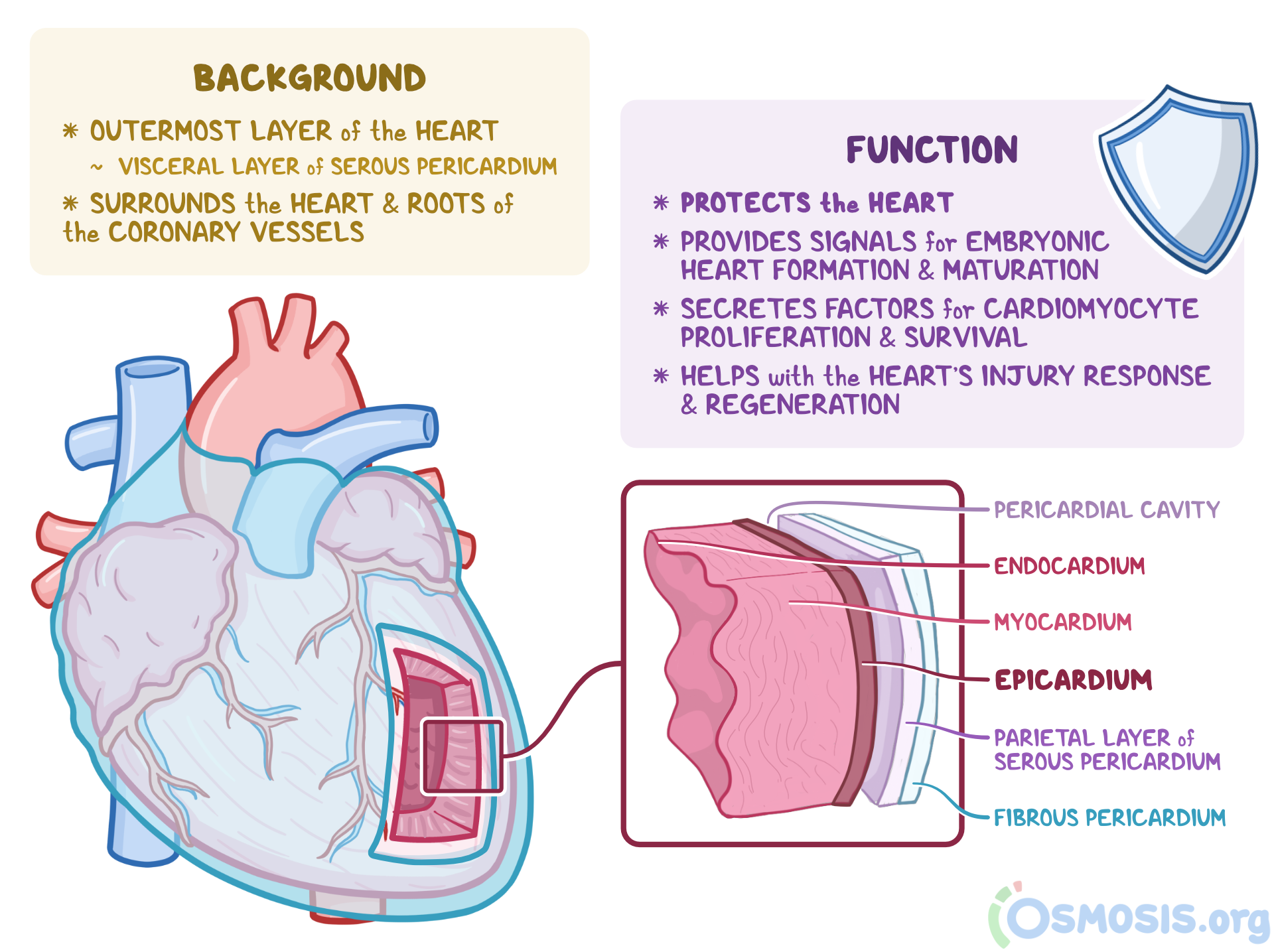
Epicardium
Outer layers of the heart. Visceral layer of serous pericardium

Myocardium
Contracting part made of cardiac muscle
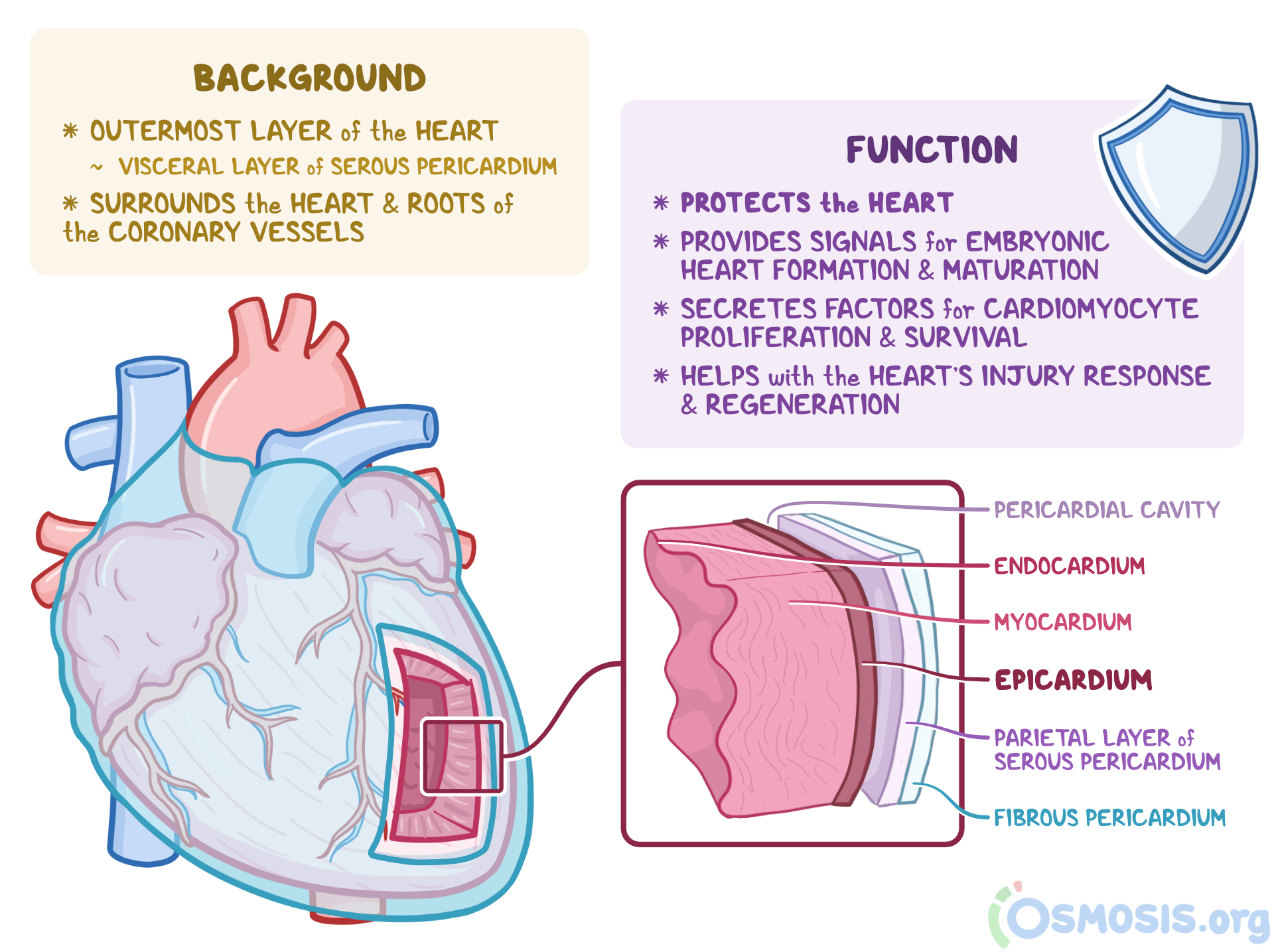
Endocardium
Inner layer lined with endothelial cells which directly contact blood. Contiguous with endothelial lining of blood vessels
Arrangement of cardiac muscle cells
Arranged in layers
Bound tightly together
Completely encircle blood chambers
Does every heart cell contract with every squeeze?
Yes! And they do so simultaneously and with little rest
Noncontracting heart cells
1% are not involved in contraction and are involved in the conducting system instead
Conducting system
Initiate heartbeat and spread electrical impulses rapidly through the heart
Heart as an endocrine organ
Can secrete atrial natriuretic peptide
Innervation of the heart
Recieves autonomic input from the sympathetic and the parasympathetic systems
What part of the heart do sympathetic neurons innervate?
Entirety of heart and nodes
NT released by sympathetic neurons and what receptor on the heart binds it
Norepinephrine binds to beta adrenergic receptors
Types of sympathetic nerves that innervate the heart
Thoracic spinal nerves
What part of the heart do parasympathetic neurons innervate?
Only the nodes
NT that parasympathetic neurons use and receptors that heart uses to respond
Acetylcholine
Muscarinic receptors
Type of parasympathetic nerves that innervate heart
Vagus nerves
Can epinephrine bind to norepinephrine receptors?
Yes and can have the same effects
What exactly does sympathetic/parasympathetic innervation of heart affect?
Affect permeability of already existing F-type Na+ channels and T-type Ca2+ channels
Conduction pathway of heart
SA node→internodal pathway→AV node→Bundle of His→Right and left bundle branches→purkinje fibers
Anatomy of an EKG, including what happens in the atria/ventricles with each part of the EKG
First bump: atrial excitation/depolarization (P wave)
Spike: ventricular excitation/depolarization (QRS complex)
Second bump: ventricular relaxation/repolarization (T wave)

Conduction pathway and P wave
SA node to AV node
Conduction pathway and QRS complex
AV node done through purkinje fibers
resting membrane potential of cardiac cells
-90 mV
Cardiac action potential steps
Voltage gated Na+ channels open
Na+ enters
Voltage gated Ca2+ and K+ chanels open (Ca2+ enters while K+ exits)
Voltage gated Ca2+ channels close
K+ exits and repolarizes cell

Function of plateau in cardiac AP
Makes AP 3x longer than neuronal AP
How long are cardiac action potentials?
~0.3 seconds (not milliseconds!)
Cardiac refractory period duration
during entire depolarization and repolarization
Purpose of cardiac refractory period
prevents another AP from firing during contraction which allows blood to refill in the heart
Node cells
cells that have automaticity that are found in the entire heart conduction system
Ultimate pacemaker
SA node
SA node action potential
No RMP so potential fluctuates
Na+ trickles in through F channels, which open at -50 mV, leading to slight and gradual depolarization
Ca2+ trickles in through T channels in order to give a depolarizing boost
Once threshold is reached L-type Ca2+ channels open and T-type Ca2+ channels close
Ca2+ enters through L channels
K+ exits

What triggers another action potential in SA node cells?
“hyperpolarization”→ no true hyperpolarization
F type sodium channels open at a more negative voltage
What type of calcium is critical for contraction?
extracellular calcium, which comes in through L channels
Ca2+ binds ryanodine receptors and lets Ca2+ exit SR
Cardiac cycle
all the events involved with blood flow through a heart during a heart beat
Systole
contraction
Steps of systole
isovolumetric ventricular contraction
ventricular ejection
Isovolumetric ventricular contraction
overall volume doesn’t change
all valves are closed
tension is building
Ventricular ejection
blood flows out of ventricles to arteries
AV valves closed
aortic and pulmonary valves open
heart size shrinks
Diastole
heart relaxation
Steps of diastole
isovolumetric ventricular relaxation
ventricular filling
Isovolumetric ventricular relaxation
no volume change
all valves closed
heart begins to relax
Ventricular filling
blood moves from atria to ventricles
AV valves open
aortic and pulmonary valves closed
Contraction-relaxation cycle duration
0.4 s: contraction
0.6 s: relaxation
What moves blood between compartments?
Pressure gradients

Pressure curve for left ventricular pressure
orange curve
half of the rise is isovolumetric ventricular contraction
top of hump is ventricular ejection
most of the fall is isovolumetric ventricular relaxation
end with a little bit of rise is ventricular filling
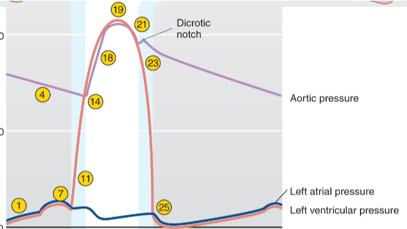

Pressure curve for left atrial pressure
blue curve
second little hump is isovolumetric ventricular relaxation
first trough is ventricular ejection
third little hump is isovolumetric ventricular relxation
second trough is ventricular filling


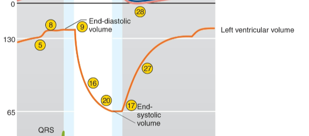
Volume curve for left ventricular volume
first peak is when heart has filled
right before drop off is isovolumetric ventricular contraction
big drop is ventricular ejection
bottom plateau is isovolumetric ventricular relaxation
rise is ventricular filling
→ space between bottom of curve and x-axis is the volume of blood remaining in heart after ejection
cardiac output
amount of blood pumped out of each ventricle in out minute
CO=HR x SV
Stroke volume
amount of blood pumped out of each ventricle in a single beat
difference between EDV and ESV
SV=EDV-ESV
normal cardiac output
5-6 L/min
If SV declines, what must occur to maintain CO?
HR must increase
regulating heart rate
chronotropic factors and ANS input
positive chronotropic factors
increase heart rate
negative chronotropic factors
decrease heart rate
ANS effect on pacemaker potentials
sympathetic stimulus speed up HR be shortening the initial depolarization
parasympathetic stimulus slows down HR by lengthening the initial depolarization

How does ANS stimulation achieve its effects on HR?
modified permeability of F-type sodium channels and T-type Ca2+ channels
end diastolic volume (EDV)
final volume in ventricle after diastole
end systolic volume
volume left in ventricle after systole
normal SV in a health human
~70 mL
60% of heart volume
key factor that controls SV
preload
preload
degree to which cardiac muscles are stretched prior to contraction→amount of blood in the ventricles influences stretch
How does cardiac muscle stretch affect contraction?
Cardiac muscle cells are held at a suboptimal resting length, so filling heart with more blood maximizes the resting tension and therefore active tension
Number one factor for controlling preload
venous return
Relationship between preload and EDV
Directly proportional
anything that increases venous return or slows HR increases EDV
afterload
pressure ventricles must pump against to force open aortic/pulmonary valves
What affects afterload?
Anything that increases systemic/arterial pressure can increase afterload (hypertension)
Frank-Starling Curve
tells us relationship between SV and EDV
→ anything that increases EDV (such as venous return), increases SV

Extrinsic controls of EDV
sympathetic and hormonal input
→ norepinephrine binds to alpha adrenergic receptors and increases force
→epinephrine and T3/T4 increase force
Cardiac contractility
inherent strength of the heart muscle (myocardium) to contract and generate mechanical force
Mechanism for increasing contractility
epinephrine/norepinephrine binds to beta-1 adrenrgic receptor
alpha subunit dissociates from G-protein
alpha subunit activates adenelyl cyclase
adenelyl cyclase makes cAMP from ATP
cAMP level increase and associate with PKA, activating it
PKA goes on to phosphorylate various things