bis102 midterm 2 concepts
 Call Kai
 Learn
 Practice Test
 Spaced Repetition
 Match
 Flashcards
 Knowt Play
1/9

There's no tags or description
Looks like no tags are added yet.
10 Terms
proximal histidine
location relative to the heme: below the heme
functions:
acts as a mechanical arm to keep the heme in plane
prevents oxidation of Fe2+ by protecting the heme’s backbone
is it in the acidic or basic form?: basic
distal histidine
location relative to the heme: above the heme
functions:
helps hold O2 when bound to the heme via an H-bond
H+ scavenger to prevent Fe2+ oxidation
forces C=O to bind to an angle
is it in acidic or basic form?: basic
what is the “packing ratio” for a protein? what is the optimal packing ratio for a protein?
the packing ratio for a protein is the (volume due to van der waals radii)/(actual volume)
the optimal packing ratio for a protein is 0.75. the protein needs to have structure but also by dynamic and be able to respond to its environment
statements that are TRUE about protein secondary structure
secondary structures of proteins are held by H-bonds between the protein backbone
the tertiary structure of a protein is the most thermodynamically stable
S cannot exist in an alpha helix because its R group is too small and lets in too many water molecules
subunits in a protein can form beta-pleated sheets where their surfaces make contact
the sequence TWECLVMN is likely to form an alpha helix
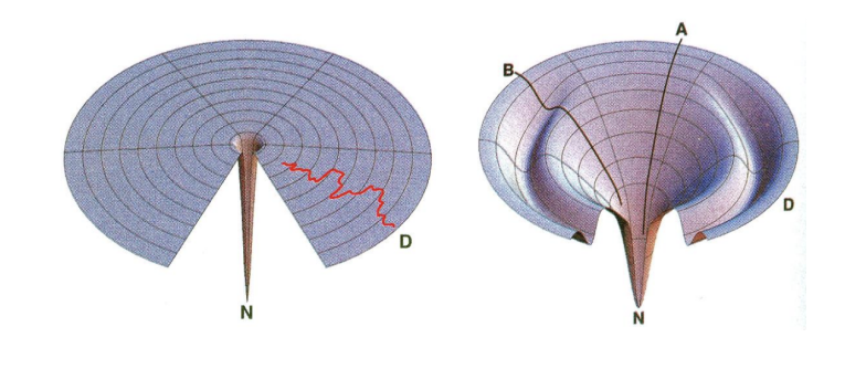
what does each letter mean? which diagram is considered to be more accurate and why?
this is a levinthal energy diagram. N is the biologically active native 3, while D is denatured (1). the diagram on the right shows ordered collapse, which is how proteins fold. A and B are two copies of a protein. the diagram on the left shows random walk, which is not how proteins fold.
what are the two parts of Professor Sanger’s strategy for sequencing a protein?
generate small fragments
look for overlapping sequences
Professor Sanger had enzymes he could use as tools to sequence a protein. there are two categories of enzymes he used. provide the definition of each category and name one specific enzyme he used that fit in each category.
endoprotease: an enzyme that catalyzes the hydrolysis of peptide bonds within the interior of a peptide chain
example:
trypsin: cleaves at the carboxyl of K and R
chymotrypsin: cleaves at the carboxyl side of the aromatic amino acids (WYF)
exoprotease: an enzyme that catalyzes the catalyzes the hydrolysis of a peptide bonds at the termini (N- and C- term) of a peptide bond
example:
carboxypeptidase A
carboxypeptidase B
Professor Sanger used a buffer at pH 8 when using FDNB. why did he specifically choose this pH? what would happen if he used a lower or higher pH?
he chose pH 8 because it is a pH where the N-term of a peptide will have 2 protons and one pair of electrons. this allows the nitrogen to attack FDNB and cause the fluoride to leave. if he used a lower pH, the N-term would be protonated and would not be able to attack FDNB. if he used a higher pH, the solution would have more OH-, which is a better nucleophile than the amino group. OH- would displace the fluoride to form dinitrophenol in a side reaction, which would also be yellow. the dinitrophenol side product is useless for sanger.
statements that are TRUE about protein sequencing
chymotrypsin cuts on the right-hand side of aromatic amino acids
fluorine is a good leaving group when FDNB is attacked by the N-terminus
when performing C- and/or N-termini analysis, you always want to use a plate that is opposite of the solvent you are using
FDNB is attacked by all amino acids
when analyzing which amino acid is at the N-terminus, only ninhydrin is used
when determining which amino acid is at the C-terminus, it is done on a TLC plate
when analyzing which amino acid is at the C-terminus, both distance and color are used
the side product no matter what pH is used when using Sanger’s reagent is dinitrophenol
it is recommended to use fluorescence to analyze on the TLC plate which amino acid is the N-terminus
