BIOL 3103: Exam 2 Study Guide
 Call Kai
 Learn
Learn Practice Test
 Spaced Repetition
 Match
Match Flashcards
 Knowt Play
1/153

There's no tags or description
Looks like no tags are added yet.
Name | Mastery | Learn | Test | Matching | Spaced | Call with Kai | Chat |
|---|
No analytics yet
Send a link to your students to track their progress
154 Terms
Dual Innervation
An organ receives input from both sympathetic and parasympathetic systems. They usually have opposite effects.
Somatic Nervous system
Skeletal muscle control
Autonomic Nervous System
Innervation of most visceral organs. Parasympathetic and sympathetic nervous systems.
Parasympathetic Nervous System
“Rest and digest”, maintains homeostasis, and has acetylcholine
Sympathetic Nervous System
“Fight or flight”, may disrupt homeostasis, and has acetylcholine and norepinephrine
Dual Innervations examples
Eye, heart, lungs, pancreas, and digestive tract.
Parasympathetic dominance: Eye
Constriction of pupil for near vision
Parasympathetic dominance: Heart
Decreased rate and force
Parasympathetic dominance: Lungs
Constriction of bronchioles (smaller airway)
Parasympathetic dominance: Pancreas
Insulin and glucagon release (as determined by blood sugar level)
Parasympathetic dominance: Digestive tract
Increased motility (most of blood needs to go here)
Sympathetic dominance: Eye
Dilation of pupil for far vision
Sympathetic dominance: Heart
Increased rate and force
Sympathetic dominance: Lungs
Dilation of bronchioles (larger airway)
Sympathetic dominance: Pancreas
Glucagon release (discharge stored glucose)
Sympathetic dominance: Digestive tract
Decreased motility
Sympathetic dominance state
Active state
Parasympathetic dominance state
Resting state
Dual innervations exceptions
Salivary glans, sweat glans, and most veins and arterioles
Sympathetic Salivary glands
Stimulatory
Parasympathetic Salivary glands
Stimulatory
Sympathetic Sweat glands
none
Parasympathetic Sweat glands
Stimulatory
Parasympathetic most Veins and Arterioles
not much change
Sympathetic most Veins and Arterioles
Construction determined by action potential firing rate
Location of nicotinic receptors in the autonomic nervous system
Neuromuscular junctions, ganglionic neurons, and adrenal medulla
Location of muscarinic receptors in the autonomic nervous system
Gut, heart, bronchioles (lung), sweat glands, and blood vessels of skeletal muscle
Vertebrate stress response
The body’s response to a stressor that activates both the sympathetic nervous system and the endocrine system.
Physiology of hormonal stress
Slower but longer-lasting stress response involving the hypothalamus, pituitary gland, adrenal cortex, and glucocorticoids.
Physiology of neutral stress
A fast, short-lasting (transient) stress response triggered by activation of the sympathetic nervous system.
How does hypothalamus mediate responses to stressors?
Activation of the sympathetic nervous system and secretion of corticotropin releasing hormones.
Glucocorticoids
Stress hormones such as cortisol and corticosterone released during the hormonal stress response.
Stress response effects on the heart
Stress causes increased heart rate and increased force of contraction.
Stress response effects on the lungs
Stress causes bronchodilation, meaning the bronchioles become larger.
Stress response effects on the liver
Stress causes the liver to make/release more glucose so energy is available.
Stress response effects on the pancreas
Sympathetic stimulation causes glucagon release, helping make stored glucose available.
Chronic stress leftover effects
Immunosuppression, hypertension, muscle loss, and inhibition of growth
Peripheral nervous system
The part of the nervous system that connects the CNS to the rest of the body through sensory input and motor output. Contains afferent and efferent pathways.
Afferent sensory tracts
Sensory information traveling towards the CNS
Efferent motor tracts
Motor information traveling away from the CNS toward muscles/organs.
Decussation
A neural pathway crossing from one side to the other, as shown in the pathway diagram.
Nerve
A bundle of axons in the peripheral nervous system.
Ganglion
A relay point in the PNS where neurons communicate; in the ANS it lies between the preganglionic and postganglionic neuron.
Collateral (corollary)
Shown as part of a sympathetic pathway
How does the peripheral nervous system and central nervous system interact with each other?
The PNS carries sensory information into the CNS and carries motor commands out of the CNS.
Dorsal root/ horn
Carries/contains afferent sensory information
Ventral root/horn
Carries/contains efferent motor information
Reflex
A rapid response that can be integrated at the spinal cord, allowing a faster response without waiting for higher brain processing.
Withdrawal reflex
A spinal reflex that causes a limb to pull away from a painful stimulus.
Crossed extensor reflex
A spinal reflex where one limb withdraws from pain while the opposite limb extends to support the body.
Enteric nervous system (ENS)
The nervous system associated with the digestive tract, controlling digestive muscle activity and secretion. “Brain in your gut”.
Submucosal plexus
Contains sensory cells that communicate with the myenteric plexus and motor fibers that stimulate fluid secretion into the lumen.
Myenteric plexus
Contains neurons that regulate the enzyme output of adjacent organs.
Lumen
Nerves do not enter this area and digestion occurs here.
Mesentery
Attaches the bowel to the body wall and contains major arteries, veins, lymphatic system, and external nerves.
Enteric nervous system complexity
Highly complex system containing about 100 million neurons and using 30+ neurotransmitters
Enteric nervous system neurotransmitters
Uses many of these to control digestive activity
Varicosities
Swollen regions along autonomic neurons.
Why are varicosities useful to the enteric nervous system?
Help spread the neural message widely across smooth muscle.
When is sympathetic dominant?
during an active/stressed state → “fight or flight”
When is parasympathetic dominant?
during the resting state → “rest and digest”
Where does the sympathetic system emerge from in the central nervous system?
Thoracic and lumbar regions
Where does the parasympathetic system emerge from in the central nervous system?
Cranial and sacral regions
Parasympathetic fiber lengths
Long preganglionic and short postganglionic
Sympathetic fiber lengths
Short preganglionic and long postganglionic
Sympathetic preganglionic neurotransmitters
acetylcholine (ACh)
Parasympathetic preganglionic neurotransmitters
acetylcholine (ACh)
Parasympathetic postganglionic neurotransmitters
acetylcholine (ACh)
Sympathetic postganglionic neurotransmitters
norepinephrine (NE)
Major types of cholinergic receptors
Nicotinic and muscarinic receptors
Nicotinic receptor
A cholinergic receptor that causes excitation through ion influx.
Muscarinic receptor
A cholinergic receptor that is G-protein coupled and is found on target tissues.
Nicotinic receptor agonists
ACh, nicotine, and carbachol
Muscarinic receptor agonists
ACh, muscarine, and carbachol
Nicotinic receptor cellular response
Binding causes excitation through ion influx.
Muscarinic receptor cellular response
Muscarinic receptors are G-protein coupled, and the response depends on the target tissue.
First step of muscle contraction:
Action potential arrives at a terminal button (of the neuromuscular junction), it then simulates the release of ACh, which then moves across the cleft, and then triggers action potential in the muscle fiber.
Second step of muscle contraction:
Third step of muscle contraction:
Second step of muscle contraction:
Fourth step of muscle contraction:
Fifth step of muscle contraction:
Sixth step of muscle contraction:
Seventh step of muscle contraction:
Eighth step of muscle contraction:

Sliding filament hypothesis
Myosin pulls actin toward the sarcomere's center. The filaments don't shorten; they slide past each other (which is how muscles shorten).

Calcium role in sliding filament
Binds troponin and initiates contraction

Troponin role in sliding filament
Changes shape when calcium binds

Tropomyosin role in sliding filament
Blocks binding sites on actin when muscle is relaxed

Actin role in sliding filament
Thin filaments pulled by myosin

Myosin role in sliding filament
Thick filament; its heads form cross-bridges and produce power strokes
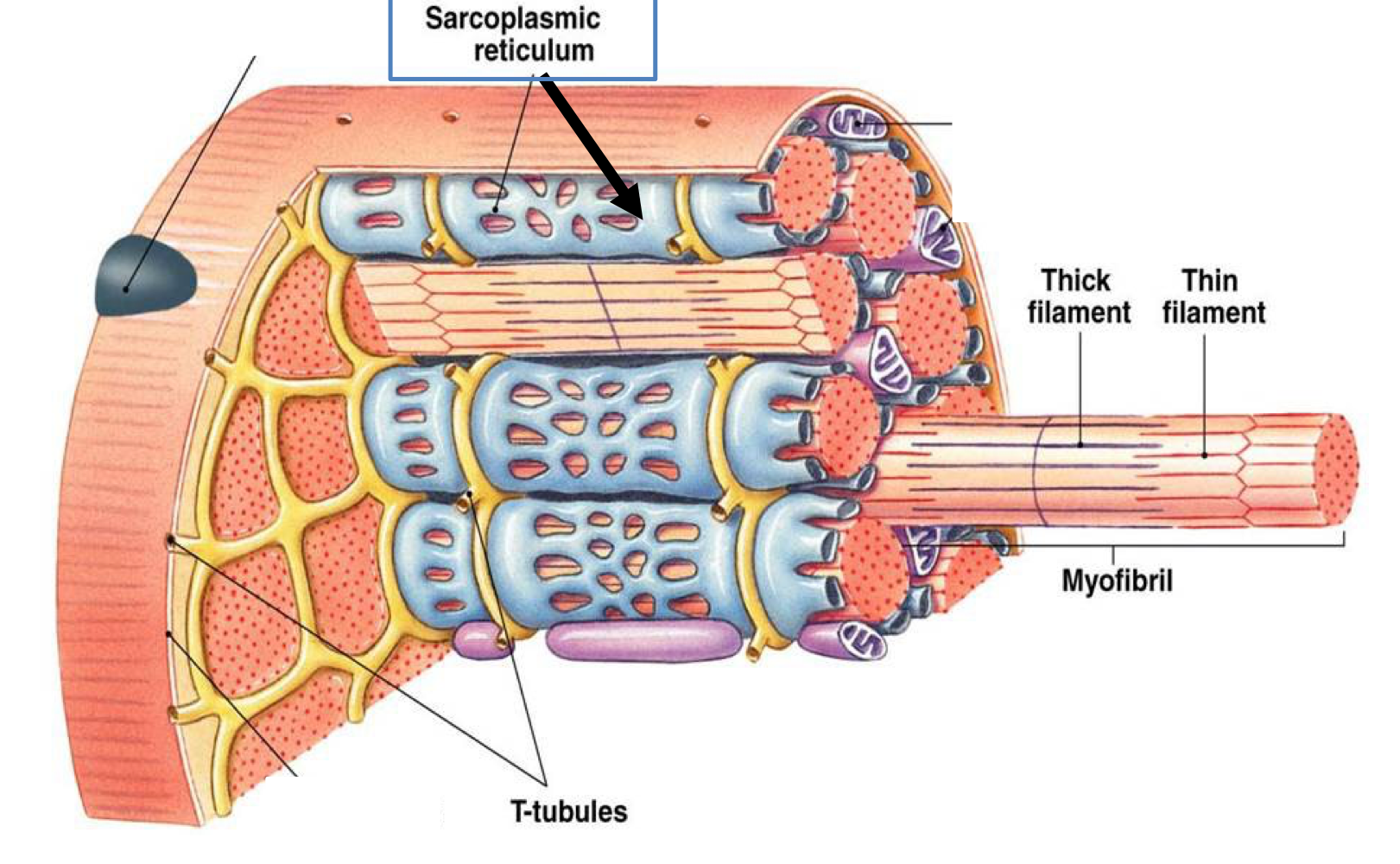
Sarcoplasmic reticulum role in sliding filament
Stores and releases calcium

Transverse tubules role in sliding filament
Conduct action potentials into muscle fibers

Ryanodine receptors role in sliding filament
Release Ca2+ from the sarcoplasmic reticulum
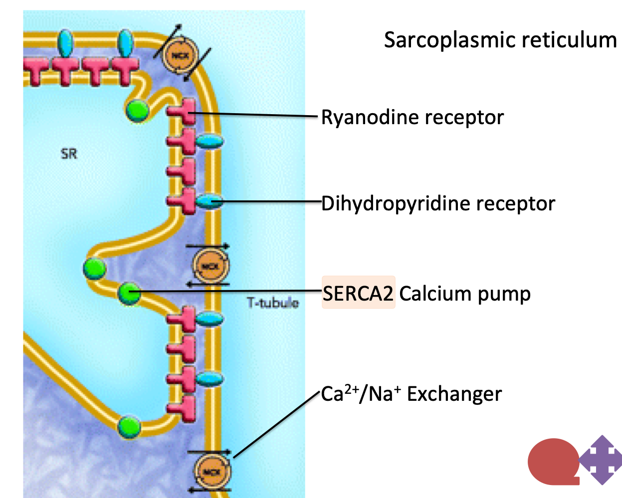
Dihydropyradine receptors role in sliding filament
Triggers adjacent ryanodine receptors

SERCA2 role in sliding filament
Uses ATP to move calcium back into the sarcoplasmic reticulum

ATP binding role in sliding filament
Causes myosin to detach from actin

ATP hydrolysis role in sliding filament
Energizes the myosin head
Phosphorylation role in sliding filament
Activates myosin in smooth muscle by adding phosphate to myosin light chains, allowing actin–myosin interaction and contraction.
Dephosphorylation role in sliding filament
Removes phosphate from myosin light chains, reducing cross-bridge cycling and promoting smooth muscle relaxation.
Release in the myosin power stroke role in sliding filament
Release of Pi and ADP during the cycle which allows myosin to pull actin toward the center of the sarcomere, producing muscle contraction.