KNES 191A Exam #3
 Call Kai
 Learn
 Practice Test
 Spaced Repetition
 Match
 Flashcards
 Knowt Play
1/107

There's no tags or description
Looks like no tags are added yet.
108 Terms
What are the function of the Cardiovascular system?
transportation
regulation
protection
What percent of the blood is plasma?
55%
What percent of the blood are cells?
45%
What is the lifespan of platelets?
5-9 days
What are the lifespan of WBC’s?
hours, days, or years
What do RBC’s do?
designed for max storage for O2 transportation
What are characteristics of RBC’s
no nucleus
no mitochondria
4-month lifespan (playability)
Where is the base of heart located?
top of heart (cause bigger)
Where is the apex of the heart located?
At bottom of the heart
What is deoxygenated blood?
high in carbon dioxide
low in oxygen (blue)
right side of body
What is oxygenated blood?
high in oxygen (red)
low in carbon dioxide
left side of body
What is the septum of the heart?
wall that divides the left & right side of heart
What valve divides the right atrium and right ventricle?
tricuspid valve
What valve stops backflow of blood in the right ventricle?
pulmonary valve
What valve divides the left atrium & left ventricle?
bicuspid valve (mitral)
What valve stops backflow of blood into the left ventricle?
aortic valve
What is the rule with valves in the heart?
When one set of valves open the other set of valves closes
What are the sets of valves?
tricuspid valve & bicuspid valve (mitral)
pulmonary valve & aortic valve
What is the flow of the pulmonary circuit?
right ventricle → lungs → left atrium
What is the flow of the systemic circuit?
left ventricle → body → right atrium
What do veins do?
carry blood away from atria
What do arteries do?
carry blood away from ventriculi
What is arrhythmia?
abnormal heart beat
What is a murmur?
abnormal sound in stethoscope
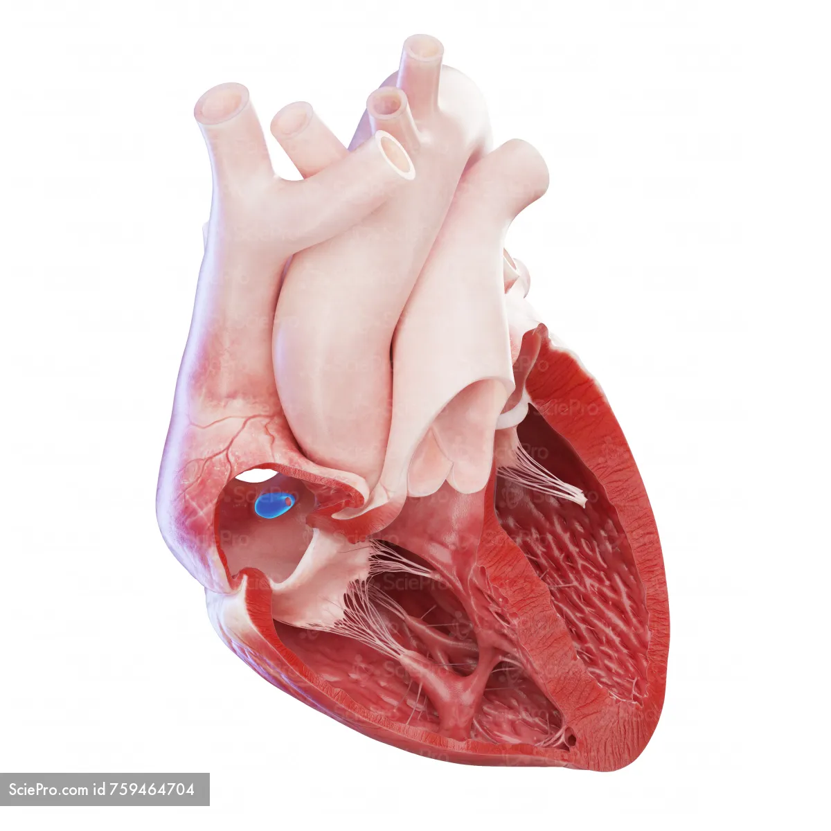
(third opening in right atrium)
Coronary sinus opening
What does the coronary sinus opening do?
bring deoxygenated blood back from myocardium (walls of heart)

(white structure coming off cusp)
Chordae tendineae
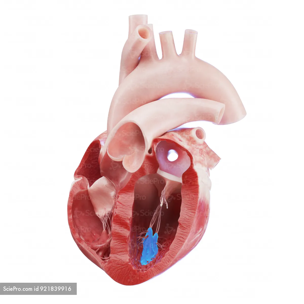
Papillary muscles
What is the function of papillary muscles?
places tension on cords of chordae tendineae & holds cusp in place
What is happening to valve when the papillary muscles are contracted?
valve is closed
What is happening to valve when the papillary muscles are relaxed?
valve is open
What is tachycardia?
more than 100 BPM resting (fast)
What is bradycardia?
less than 60 BPM resting (slow)
What is the cardiac cycle?
one complete heartbeat
What makes the lubb-dupp sound (blood turbulence)?
blood hitting the valves
What makes the lubb sound?
when the AV node closes & blood hits valve (slower sounding)
What makes the dupp sound?
When aortic & pulmonary valves close & blood hits valve (faster sounding)
What is arrhythmia?
abnormal heart beat
What is a heart murmur?
abnormal sound in stethoscope
What do pressure differences control?
control valve open & closure
When a heart chamber is empty?
pressure decreases, Large (diastole)
When a heart chamber is full?
pressure increases, smaller (systole)
What happens with the myocardium when the heart is in diastole?
myocardium is relaxed
What happens with the myocardium when the heart is in systole?
myocardium is contracted
What do ECG waves measure?
measures electrical activity (changes in myocardium)
What do P-waves (first wave) mean?
depolarization of atrial myocardium (small contraction)
What is the QRS complex?
very fast depolarization of a lot of myocardium (left ventricular myocardium) (very fast & strong contraction)
What does the T-wave indicate?
repolarization of same ventricular myocardium (delay long enough to fill up or slower)
What is coronary circulation?
blood that goes back to the wall of the heart
In coronary circulation where does all the deoxygenated blood funnel to?
coronary sinus
What does atherosclertic plaque do?
partially obstructed arteries
What is syncytium?
depolarization spreading from cell to cell to cell (set up of cardiac muscle)
What are gap junctions in cardiac muscle?
relay station from depolarization (transmits to next fiber)
Characteristics of nucleus in cardiac muscle?
one-three nucleus, big & centralized
What does the flatline of ECG waves indicate?
depolarization stops (systole, contraction)
What happens when there is an obstruction in arteries?
not enough blood flow getting to tissues
smaller chamber
less elastic tissue in artery
What are the risk factors of obstructed arteries?
lack of physical activity
diet
smoking
(lifestyle diseases)
What is true about atrial syncytium and ventricular syncytium?
they are separate muscle fibers in the heart
What are intercalated disc?
line shows difference between cardiac muscle fibers ending & starting (stair stepping design)
What is the cardiac conduction system?
muscle cells but do not produce force instead spread signal through heart (heart’s electrical system)
What is autorhythmic?
muscle that create signal & spread through myocardium
What are the five sections of the cardiac conduction system?
sinoatrial node (SA node) (pacemaker)
atrioventricular node (AV node)
bundle of his
right & left bundle branches
purkinje fibers
When is there a need for an artificial pacemaker?
when the SA node starts failing
Sinoatrial node (SA node) (pacemaker)
Electrical system all by itself (start to see P-wave)
embedded in myocardium on top right atrium
Atrioventricular node (AV node)
holds onto signal for a sec (creates flatline)
embedded in myocardium on bottom of right atrium
Bundle of His
transferring signal into ventricular myocardium creating QRS complex
right & left bundle branches
Go down to apex and carry signals to lower chambers of heart
Purkinje fibers
forms final link of heart electrical conduction (forms synchronized heartbeat)
When is the parasympathetic nervous system (PNS) most active?
during rest
What neurotransmitter is released from the PNS?
acetylcholine (AcH)
When is the sympathetic nervous system (SNS) most active?
during the process of getting ready to expand energy
What is the neurotransmitter that is released from the SNS?
norepinephrine (NE)
What is extrinsic regulation of the cardiac cycle?
regulation from outside of the system
What is intrinsic regulation of the cardiac cycle?
regulation within the system itself
What is stroke volume?
amount of blood pumped out of heart per beat
What is cardiac output?
amount of blood pumped out of heart per min. (SV x BPM)
What are the extrinsic regulation of cardiac cycle?
ANS & hormones
How does the ANS regulate HR?
SNS releases NE (speeds up)
PNS releases AcH (slows down)
How does hormones regulate HR & SV
by increasing HR & SV
What are the three hormones learned in class?
epinephrine, norepinephrine, & thyroid hormone
Hormones
released from glands & taken to target cells (takes longer to get to cells then neurotransmitters but effects last longer)
How does ANS regulate SV?
SNS release NE (more forceful contractions of ventricular myocardium
PNS release AcH (less forceful contractions of ventricular myocardium)
What is the starling’s law of the heart?
more stretch out of myocardium the harder the myocardium will contract
What is the flow of blood?
heart → arteries → arterioles → capillaries (site of exchange) → venules → veins → heart
Are arteries deep or superficial?
arteries are found more deeper
Are veins deep or superficial?
veins are found more superficial
How are blood vessels regulated?
only regulated by SNS
How does the SNS regulate blood vessels?
increased SNS - vasoconstriction
decrease SNS - vasodilation
How are smooth muscle regulated?
regulated by autonomic nervous system
What are the layers of blood vessels?
outer layer (tunica externa)
medial layer (tunica media)
inner layer (tunica intima)
What walls do arteries have?
All three (externa, media, & intima)
What is needed in arteries that aren’t needed in veins?
arteries need elastic tissue due to the higher pressure in arterial blood
What walls do arterioles have?
tunica media
tunica intima
(No tunica externa)
What walls do capillaries have?
tunica intima
(No tunica media or externa)
What is the rule of walls in the arteries?
further you go walls come off & smaller diameter
What is the rule of walls in the veins?
further you go put walls back on & bigger diameter
What do precapillary sphincters do?
open & close to what capillaries need blood (round circles of smooth muscle)
What is the function of arterioles?
deliver blood to capillaries & greatest collective influence on both local blood flow & overall BP
continuous capillaries
capillaries that can hold small particles (CO2 & O2)
Fenestrated capillaries
capillaries that can hold medium particles (H2O) (pores)