BME 311: Lecture 1
 Call Kai
 Learn
Learn Practice Test
 Spaced Repetition
 Match
Match Flashcards
 Knowt Play
1/42

There's no tags or description
Looks like no tags are added yet.
Name | Mastery | Learn | Test | Matching | Spaced | Call with Kai | Chat |
|---|
No analytics yet
Send a link to your students to track their progress
43 Terms
bioimaging
any imaging of biological tissue-living or not (CT, MRI, PET, ultrasound)
also non-human organisms
can be invasive, including excised tissue/biopsy
can be used purely for research
can be very high resolution - light or electron microscopy
different purposes/different techniques
detection/screening-lesion (cancerous)?
diagnosis-invasive or benign?
staging-how bad, metastasized?
monitoring of treatment-shrinking?
surveillance-returned?
research-understand structure/function
organs
organization of several different kinds of tissues arranged together to perform a specific function
medical imaging of human body requires some form of energy
capable of penetrating tissue
all imaging techniques (except nuclear imaging) require energy-tissue interaction
visible light has limited penetration
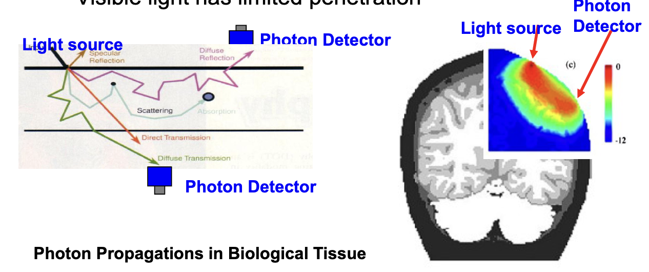
electromagnetic (EM) radiation is used for
nuclear imaging (ionizing)
x ray imaging (ionizing)
magnetic resonance imaging (non-ionizing)
optical/infrared imaging (non-ionizing)
NOT ultrasound


radiography
2D imaging with x-rays

computer tomography (CT)
3D imaging with x-rays

nuclear imaging (PET, SPECT)
mostly 3D imaging with gamma-rays

magnetic resonance imaging (MRI)
3D imaging with radio-waves

ultrasound
mostly 2D imaging with sound waves
morphology in biology
outward appearance (shape, structure, color) of an organism and its component parts
microscopic vs macroscopic
micro-cells macro-tissues, organs, systems
superior or cranial
toward the head end of the body
upper (the arm is a superior extremity)
inferior or caudal (tail)
away from the head
lower (the leg is an inferior extremity)
anterior or ventral
front (knee cap is located on the anterior side of the leg)
in human (bipedal) brain, ventral has different meaning:towards body
posterior or dorsal
back (spine located on the posterior of the body)
in human (bipedal) brain, dorsal has different meaning:towards top of head
medial
toward the midline of the body or body part
(middle toe is located at the medial part of the foot)
lateral
away from the midline of the body or body part
(little toe is located at a lateral part of the foot)
proximal
toward or nearest the trunk or the point of origin of a part
(proximal end of the femur joins with the pelvic bone)
distal
away from or farthest
(hand is located at the distal end of the arm)
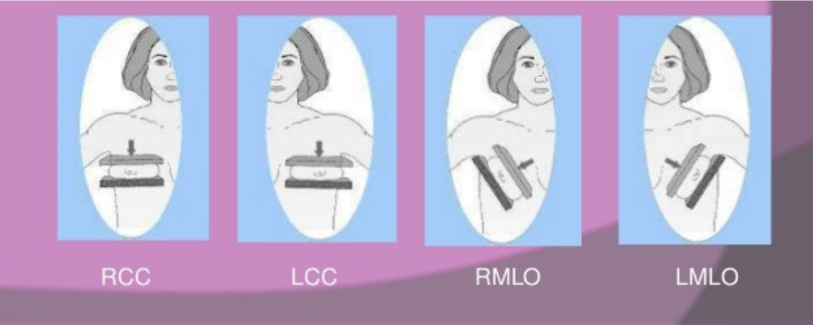
mammography views
cranio-caudal (CC) view - direction of x-rays
medio-lateral (ML) view
medio-lateral oblique (MLO) view

supine vs prone
supine = face up (most common)
prone = face down (breast imaging)
neurological = patient left on image right = view from feet when supine (nose up)
radiological = patient left is on image left = view from top of head when supine
coronal (frontal)
if standing, vertical plane running from side to side
divides the body or any of its parts into anterior and posterior portions
sagittal (lateral)
if standing, vertical plane running from front to back
divides the body or any of its parts into right and left sides

transverse (transaxial/axial)
if standing, horizontal plane divides the body or any of its parts into upper and lower parts
physiology
study of the functions of living organisms
general-blood pressure, heart rate, body temp
brain-cerebral blood flow, tissue oxygenation
gantry
main part of scanner, as opposed to the bed and any ancillary devices
phantom
device with known geometry/characteristics used to test or calibrate an imaging system
source
usually a source of radioactivity (often a small “point”)
subject/object
thing being imaged
image reconstruction
creating images from raw data
artifact
a feature in an image that does not represent the anatomy or physiology being measured (ring or streak)
pixel
the smallest 2D area represented in an image, usually has a single intensity value
voxel
the smallest 3D volume represented in an image, usually has a single intensity value
image matrix
the 2D or 3D array of pixels/voxels/numbers that make up a single 2D or 3D image
projection
2D representation of 3D volume
-some modalities are inherently 2D
-3D modalities can be viewed as projections too using sum or maximum intensity projection = MIP
slice/plane
2D representation of 3D volume at a certain depth only
ROI/VOI
region of interest in an image, often drawn by hand, used to determine parameters for the region such as mean or average
segmentation
process of delineating regions in an image that correspond to anatomy (often using automated computer algorithm)
FOV
field of view = physical space represented by the whole image
image fusion
viewing of 2 images of the same part of the body overlaid on each other (often complementary structural and functional images)
noise vs background
-noise is randomness in pixel/voxel (repeat same measurement and get a different value)
-background is more like a bias or offset in the pixel/voxel value, does not represent the thing you’re trying to measure (repeat the same measurement and will get the same bias)