Cardiovascular System
 Call Kai
 Learn
 Practice Test
 Spaced Repetition
 Match
 Flashcards
 Knowt Play
1/61

There's no tags or description
Looks like no tags are added yet.
62 Terms
Arteries
Transports oxygenated blood away from the heart towards distal organs/tissues under high pressure

Veins
Transport deoxygenated blood towards the heart away from the distal organs/tissues under low pressure

Capillaries
Connect arteries to veins, responsible for gaseous exchange

Heart rate
Number of complete cardiac cycles in one minute (70 bm at rest)
Stroke volume
Volume of blood leaving the heart each beat (70 ml at rest)
Cardiac output
Volume of blood leaving the heart per minute (5l at rest) - Q = SV x HR
What are the two atrioventricular valves?
Tricuspid + mitral/bicuspid

What are the two semilunar valves?
Aortic + pulmonary valves

2 aspects of semilunar valves
No attachments
Work based on pressure of blood flow
What are the three layers of the heart?
Pericardium
Myocardium
Endocardium

Pericardium fluid function
Fluid in the pericardial space reduces friction created by movement of the heart pumping blood
3 aspects of myocardium
Contain cardiomyocytes
Thickest layer
Contain coronary arteries which provide blood supply
3 aspects of cardiomyocytes
Striated
Contain many mitochondria
Have auto-rhythmicity
2 aspects of endocardium
Single celled
Contain endothelial cells
How does the cardiac cycle start?
Blood enters the right atrium via the inferior and superior vena cava.
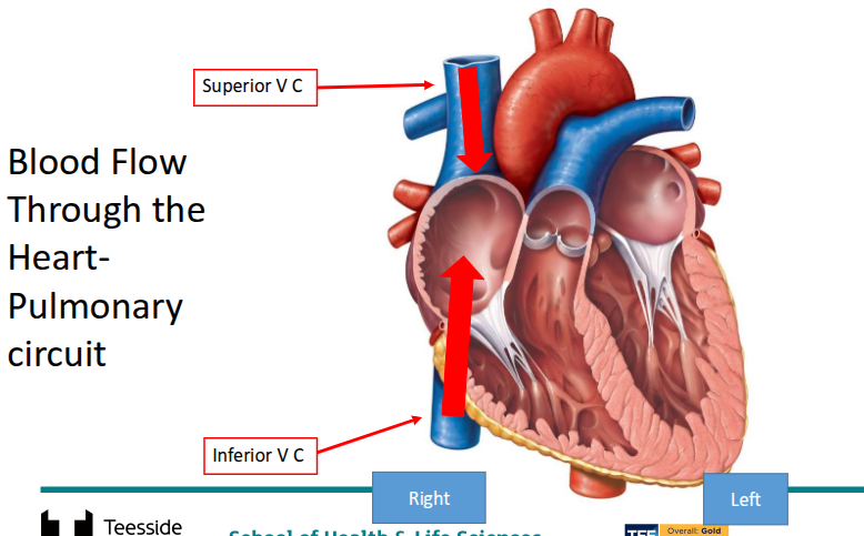
What happens after blood enters the right atrium?
The right atrium fills and blood begins to passively flow into the right ventricle

What happens after the passive flow into the right ventricle?
The right atrium contracts and the tricuspid valve closes to prevent blood backflow.
What happens after right atrium contraction?
The right ventricle contracts and blood leaves the ventricle via the pulmonary valve into the pulmonary artery, and to the pulmonary circulatory system (lungs).

What happens after the right ventricle contracts?
Oxygenated blood returns to the left atrium via the pulmonary vein.

What happens after blood returns to the left atrium?
Blood passively flows into the left ventricle.
What happens after blood flows into the left ventricle?
The left atrium contracts and the bicuspid/mitral valve closes to prevent blood backflow.
What happens after the left atrium contracts?
The left ventricle contracts and blood leaves the ventricle via the aortic valve into the aorta, and then to the systemic circulatory system (rest of body).

How does the conduction system start?
The sino-atrial node transmits the impulse across the atria.
What happens after the impulse reaches the atria?
The impulse travels across the atria via the intermodal tracts, Brachmann’s Bundle and atrioventricular node which delays the impulse for atrial contraction + ventricular filling.
What happens after the AV node delays the impulse?
The impulse reaches the Bundle of His which splits the impulse in two into the left and right bundle branches.
What happens after the impulse is split?
The impulse then reaches the base of the ventricles at the Purkinje fibres which causes a contraction from the apex of the heart upwards.

Preload
Level of stretch of the ventricles before contraction, caused by blood returning and filling the heart (increased in heart failure and regurgitation of cardiac valves)
Contractility
Strength at a given length of muscle, dependant of amount of calcium present
Afterload
Force that impedes ventricular contraction, caused by vessel pressure (increased in hypertension and vasoconstriction – leads to increased cardiac workload)
Receptors during exercise 2 parts
Chemoreceptors detect increased carbon dioxide + lactic acid levels
Baroreceptors detect increased stretch on vessel walls
Receptors during rest 2 parts
Chemoreceptors detect increased oxygen and decreased lactic acid/carbon dioxide
Baroreceptors detect a decreased stretch on vessel walls
Sympathetic nervous system during exercise 3 parts
Sympathetic release of adrenaline and noradrenaline
Information is relayed to the cardiac control centre in the medulla oblongata
Sympathetic nervous system increases stimulation of the sino-atrial node via the accelerator nerve to increase heart rate
Parasympathetic nervous system during rest 3 parts
Parasympathetic inhibition of adrenaline and noradrenaline
This information is related to the cardiac control centre in the medulla oblongata
Parasympathetic nervous system decreases stimulation of the sino-atrial node via the vagus nerve to decrease heart rate
Preload during exercise
Preload increases resulting in more blood filling the ventricle before contraction increasing stroke volume
Preload during rest
Preload decreases resulting in less blood filling the ventricle before contraction decreasing stroke volume
Contractility during exercise
Contractility increases causing the heart muscle to have stronger contractions increasing stroke volume
Contractility during rest
Contractility decreases causing the heart muscle to have weaker contractions decreasing stroke volume
Afterload during exercise
Afterload increases which creates more cardiac resistance decreasing stroke volume
Afterload during rest
Afterload decreases which creates less cardiac resistance increasing stroke volume
Overall result during exercise
HR + SV increase - increasing Q
Overall result during rest
HR + SV decrease - decreasing Q
Blood pressure
Driving force that moves blood throughout the circulatory system and allows it to reach vital organs
3 primary functions of BP
Nutrient + oxygen delivery, waste removal and immune response
Healthy adult BP
120 (systole)/80 (diastole)
End diastolic volume
Amount of blood in the ventricles before contraction
End systolic volume
Amount of blood in the ventricles after contraction
Ejection fraction
Percentage of EDV ejected during each contraction (70ml SV/120ml EDV) = 58%
P wave
Indicates atrial depolarization

PR interval
Time between atrial and ventricular contraction

QRS complex
Reflects ventricular contraction
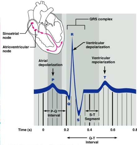
ST segment
Period between ventricular contraction and repolarization
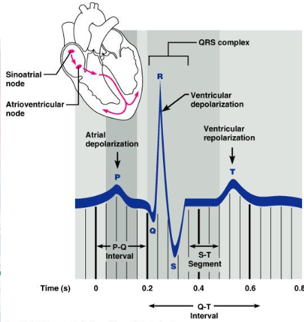
T wave
Ventricular recovery

4 things an ECG can detect
Arrythmias
MIs
Structural problems
Electrolyte imbalances
4 modifiable risk factors for CVD
Smoking
High BP
Obesity
Excess Alcohol
4 non-modifiable risk factors for CVD
Age
Sex
Type 1 diabetes
Ethnicity
Atherosclerosis
Thickening of the arteries caused by a build-up of plaque in artery lining + vessel walls
What can atherosclerosis cause? 2 parts
Angina/unstable
NSTEMI/STEMI
Percutaneous Coronary Intervention (PCI)
Using a balloon and stent to increase lumen of the narrowed artery + is non-invasive
Primary PCI (PPCI)
More specialised treatment for an active MI(STEMI)
Coronary Artery Bypass Graft (CABG)
A graft is taken from the radial artery (arm), the saphenous vein (leg) or chest left internal mammary artery (LIMA)
When is CABG performed?
When 3 main coronary arteries are blocked
What is used to create blood flow past the narrowing (CABG)
Blood vessels