Cell bio lab midterm
 Call Kai
 Learn
Learn Practice Test
 Spaced Repetition
 Match
Match Flashcards
 Knowt Play
1/129

There's no tags or description
Looks like no tags are added yet.
Name | Mastery | Learn | Test | Matching | Spaced | Call with Kai | Chat |
|---|
No analytics yet
Send a link to your students to track their progress
130 Terms
Iodine test
This test for carbohydrates is used to detect starch with I2KI solution. In the presence of starch, an iodine-starch complex is formed, causing a color change from reddish/yellow/brown to violet-black
Benedicts test
This test for carbohydrates is used to detect reducing sugars which are small sugar subunits and individual sugar monomers. The reagent used is a solution of sodium citrate and CuSO4, and it produces an insoluble orangish CU2O precipitate in the presence of reducing sugars (aldoses and some ketones) and heat
Sudan IV dye technique
The reagent used for this technique will only dissolve in hydrophobic compounds such as lipids. When in contact with lipids, the dye will lend a red-pink color to the solution.
Biuret test
In this test for proteins, NaOH and CuSO4 are added to test solutions. In alkali environments, the Cu^2+ ions will oxidize peptide bonds in protein. In the process, Cu^2+ ions are reduced to Cu^+1 producing a purple color whose intensity directly correlates with the amount of protein present
Principle
This term describes the chemical reaction that is occurring
Reducing sugars can be either a mono- or disaccharide which contains a hemiacetal group with one OH group and one OR group attached to the same carbon. Simple (reducing) sugars have free aldehyde and ketone groups. The OH group is able to reduce other compounds.
Starch is a polysaccharide with many glucose monomers joined together by glycosidic bonds.
What is the difference between starch and reducing sugars?
The color changes from a yellow/brown color (which is seen as the reagent or in a negative test) to a violet/black color
What is the observable change in a positive starch test?
If starch is present, the iodine in the reagent reacts with starch to create an iodide complex
What is the chemical basis of the change in a positive iodine test?
The color changes from a light blue (seen in negative test and the reagent itself) to a green/orange/brick red color when placed in heat and if reducing sugars are present.
What is the observable change in a positive Benedict's test?
If reducing sugars are present, Cu^2+ in the form of sodium citrate and CuSO4 (Benedict's Reagent) is reduced to Cu^1+ as an insoluble Cu2O precipitate is formed
What is the chemical basis of a positive Benedict's test?
Reducing sugars react because the simple carbohydrates and their OH groups have the ability to reduce compounds while starch cannot. Benedict's test only tests for simple sugars which have free aldehyde and ketone groups
Why do reducing sugars react to the Benedict's test while starch does not?
Red sudan IV crystals dissolve and create a red solution
What is the observable change in a positive Sudan IV test?
Sudan IV dissolves in a hydrophobic/lipophilic solution due to its solubility in lipids. Once dissolved it stains the lipids with a red color
What is the chemical basis of a positive Sudan IV test?
The other tests form a complex or undergo a redox reaction, so a chemical reaction and change is occurring. The Sudan IV test, however, only stains lipids if they are present due to its solubility in them
How is the Sudan IV test different from the other tests mentioned?
The color changes from blue (negative result and reagent color) to purple
What is the observable change in a positive Biuret test?
The change in color occurs due to the reduction of Cu^2+ ions to Cu^1+. In alkali environments, Cu^2+ ions oxidize peptide bonds in proteins and the Cu^2+ ions are reduced to Cu^1+
What is the chemical basis of a positive Biuret test?
Negative controls are used for (1) seeing the color of reagents when negative and/or by themselves and (2) so that one can make sure the solvent is pure.
Positive controls are used to make sure the reagents used are working as they should be
What is the significance of the use of negative control and positive control in all of the tests mentioned?
Body tube
Separates objective and eyepiece, allows alignment of optics, and brings the image to your eye
What is #1, and what is its function?

Revolving nosepiece
Holds objective lenses and is rotated to switch between them
What is #2, and what is its function?

Low objective
Likely to provide 4x magnification of the specimen. When combined with the eyepiece, it produces 40x magnification and is used to see larger structures
What is #3, and what is its function?

Medium objective
Likely to provide 10x magnification of the specimen. When combined with the eyepiece, it produces 100x magnification and is used to see specimen components closer than the low objective, but still zoomed out enough compared to the high objective
What is #4, and what is its function?

High objective
Likely to provide 40x magnification of the specimen. When combined with the eyepiece, it provides 400x magnification and provides more magnification than the medium objective, but not enough as an oil-immersion objective lens.
What is #5, and what is its function?

Stage clips
Holds the slide in place
What is #6, and what is its function?
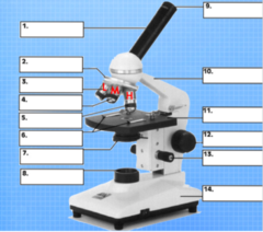
Condenser
Focuses/concentrates light onto the specimen
What is #7, and what is its function?

Light source (lamp)
Illuminates the specimen and allows light to pass through it, enabling an image to be produced
What is #8, and what is its function?

Eyepiece
The lens the viewer looks through to see a specimen which also provides 10x magnification
What is #9, and what is its function?

Arm
Connects the body tube to the base of the microscope and acts as a structure to carry the scope by
What is #10, and what is its function?
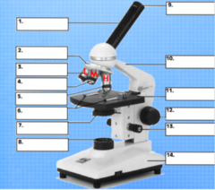
Stage
The flat platform that supports the slide
What is #11, and what is its function?

Fine adjustment
Brings specimen into sharp focus under low power and used for all focusing with high power objectives
What is #12, and what is its function?
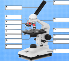
Coarse adjustment
Moves the stage up and down to bring the specimen into focus
What is #13, and what is its function?

Base
Bottom of the microscope used for support
What is #14, and what is its function?

Bright field microscope (lab - light compound microscope)
This type of microscope is (1) the most widely used, (2) it forms its image when light is transmitted through the specimen, (3) the specimen produces an image that is darker than the surrounding illuminated field, and (4) it can be used with live, unstained and preserved, stain specimens
Phase contrast microscope
This type of microscope is (1) used for live specimens, (2) the specimen is contrasted against a gray background, and (3) it is excellent for internal cell details
Differential-interference microscope
This type of microscope (1) allows for detailed view of live, unstained specimens, (2) it includes two prisms that add contrasting colors to the image, and (3) the image is colorful and three-dimensional
Fluorescence microscope
This type of microscope (1) includes a UV radiation source and a filter that protects the viewer's eyes, (2) it is used with dyes that show fluorescence under UV rays, (3) it forms a colored image against a black field, and (4) it is used in diagnosing infections caused by specific bacteria, protozoa, and viruses using fluorescent antibodies
Confocal microscope
This type of microscope (1) allows for viewing cells art higher magnifications using a laser beam of light to scan various depths in the specimen, and (2) it is most often used on fluorescently stained specimens.
Electrons
This energy source used in microscopy (1) travels in wavelike patterns 1000x shorter than visible light waves, (2) it increases resolving power tremendously, (3) allows for magnification between 5000x and 1000000x for biological specimens, and (4) allows scientists to view the finest structure of cells
Transmission electron microscope (TEM) (up to 100,000x)
This type of microscope (1) is often used to view structures of cells and viruses, (2) it transmits electrons through the specimen, (3) the specimen must be very thin and stained to increase image contrast, and (4) the dark areas of this microscope's image represents thicker or denser parts.
Scanning electron microscope (SEM) (up to 650,000x)
This type of microscope (1) creates an extremely detailed 3D view of all kinds of objects, (2) its electrons bombard the surface of a whole metal-coated specimen, (3) electrons deflected form the surface are picked up by detectors(s), (4) the electron pattern is displayed as an image on a television screen, (5) contours of specimens resolved with this are very revealing and surprising, and (6) the images produced from this may be computer enhanced to give them color.
Actin
This is a structural component of the cells involved in forming the contractile filaments of muscle cells and is a critical feature responsible for movement in other cells
Breast epithelial (MCF-10A) and breast cancer (MDA-MB-231) cells
What types of cells did we examine in this experiment?
Excitation
This is an increased state of energy compared to a molecules ground state which causes it to absorb a certain wavelength of light
Emission
This occurs when an excited molecule returns to its ground state and releases a wavelength of light that can be detected
Fluorescence
This is the emission of light by a substance that has absorbed light or other electromagnetic radiation
Fluorophore
This type of molecule can absorb and emit photons with a range of wavelengths (often in the excitation and emission spectra). These become excited, absorbing a certain wavelength of light, and when the molecule goes from its excited state to its ground state, this emits a different wavelength of light.
Photobleaching
This is when a fluorophore loses its fluorescence due to damage induced by light (often is white light)
Fluorophores have specific excitation or emission spectra which determine the intensity and production of fluorescence
Excitation is similar to the specific wavelengths the molecules can absorb, and emission refers to the specific wavelengths of light that are released. The spectra between the two corresponds with certain wavelengths of light
What do you understand by excitation or emission spectra in fluorescence microscope?
1. A phase contrast microscope uses different phases that light passes through unlike basic microscopy
2. Phase contrast microscopy uses a condenser annulus, a -90° phase shift ring, and a gray filter ring. These components are not present in basic light microscopes.
3. Phase contrast is used for live specimens, for internal cell details, and if it is against a gray background - basic microscopy can have dead, live, stained specimens and views them against a bright background
How is phase contrast microscopy different from basic microscopy?
DAPI stains DNA and can pass through cell membranes. DAPI is a fluorescent stain that binds to DNA and makes it appear blue under UV light, creating an image depicting the stained DNA under the microscope
What is the use of DAPI in fluorescent microscopy?
Paraformaldehyde has the purpose of fixing cells to the plate
Why was paraformaldehyde used in the protocol?
Triton-X-100 is a detergent for permeabilizing the cells so that they are able to accept the dye. This allows dye to enter the cells, stain them, and be visible under UV light
Why was triton-X-100 used in the protocol?
PBS - phosphate buffer saline
What buffer solution was used in the experiment?
430 nm (blue) and 662 nm (red)
What wavelengths of light do chlorophyll a pigments absorb the most? What colors do these wavelengths correspond to?
453 nm (blue) and 642 nm (red-orange)
What wavelengths of light do chlorophyll b pigments absorb the most? What colors do these wavelengths correspond to?
Wavelengths between 460 nm (blue) and 510 (blue-green)
What wavelengths of the visible light spectrum do carotenoid pigments absorb the most? What colors do these wavelengths correspond to?
Wavelengths between 500 nm and 620 nm. These wavelengths correspond to the colors green and yellow
What wavelengths of the visible light spectrum do plants utilize the least for photosynthesis? What colors do these wavelengths correspond to?
Photosynthesis consumes CO2
Does photosynthesis produce or consume carbon dioxide?
Red
A bicarbonate indicator solution may be used to show changes in the pH of a solution. When CO2 from the atmosphere dissolves into water containing a bicarbonate indicator at equilibrium, the pH value of the solution is measured at 8.6. What color is the indicator solution at a pH value of 8.6?
Yellow
Cellular respiration
When CO2 level increases, the indicator solution changes as CO2 dissolves in water to form carbonic acid. What color would the indicator solution change to in response to increased levels of CO2? During what process does this occur?
Purple
Photosynthesis
When CO2 levels decrease, the solution becomes more basic. What indicator color will be observed if the concentration of CO2 decreases in the control solution? During what process does this occur?
Purple. Photosynthesis removes CO2 from the environment, so there is a lower CO2 concentration than the control and it is less acidic (higher pH). Therefore, the color changes from red (control) to purple (low CO2 concentration)
Algae and plants perform both photosynthesis and respiration. Assume that algae are placed into a culture tube containing bicarbonate indicator solution with an initial pH value of 8 .6. After 24 hours, what color will the bicarbonate indicator become if photosynthesis is occurring at a greater rate than algal respiration? Explain why this may occur.
Yellow. Respiration produces CO2 as a by-product, so it will be released into the solution. The higher CO2 concentration compared to the control causes the solution to become more acidic (lower pH). Therefore, an increase in CO2 causes the color to change from red (control) to yellow (high CO2 concentration)
Assume that a glass culture tube containing algae beads and bicarbonate indicator solution is kept in the dark. After 24 hours, what color will the bicarbonate indicator become if algal respiration is occurring at a greater rate than photosynthesis? Explain what differences in dissolved carbon dioxide concentrations may cause this color change.
Wavelengths around 450 nm (blue) and 660 nm (red)
What wavelengths/colors of light do photosynthetic pigments absorb the most?
Red and Blue
Which colored filters provide the wavelengths of light necessary for maximum absorption by photosynthetic pigments?
Purple
If photosynthesis is occurring at a greater rate than respiration, what color should the indicator change to?
Yellow
IF there are not sufficient, appropriate wavelengths of light to perform the light-capturing process of photosynthesis, plant respiration will exceed plant photosynthesis. What color should the indicator change to when photosynthesis is compromised due to less than ideal inputs of light?
Respiration. Respiration releases CO2 as a by-product, so CO2 concentration increases. This makes the environment more acidic and causes the pH to decrease. These things are indicated by the yellow-colored solution
- Low pH (more acidic) = yellow
What process dominates if the solution is yellow? Why?
Photosynthesis. Photosynthesis consumes CO2 in the reaction, so CO2 concentration in the solution decreases. This makes the environment less acidic and raises the pH. These are indicated by the purple-colored solution
- High pH (less acidic) = purple
What process dominates if the solution is purple? Why?
Sodium alginate + algae was dropped into ice chilled calcium chloride to form the beads
What components were used to create the algae beads?
The tube with no covering and no algae beads, only the bicarbonate indicator solution. This ensured that neither respiration nor photosynthesis would occur without algae beads
Which tube was the negative control for the experiment? What was the purpose of this?
The tube with no covering but contained algae beads and the bicarbonate indicator solution. The tube was completely exposed to light and had the necessary components for photosynthesis to occur, so it should dominate the reaction process
Which tube was used for the positive control for photosynthesis? Why?
The tube with aluminum foil covering that contained algae beads and the bicarbonate indicator solution. This allowed the reaction to occur in the complete absence of light, so only respiration would occur. Photosynthesis could not possibly occur because of the lack of light
Which tube was used for the positive control for respiration? Why?
pH = 8.6
red
What was the pH and color of the bicarbonate indicator solution?
6CO2 + 6H2O ―(Light)→ C6H12O6 + 6O2
What is the net reaction for photosynthesis?
C6H12O6 + 6O2 → 6CO2 + 6H2O + Energy
What is the net reaction for cellular respiration?
Chloroplasts. The light reaction occurs in the thylakoid membrane and the dark reaction occurs in the stroma
Where does photosynthesis occur in plant cells?
Mitochondria
Where does cellular respiration occur in plant cells?
What is the equation that shows the production of bicarbonate from CO2 and H2O?
CO2 + H20 ⇄ H2CO3 → HCO3- + H+
Photosynthesis
What process in aquatic algae and plants removes dissolved carbon dioxide from the environment?
The control bicarbonate solution has a pH of 8.6 and is the color red. The change in color is dependent on the concentration of CO2 in the environment, so cellular respiration and photosynthesis both affect CO2 concentration which causes a change in the color. Photosynthesis removes CO2 from the environment, lowering its concentration, raising the pH, decreasing acidity, and changing the color to purple. Cellular respiration releases CO2 into the environment, increasing its concentration, decreasing the pH, increasing acidity, and changing the color to yellow. The resulting color determines to what extent photosynthesis or cellular respiration occurs.
Explain how the color change in the bicarbonate indicator solution allows an experimenter to determine differences in the rate of photosynthesis between culture tubes
Diffusion
This is the net passive movement of molecules down their concentration gradient; molecules are in constant motion and tend to move from regions where they are in higher concentration to regions where they are less concentrated
Facilitated Diffusion
This is the passive movement of molecules along their concentration gradient, guided by the presence of another molecule that is usually an integral membrane protein forming a pore or channel
Osmosis
This is the diffusion/passive transport of water as it moves through a selectively permeable membrane from a region of its higher concentration to a region of its lower concentration
Water potential
- Water potential is the potential energy of water in a system compared to pure water, when both temperature and pressure are kept the same
- Water always moves from an area of higher water potential to an area of lower water potential
This is the tendency of water to leave one place in favor of another. It is the ability of any biological cell to take in water, creating pressure exerted on the cell wall.
Pressure and amount of solute
What are the factors affecting water potential?
Water potential (Ψ) = pressure potential (Ψp) + solute potential (Ψs)
What is the equation used to calculate water potential?
Into the cell
Where does water move if it has a negative water potential?
Solute potential
In lab, pressure potential is equal to zero, so what is water potential equal to?
(final weight - initial weight) / (Initial weight) x 100
What is the formula for percent weight change?
Ψs = -iCRT
What is the formula used to calculate solute potential?
The ionization constant which depends on what solute is provided
- e.g. sucrose: i = 1 ; NaCl: i = 2 ; MgCl2: i = 3
For the solute potential equation, what does "i" stand for?
The concentration fo solute as determined
For the solute potential equation, what does "C" stand for?
The pressure/gas constant which is equal to 0.0831 (L)(bar)/(mol)(K)
For the solute potential equation, what does "R" stand for?
Temperature in degrees Kelvin which is 273 + °C of the solution
For the solute potential equation, what does "T" stand for?
The cell is hypertonic, so it takes up water from the solution. Negative water potential draws water up into the plant stem
If the water potential of a plant cell is negative, is the cell hypertonic or hypotonic compared to its surrounding solution? Will this cause water to rise through the plant stem?
Physical rupture and chemical lysis
What are the two general methods available to lyse cells?
Physical rupture
This method of lysing cells includes mechanical disruption, liquid homogenization, sonification, freeze/thaw, and manual grinding
Chemical lysis
This method of lysing cells uses detergents like sodium dodecyl sulfate (SDS), Triton X-100, NP40, Tween 20, and osmotic agents such as mannitol (where a high mannitol concentration ruptures membranes)
- Variable yields
- Variable viscosities
- Expensive instruments
- Subsequent heating of cell sample
What are the disadvantages of lysing cells through physical rupture?
It is very effective, however, there are many disadvantages
What is an advantage of lysing cells through physical rupture?