UNIT 5.2. MEMBRANE0-BOUND ORGANELLES: CYTOSKELETON
 Call Kai
 Learn
 Practice Test
 Spaced Repetition
 Match
 Flashcards
 Knowt Play
1/60

There's no tags or description
Looks like no tags are added yet.
61 Terms
Microfilament
3 Cytoskeletal Fibers:
● Thinnest filament present in the cytoskeleton in cytoplasm of eukaryotic cell.
● Give the cell its shape and enable cell movement.
G-actin (globular actin)
Microfilaments are made of actin monomers called
F-actin (filamentous actin) ;
7 nm in diameter
G-actin polymerizes into ____________ forming thin filaments about ____________
Nucleation
Elongation
Treadmilling (Steady State)
3 Stages of Microfilament Assembly
Nucleation
3 Stages of Microfilament Assembly:
Initial and rate-limiting step.
Three G-actin monomers come together to form a stable “nucleus.”
This process is regulated by nucleation-promoting proteins.
Elongation
3 Stages of Microfilament Assembly:
Rapid addition of ATP-actin monomers at the plus (+) end of the filament.
Filament grows as monomers add in a helical/thread-like structure.
Treadmilling (Steady State)
3 Stages of Microfilament Assembly:
At equilibrium, monomers add at the plus end and dissociate at the minus (-) end.
allowing filament remodeling.
amoeboid movement ;
lamellipodia
Cell motility: Actin filaments are involved in cell movement, like in ____________ and the formation of __________during cell migration.
actin ; myosin
Muscle contraction: ______ works with _______ to enable muscle contraction
Cell division: During cytokinesis, actin forms a ___________ to separate daughter cells.
Microfilaments
play a role in structure,
cell motility, and
cell division.
involved in muscle contraction
as well as cell division.
8 to 12 nanometer
Composed of two antiparallel dimers
Intermediate Filaments
3 Cytoskeletal Fibers:
Made of variety of proteins, including keratin, desmin, and vimentin
keratin
Intermediate Filaments:
a protective barrier in our tissues like skin, hair and nails so it gives a mechanical strength and resilience on our tissues, subject to friction and your mechanical stress.
desmin
Intermediate Filaments:
crucial in maintaining the integrity and the alignment of the muscle fiber, so, it forms a scaffold around your z-disc in muscle cell helping in force the transition during your muscle contraction
defects will cause you myopathies leads to your muscle weakness and dysfunction
Vimentin
mediate Filaments:
found on your mesenchymal cell that gives flexibility to cells that undergo shape changes and migration, it is also used for your wound healing, cell migration and maintaining cell integrity.
Dimer formation
Tetramer formation
Protofilament formation
Protofibril formation
Intermediate filament formation
5 Steps of Intermediate Filament Assembly:
Dimer formation
5 Steps of Intermediate Filament Assembly:
Two monomers align in parallel to form a coiled-coil structure.
The N-terminus and C-terminus of each monomer point in the same direction
Tetramer formation
5 Steps of Intermediate Filament Assembly:
Two dimers align in antiparallel (opposite orientation), staggered arrangement
Polarity is lost at this stage because of the antiparallel arrangement.
Protofilament formation
5 Steps of Intermediate Filament Assembly:
Tetramers align end-to-end
a linear bundle of tetramers.
Protofibril formation
5 Steps of Intermediate Filament Assembly:
Pairs of protofilaments associate laterally
Intermediate Filament formation
5 Steps of Intermediate Filament Assembly:
Eight protofibrils twist together into a rope-like filament
~10 nm in diameter
provides mechanical strength and flexibility
antiparallel tetramer arrangement
The _______________________ makes IFs highly stable and resilient.
Intermediate Filaments
3 Cytoskeletal Fibers:
● Provide mechanical support for the plasma membrane where it comes into contact with other cells or with extracellular matrix
● It does not participate in cell motility
Microtubules
3 Cytoskeletal Fibers:
the thickest fibers
are hollow rods about 25 microns in diameter
made up of the globular protein, tubulin, and they grow or shrink as more tubulin molecules are added or removed.
alpha and beta tubulin
Monomers of Microtubules
Tubulin Heterodimer Formation
Protofilament Formation
Lateral Association
Helical Lattice Formation
Dynamic Instability
Microtubule Assembly
Tubulin Heterodimer Formation
Microtubule Assembly:
Alpha-tubulin and beta-tubulin bind together
Each is the building block of microtubules.
heterodimer
alpha tubulin + beta tubulin =
Protofilament Formation
Microtubule Assembly:
Tubulin heterodimers polymerize head-to-tail to form linear chains
Polarity is established
Minus (-) end = alpha tubulin = grows slowly = depolymerization
Plus (+) end = beta tubulin = grows fast = polymerization
Minus (-) end vs. Plus (+) end of microtubules
Lateral Association
Microtubule Assembly:
Multiple protofilaments (typically 13) associate side by side to form a hollow tube
25 nm in diameter
Helical Lattice Formation
Microtubule Assembly:
Protofilaments are slightly offset, creating a helical spiral.
left-handed helical pitch ;
helical lattice
Each heterodimer binds to the next in a ___________________ forming a _______________.
helical arrangement
The ________________ gives the microtubule rigidity and flexibility.
helps in spindle formation, chromosome segregation, and motility of cilia/flagella.
Dynamic Instability
Microtubule Assembly:
Microtubules can grow (polymerize) or shrink (depolymerize) rapidly.
This dynamic behavior allows remodeling, intracellular transport, and mitotic spindle formation.
(MTOC) Microtubule-Organizing Center
Microtubules are often anchored at the _________________ — a cellular structure that nucleates and organizes microtubules.
MTOC of animal cells = centrosome
MTOC of plant cells = nuclear envelope
MTOC of cilia/flagella = basal body (Kinetosome)
MTOC of animal cells, plant cells, cilia/flagella
Microtubules
They provide tracks for motor proteins
Kinesin
Motor Proteins:
Moves toward plus (+) end (cell periphery)
away from center
Dynein
Motor Proteins:
Moves toward minus (-) end
toward center
Microtubules
3 Cytoskeletal Fibers:
It participates in the intracellular transport of organelles and vesicles.
participate in maintaining cellular organization
facilitates also your communication between the other parts of the cell
Cilia
A hair;ike organelles, identical in structure to flagella, that line the surfaces of certain cells and beat in rhythmic waves, providing locomotion to ciliate protozoan and moving liquids along internal epithelial tissue in animals.
Power Stroke (Propulsive Phase)
Ciliary and Flagellar Movement:
Generates maximum force to move fluid or the cell in a desired direction.
The axoneme bends toward the baseline.
The bending is rapid, creating thrust that pushes mucus, eggs, or microorganisms forward.
Dynein “walks” along microtubules, producing the bending motion.
Power Stroke (Propulsive Phase)
Ciliary and Flagellar Movement:
Respiratory tract: Moves mucus.
Female reproductive tract: Moves egg cells.
Protozoans: Allows swimming in water.
Return Stroke (Recovery Phase)
Ciliary and Flagellar Movement:
Resets the cilium/flagellum to its original position for the next stroke.
Less forceful, low-resistance movement.
Prepares cilia for coordinated, rhythmic beating.
Ensures efficient fluid movement without backflow.
Flagella
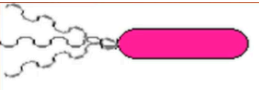
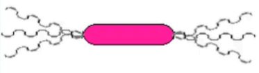
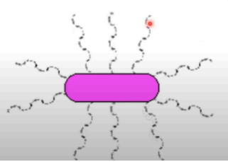
A slender threadlike structure, especially a microscopic whiplike appendage that enables many protozoa, bacteria, spermatozoa, etc., to swim.
Monopolar Flagella
Bipolar FLagella
Peritrichous Flagella
Cancer
Diseases of Cytoskeleton:
target microtubules to disrupt cell division
taxanes (paclitaxel)
stabilize microtubules, preventing their disassembly and thereby blocking cancer cell division
Neurodegenerative Disorder:
Alzheimer’s Disease
Diseases of Cytoskeleton:
Tau protein, a microtubule-associated protein, becomes hyperphosphorylated and forms tangles, disrupting microtubule stability and transport within neurons.
Neurodegenerative Disorder:
Huntington’s Disease
Diseases of Cytoskeleton:
Mutations in the huntingtin protein affect microtubule dynamics and neuronal transport, contributing to neurodegeneration.
Ciliopathies:
Primary Ciliary Dyskinesia (PCD)
Diseases of Cytoskeleton:
A genetic disorder that affects the structure and function of cilia, leading to respiratory issues due to impaired mucociliary clearance.
Ciliopathies:
Bardet-Biedl Syndrome
Diseases of Cytoskeleton:
A condition affecting cilia and microtubule function
resulting in obesity, retinal degeneration, and kidney abnormalities.
Cardiomyopathies
Diseases of Cytoskeleton:
Abnormalities in microtubule dynamics can affect cardiac muscle contraction and heart function
Cardiomyopathies:
Familial Hypertrophic Cardiomyopathy
Diseases of Cytoskeleton:
Mutations in the gene encoding tropomyosin, a microtubule-associated protein
Neuromuscular Disorders:
Charcot-Marie-Tooth Disease
Diseases of Cytoskeleton:
Certain forms of this inherited neuropathy are linked to defects in proteins involved in microtubule dynamics, affecting peripheral nerve function.
Neuromuscular Disorders:
Spinal Muscular Atrophy (SMA)
Diseases of Cytoskeleton:
loss of the SMN protein affects microtubule stability and axonal transport in motor neurons, leading to muscle weakness and atrophy.
Infertility
Diseases of Cytoskeleton:
Microtubules are crucial for sperm motility and oocyte division.
Abnormalities can lead to issues in sperm function or embryo development.
Defects in axonemal microtubules in sperm can result in reduced motility and effectiveness.
Developmental Disorder:
Syndrome of Microcephaly with Distal Limb Abnormalities
Diseases of Cytoskeleton:
inked to mutations affecting microtubule dynamics during neuronal development, leading to brain size reduction and limb deformities