The Brain
 Call Kai
 Learn
 Practice Test
 Spaced Repetition
 Match
 Flashcards
 Knowt Play
1/51

There's no tags or description
Looks like no tags are added yet.
52 Terms
Basic Parts of the Brain
Cerebrum (2 hemispheres)
Cerebellum
Brainstem
Diencephalon
Ventricles
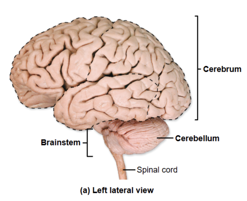
Cerebellum
Right and left hemispheres like cerebrum
Folia- folds on surface of cerebellum
Refines skeletal muscle movement so movement is smooth and coordinated
Stores “muscle memory”
receives rough draft of motor commands from cerebrum (via pons)
receives proprioceptive input from skeletal muscles (via medulla oblongata)
sends adjustments of motor commands back to cerebrum
Cerebellum function
motor commands sent from primary motor cortex → spinal cord
pons relays motor commands to cerebellum
cerebellum compares desired movements with real-time position of body parts
cerebellum makes adjustments to plan and sends those adjustments to cerebrum
Parts of Brainstem
midbrain
pons
medulla oblongata
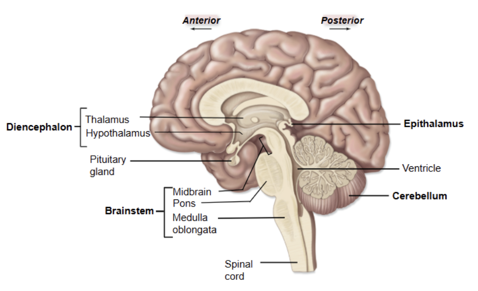
Brainstem functions
passageway for projection tracts between spinal cord and cerebrum
gives rise to 10 cranial nerves
responsible for many critical automatic behaviors
integrates auditory and visual reflexes

Parts of Midbrain
Cerebral peduncles
descending motor fibers (pyramidal motor tracts)
Colliculi
Substantia nigra: assists with control of voluntary movement
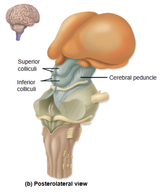
Colliculi
4 bumbs in posterior side
2 superior colliculi- visual tracking of moving objects
2 inferior colliculi- reflexive response to sound

Pons
Middle part of brainstem, anterior to cerebellum
Nuclei act as a relay center for motor commands:
send copies of motor commands from cerebrum to cerebellum
coordination of voluntary movement
Contains pontine respiratory center (for automated control of breathing)
Medulla oblongata
Nuclei act as relay center for sensory information:
Relay sensory & proprioceptive information to cerebrum and cerebellum
Pyramidal motor tracks
Decussation (crossing over) of motor axons happens here
Reticular formation- 3 nuclei that act as:
Cardiac center- controls heart rate and force of contraction
Respiratory center- controls basic breathing rhythm
Vasomotor center- regulates blood pressure via smooth muscle in blood vessels

Diencephalon
mostly gray matter, forms walls of third ventricle
hypothalamus
epithalamus
thalamus

Hypothalamus
main control of autonomic nervous system (cardiac/smooth muscle and glands)
main control of endocrine system through pituitary gland
regulates visceral sensations:
body temperature (body’s thermostat)
regulates hunger and thirst
connections with limbic system to control emotions
regulate sleep-wake cycle, directs pineal gland when to secrete melatonin
Epithalamus
Small group of nuclei and pineal gland
Pineal gland- secretes melatonin for circadian rhythm (sleep-wake cycle)
Influenced by hypothalamus
Thalamus
Gateway to the cerebral cortex
Contains many thalamic nuclei
Receives, filters, and relays somatic sensory information to the cerebral cortex
Development of the brain: primary vesicles
Develops from the rostral (head) end of the neural tube
Form 3 primary vesicles:
1. Prosencephalon (forebrain)
2. Mesencephalon (midbrain)
3. Rhombencephalon (hindbrain)
Caudal end of neural tube becomes spinal cord
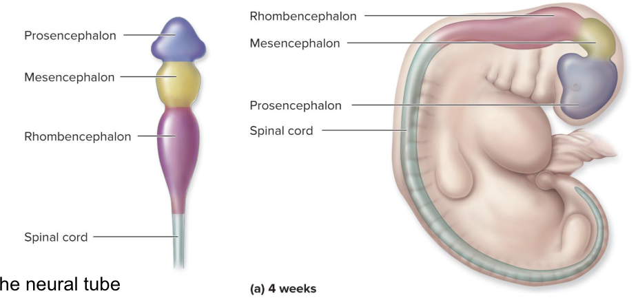
Development of the brain: secondary vesicles
Primary vesicles divide into secondary vesicles
Prosencephalon (forebrain)
1. Telencephalon (cerebrum)
2. Diencephalon (thalamus-y parts)
Mesencephalon stays the same (midbrain)
Rhombencephalon (hindbrain)
1. Metencephalon (pons, cerebellum)
2. Myelencephalon (medulla oblongata)

Meninges
Dura mater: most superficial
2 layers: periosteal and meningeal
Arachnoid mater:
Pia mater: most deep, follows contour of brain
Subarachnoid space: between arachnoid and pia mater (location of CSF)

Dural folds
Meningeal layer of dura produces (dural) folds that travel between fissures (deep grooves) of the brain

Falx cerebri
dural fold that separates left and right cerebral hemispheres
Falx cerebelli
dural fold that separates left and right sides of cerebellum
Tentorium cerebelli
dural fold that separates cerebrum and cerebellum
like a “tent” over cerebellum
Dural venous sinuses
Spaces (or sinuses) are present between dural layers where folds occur
Sinuses carry venous blood away from brain
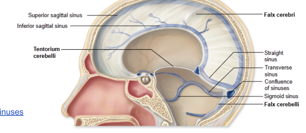
Superior sagittal sinus (SSS)
located in falx cerebri
Inferior sagittal sinus (ISS)
located in falx cerebri
Straight sinus
drains ISS and great cerebral vein from brain
Confluence of sinuses
junction of SSS and straight sinus
Transverse sinus
in tentorium cerebelli
Sigmoid sinus
carries blood from transverse sinus to jugular foramen
Ventricles
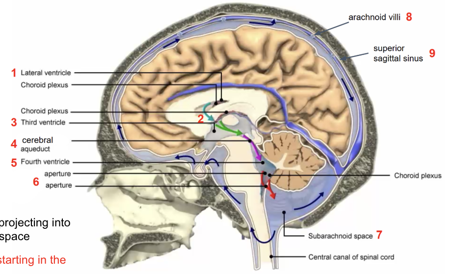
spaces in brain, continuous with central canal of spinal cord
filled with cerebrospinal fluid (CSF), lined my ependymal cells that produce CSF
CSF is produced from blood, nourishes and protects brain and spinal cord
2 lateral ventricles, third ventricle, fourth ventricle

Circulation of CSF
Lateral ventricles
interventricular foramina
third ventricle
cerebral aqueduct
fourth ventricle
apertures
subarachnoid space
arachnoid villi
superior sagittal sinus
CSF returned to blood by arachnoid villi projecting into superior sagittal sinus from subarachnoid space
Features of the Cerebrum
wrinkles: gyri
valleys: sulci
lobes name after associated skull bones
Fissures of brain
deep grooves that divide brain into distinct hemispheres and lobes
longitudinal fissure: separates right & left hemispheres
transverse cerebral fissure: separates cerebrum from cerebellum
Sulci of brain
lateral sulcus: separates temporal lobe from frontal & parietal lobes
central sulcus: separates frontal and parietal lobes
Parieto-occipital sulcus: separates parietal and occipital lobes

Gyri of brain
Pre-central gyrus: sends motor commands
Post-central gyrus: receive sensory information

Gray matter in Brain
cell bodies and non-myelinated axons
Cerebral cortex- outer layer of gray matter
Cerebral nuclei- concentrations of cell bodies
Perform complex neural functions

White matter in brain
mostly myelinated axons
Deep to cortex, surround nuclei
Allow for rapid processing between areas
carries information between gray matter regions

Sensory and posterior association areas
Primary somatosensory cortex
Primary visual cortex
Primary auditory cortex
Primary olfactory cortex
Gustatory cortex & visceral sensory cortex
Posterior association areas
Wernicke’s area

Primary somatosensory cortex
postcentral gyrus (parietal)
receives and processes sensory information from skin on opposite side of body (touch, pressure, pain, proprioception)
Primary visual cortex
occipital lobe
receives visual info from retina
Primary auditory cortex
superior edge of temporal lobe
conscious awareness of sound
Primary olfactory cortex
medial side of temporal lobe
conscious awareness of smell
Gustatory cortex & visceral sensory cortex
Located in insula
awareness of taste, pain, pressure, hunger, etc. (visceral sensations)
Posterior association areas
Integrates all types of sensory information for a unified perception of sensory input
Wernicke’s area
Left hemisphere, involved with understanding written and spoken language
Motor and anterior association areas
Primary motor cortex
Motor speech area (Broca’s area)
Anterior association area (prefrontal cortex)

Primary motor cortex
precentral gyrus (frontal lobe)
control skeletal muscle activity on opposite side of body
axons descend pyramidal tracts
Motor speech area (Broca’s area)
control ability to speak (and write)
Anterior association area (Prefrontal cortex)
receives highly processed sensory info, coordinate motor responses
thinking, remembering, reasoning, planning, memory, problem solving
White matter tracts in Cerebrum
tracts are how areas of cerebral cortex communicate with each other and other parts of brain and spinal cord
Commissural tracts
Association tracts
Projection tracts
Commissural tracts
cross from one hemisphere to the other
corpus callosum-above lateral ventricles

Projection tracts
link the cerebral hemispheres to the brainstem and spinal cord

Association tracts
connect different parts of same hemisphere
