(2) Blood Smear Evaluation, RBC indices, PCV/TP, & Abnormalities in the CBC
 Call Kai
 Learn
Learn Practice Test
 Spaced Repetition
 Match
Match Flashcards
1/59

Earn XP
Description and Tags
Vocabulary flashcards covering blood collection equipment, anticoagulants, CBC parameters, RBC indices, stain mechanisms, and erythrocyte morphological variations from veterinary hematology lecture notes.
Name | Mastery | Learn | Test | Matching | Spaced | Call with Kai | Chat |
|---|

60 Terms
Describe what a vacutainer system is?
A specialized vacuum device consisting of a special needle, needle holder, and vacuum-filled tubes, used to fill multiple blood collection tubes with a single venipuncture.
Describe what a Serum Separator Tube (SST) is and what is its cap color.
A tiger-topped (gray and red capped) blood collection tube with no anticoagulant that contains a gel. After allowing the blood in the tube to sit and clot (15 min), the tube is centrifuged. The gel ends up between (and physically separating) the serum (on top) from the clot (cellular components and clotting factors). This collection tube is designed strictly to collect serum for serum biochemistry profiles of various types.
Describe what an EDTA blood collection tube is and what is its cap color.
A lavender-topped blood collection tube containing ethylenediaminetetraacetic acid (EDTA) that binds to blood calcium, preventing clotting (coagulation) from occuring. These tubes are selected when you need a complete blood count (CBC), white blood cell differentials, PCR tests, or want to make blood smears. Since it has an anticoagulant, it preserves blood in the most natural in-situ state possible. If centrifuged, you will end up (from top-to-bottom) with plasma, a buffy coat, and cellular components.
Describe a Plain Red Top Tube (RTT) and its cap color.
A white-capped or red-capped sterile tube with nothing in it (no additives). It is used to collect urine, fluid samples, or samples for culture, cytology, and toxicant analysis. Essentially, it’s a plain, sterile tube for collecting anything you want to simply keep in a sterile tube for transport to the lab. For example, you can place urine, body cavity fluids (from ascites or effusions, etc.), or any other liquid or tissue samples that need to be analyzed without interference from anticoagulants or preservatives.
Describe a variaton of the plain red-capped collection tube.
If the collection tube has a red cap with a solid black circle in the middle of the cap, it is a plain sterile tube with a clot-activating ingredient coating the inside of the tube.
Describe a Blue Top collection tube.
A blue-capped blood collection tube contains the anticoagulant, sodium citrate . It is used for coagulation assays (tests) like ACT, PT, PTT, ACT, and platelet function tests.
How are platelet counts different than platelet function tests?
Platelet counts are simply a numerical tally of how many platelets are circulating in your bloodstream - You can do this from the blood sample in an EDTA collection tube. You can also make a blood smear from the EDTA blood mixture and estimate the number of platelets manually by using a microscope. The platelet count is a normal analyte reported in a CBC so a lavendar capped collection tube would be utilized.
Platelet function tests are testing how the platelets are “working” or interacting with each other and with blood vessel walls to stop bleeding. These various tests determine how well platelets are doing their job and are not intended to be tests that “count” how many platelets there are….rather if the ones that are present are functioning normally.
Describe a green top tube and what it is used for.
A collection tube containing sodium or lithium heparin, used for toxicology tests, ammonia levels, blood gases, and plasma biochemistry profiles (usually in-house blood machines).
Describe a blood film (blood smear) and where the blood to make one should come from?
A thin film of blood smeared on a glass slide using a wedge technique. Ideally, the blood should come directly from the syringe used to draw blood to avoid artifacts from anticoagulants in collection tubes. If this is not possible, then getting blood from an EDTA tube is the second best source. Blood films are used under the microscope to evaluate cellular morphology and obtain white blood cell counts/differentials.
What are the three most common anticoagulants used in blood collection tubes (and their corresponding cap color)?
EDTA (lavender)
sodium or lithium heparin (green)
sodium citrate (blue)
Which blood collection tubes do not have any anticoagulant in them?
Serum separator tubes (tiger-top) and plain red-top or white-top tubes.
The single-cell thick region of a blood film located between the thick area and the feathered edge where cell count and morphology evaluations are conducted is called the ____________.
monolayer (count area)
Describe Diff-Quik®.
A Romanowsky-based stain kit that mimics Wright Stain but is less expensive and faster, consisting of a fixative, an eosinophilic (red/acidic) stain, and a basophilic (blue-purple/basic) stain.
Describe the packed cell volume (PCV). What is it? What does it measure? What unit is it reported in?
It’s the percentage of whole blood that is composed of red blood cells, measured by centrifuging blood in a microhematocrit tube. For example, if the PCV = 45%, then RBCs make up 45% of your blood volume (think of a column of centrifuged anticoagulated blood).
What’s the difference between the PCV and HCT (hematorcrit)?
They both measure the same thing in the same units…the % of RBCs from a whole blood sample. The difference is in the method used to obtain this value. If you spin whole anticoagulated blood in a microhematocrit tube and then read the %RBC from a microhematocrit card reader, then it’s reported as the “PCV”. If you have a countertop blood machine that measures the blood’s electrical conductivity and uses that in a formula to obtain the % or RBCs, then the result, although the same (45% in this case) is called the HCT (hematocrit).
Give your best definition of what a CBC is.
CBC = Complete Blood Count = a commonly performed venous blood test that “counts” how many red blood cells, white blood cells, and platelets are present in the blood, along with measures of hemoglobin, hematocrit, and other red blood cell qualities and measurements (RBC indices) that provide information about the overall health of the animal. It is an analysis and count of the cellular components!
Give your best definition of total protein (TP).
The measure of plasma or serum protein concentration reported in g/dL and obtained using a refractometer.
Specifically, the “total protein” can be broken down into what two protein groups? How do these two groups relate to a formula that describes what total protein equals?
albumin and globulins
TP = albumin + globulins
How do we define “globulins”?
globulins = liver proteins (other than albumin) and antibodies.
What are the 3 values we use to assess the condition or health of the red blood cells, otherwise known as “red blood cell indices”?
MCV = mean corpuscular volume
MCH = mean corpuscular hemoglobin
MCHC = mean corpuscular hemoglobin concentration
What does the mean corpuscular volume (MCV), reported in femtoliters, describe?
The average volume or size of individual red blood cells.
If the MCV is elevated above normal, we describe the RBCs as ______________ whereas if the MCV is reported below normal volume (smaller), we describe the RBCs as ____________,.
macrocytic (larger than normal RBCs)
microcytic (smaller than normal RBCs)
What does the MCH describe? Explain.
The mean corpuscular hemoglobin which is the mean quantity (in weight—measured in picograms) of hemoglobin present per red blood cell. It is essentially the average amount of Hgb present in one cell…hence the tiny unit of picograms!
Describe what MCHC refers to.
Mean corpuscular hemoglobin concentration is the average amount of hemoglobin present in a given volume (liquid) of packed red blood cells (not including plasma), expressed in grams per deciliter. It indicates the concentration of hemoglobin relative to the volume of red blood cells, regardless of their size.
If the MCHC is elevated above normal, we describe the RBCs as ______________ whereas if the MCVHC is reported below normal, we describe the RBCs as ____________,.
hyperchromic (darker than normal RBCs) hypochromic (lighter than normal RBCs).
Specifically, the MCV and MCHC are used, along with any positive finding of anemia, to further describe the anemia. Give an example of how you would describe an anemia with a reported MCV higher than normal and MCHS lower than normal.
The patient would have a macrocytic, hypochromic anemia.
What is more important, the % present of each type of white blood cell (e.g. 45% neutrophils from the total count of WBCs) or the absolute number of WBCs reported (e.g. 35,000 neutrophils/mcL reported)? Explain.
The absolute number of each type of white blood cell is more important because it tells you exactly how many of that particular type of WBC is present per mcL of whole blood whereas the % may be misleading. The % reported for each type of WBC represents what percent from the total number of WBCs (not per mcL of whole blood). For example, if you only had 10 WBCs in your entire body, but we reported that the neutrophils accounted for 40% of the total WBC count, the “40% neutrophils” might appear normal when in actuality, it represents 4 neutrophils in your entire body.
When would we want to apply the Corrected White Blood Cell Count formula to a CBC result?
When the patient had more than 5 nucleated RBCs reported for every 100 WBCs. In these cases, the machines would report an artifactually increased number of WBCs because many of the nucleated RBCs would be accidentally counted as a WBC (e.g. a lymphocyte) because of their nucleus. So, this formula decreases the reported WBC total count to a more accurate (true) number of total WBCs present.
An arrangement of red blood cells grouped like a stack of coins, normal in horses, cats, and pigs, or associated with high immunoglobulin levels is called ___________ ____________.
rouleaux formation

Irregular, three-dimensional abnormal clumping of red blood cells caused by antibody cross-linking, commonly observed in immune-mediated (autoimmune) hemolytic anemia is called _____________.
agglutination


What do we call the condition where red blood cells have decreased (abnormal) hemoglobin content and an enlarged area of central pallor (broken arrows)?
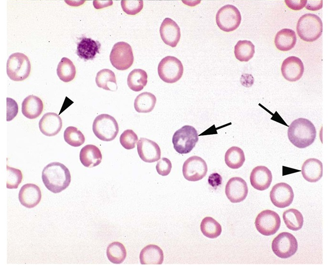
hypochromasia
What do we call the condition where some RBCs might stain light blue instead of red like other (normal) RBCs?
Polychromasia; we can also say that these particular RBCs are “polychromatophilic RBCs” (unbroken, whole arrows).

A visible variation in the size of red blood cells on a blood smear; normal in bovine blood but indicative of pathology or regeneration in other domestic species.
anisocytosis
If some RBCs are smaller than normal (MCV lower than normal), we say _____________ is present. If some RBCs are larger than normal, we say there is ________________ present.
microcytosis; macrocytosis
A term that means that more than 10% of the red blood cells on a blood smear display abnormal shapes (of various types). In other words, there is variation (abnormal) in the shapes of RBCs present.
poikilocytosis
RBC fragments on the blood film and in circulation which result from shearing (tearing) trauma within blood vessels, possible from conditions like vascular neoplasms, iron deficiency, and disseminated intravascular coagulation (triangles) are called _________________.

schistocytes
Abnormal RBCs featuring spiky, unevenly spaced surface projections of variable length and diameter, associated with altered membrane lipid concentrations are called _____________.

acanthocytes
Abnormal RBCs that have short, uniform, evenly spaced spiky surface projections, frequently occurring as an artifact of slow drying or underfilled EDTA tubes are called ___________.
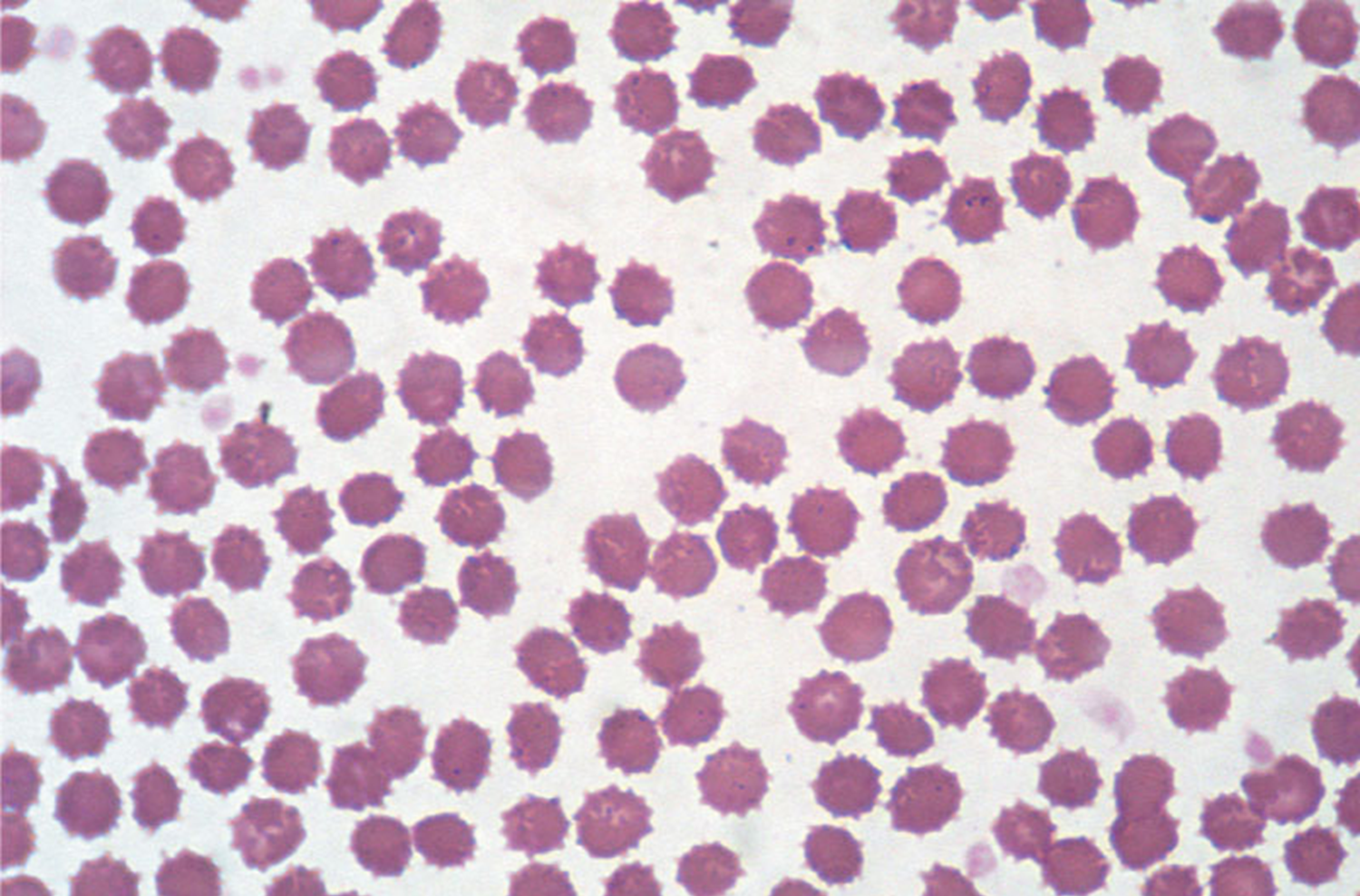

echinocytes
These abnormal RBCs have a vacuole or blister-like membrane lesion that ruptures, leaving two horn-like projections. Shaped like Manta-rays! See complete arrows in the image below.

keratocytes
Small, dark-staining, spherical red blood cells lacking central pallor, formed when antibody-coated membrane parts are phagocytized; characteristic of immune-mediated hemolytic anemia in dogs (short arrows).

spherocytes
RBCs that have hemoglobin shifted to one side and a pale crescent-shaped zone on the opposite side, caused by oxidative membrane damage from toxic ingestions like onions (dogs) or acetaminophen (cats). The look like an open-mouth smiley face.

eccentrocytes
This term refers to two presentations of RBCs that have excess membrane relative to volume, so depending on which side of the RBC you look at, they can look like the Target icon or a folded cell.

leptocytes (includes folded cells and target cells).
Artifactual red blood cells ("punched-out" cells) showing a sharply defined, circular central pallor surrounded by a thick rim of hemoglobin, caused by improper smear technique.

torocytes
These uniconcave RBCs have a mouth-like or slit-shaped central clear zone, seen in hereditary conditions in specific dog breeds.

stomatocytes
When a reticulocyte has multiple, intracellular, small, purple staining structures representing residual RNA which can occur with anemia or lead toxicity, is called ________________ ________________.
basophilic stippling
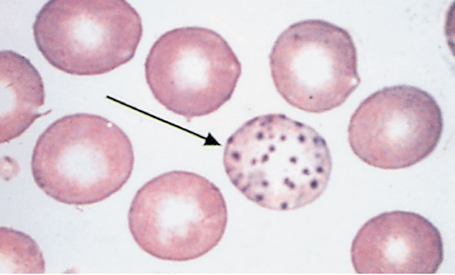
Small, light blue staining, round structures of denatured hemoglobin located at or protruding from the RBC membrane, caused by oxidative damage from agents such as onions, garlic, or acetaminophen.
Heinz Bodies (complete arrow)

The nuclear remnant of an RBC, usually concentrated into one dark staining spheroid structure within the cell.
Howell-Jolly Body(ies)

What do basophilic stippling and Howell-Jolly bodies have in common?
They both occur in immature RBCs and are therefore indicative of an anemia because that’s when the bone marrow spits out a lot of immature RBCs as a response to an anemia and some of these immature RBCs can still have a nucleus (Howell-Jolly body) and residual RNA (basophilic stippling).
Immature erythrocyte precursors retaining a nucleus, released early into peripheral circulation by bone marrow due to severe anemia, hypoxia, or marrow disease.
Nucleated Red Blood Cells (nRBCs)
Make a list of features/characteristics you could see on a blood film under the microscope if the patient is moderately or severely anemic.
basophilic stippling
Howel-Jolly bodies
Heinz bodies
polychromasia
hypochromasia
macrocytosis or microcytosis
nucleated RBCs
reticulocytosis
poikilocytosis
anisocytosis
What does it mean to say there is a regenerative anemia?
It means there is a decrease in the RBC count (PCV, HCT, ± Hb) and the red bone marrow has responded appropriately and adequately by releasing more RBCs, including immature forms (reticulocytes). This “regenerative” response is normal and expected to counteract the anemia. There is usually polychromasia, macrocytosis, hypochromasis in the blood film of these anemic patients if the bone marrow is capable of responding.
W/hat does it mean to say there is a non-regenerative anemia?
An anemia in which the red bone marrow fails to respond or produce additional red blood cells to compensate, marked by an absence of reticulocytes despite decreased PCV and hemoglobin.
Name the items in this image

microhematocrit tubes

Name the item in this image
clay sealant
What is the name of this instrument:

A centrifuge (to spin blood collection tubes).
What is the name of the item shown here:

A microhematocrit card reader.
Can you name the tubes from left to right in this image:

lithium heparin (green) tube
EDTA (lavender top) tube
EDTA (lavender top) tube (small volume version).
sodium citrate (blue) tube
plain, sterile, red top tube
plain, sterile, red top tube
serum separator (tiger top) tube
Name this instrument

refractometer
What is the name of the region we focus on when looking at blood cells?

The monolayer counting area.
What is the name of this instrument and what is it used for?

A differential cell counter and is used to tabulate (add) all the different white blood cells you are seeing (counting) at the monolayer. When you reach a total of 100 cells counted, a chime is heard.