Pathology of the Lymphoid System
 Call Kai
 Learn
 Practice Test
 Spaced Repetition
 Match
 Flashcards
 Knowt Play
1/85

There's no tags or description
Looks like no tags are added yet.
86 Terms
primary lymphoid organs
-thymus
-bone marrow
-bursa of fabricius
secondary lymphoid organs
-spleen
-LN
-lymph nodules (MALT, GALT)
lymphoid system is…
-interconnected
-lymphocytes continually recirculate
-connected to the blood circ via the thoracic duct
high endothelial venules
specialised cuboidal endothelial cells that facilitate receptor mediated transmigration of lymphocytes from blood
spleen lymphatic drainage
none
lymph node structure
-multiple afferent lymphatic vessels drain into subscapular sinus into trabecular sinuses into medullary sinuses and out into efferent lymphatic vessels

how do lymphocytes enter LN
-most through HEVs
-some through lymphatic circulation
lymphosomes
can look at where closer lymph node is to lump to see if lymph affected
the region of skin that is drained by the ln
can go back to the area from the enlarged ln to see if problem is there

1.sub-mandibular
2.parotid
3.dorsal superficial cervical
4.axillary
5.medial iliac
6.lateral sacral
7.hypogastric
8.popliteal
9.superficial inguinal
10.ventral superficial cervical
when indicated to investigate LN
-LN enlargement
-suspect underlying infectious disease e.g. leishmania, FIP, fungi
-stage malignant neoplastic disease
-provide material for molecular testing
label pic

-left is normal
-middle is hyperplastic
-right is hyperplastic only on one side so shows importance of sampling all over LN
ln gets lymphatic material from diff afferent lymphatic vessels so cna be compartmentalised
how to FNA LN well
-avoid aspiration of centre of markedly enlarged LN→ often necrotic
-spread gently→ lymphocytes are fragile
-non suction to minimise blood contam
-keep cytologic prep away from formalin fumes
-ideally take sample before giving steroids (sus lymphoma)

what is this sample
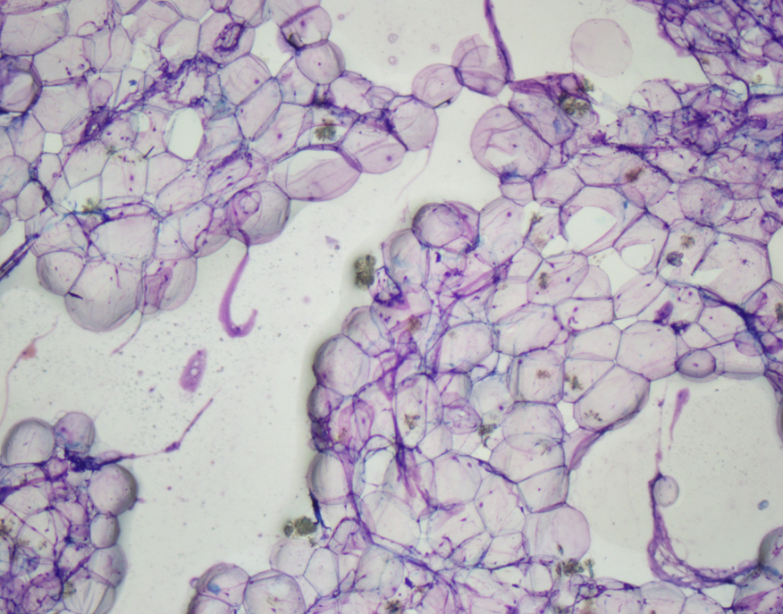
perinodal fat
can be accidentally sampled
what is this sample

slaivary gland
-epithelial cells
-cells in rows which is due to proteinaceous viscous saliva (windrowing)
-can also be accidentally sampled
normal LN look like
-small lymphocytes mostly
-medium and large less than 5-10%
-plasma cells, macrophages, mast cells, neutrophils are rare
-lymphoglandular bodies- fragile, fragments of cytoplasm
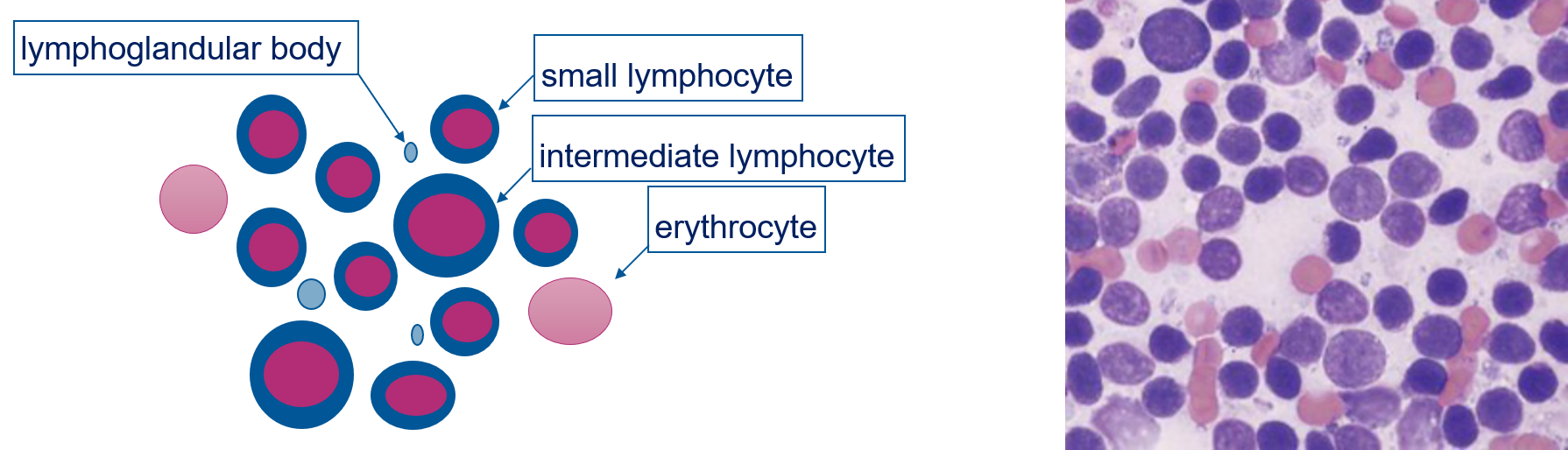
compare lymphocyte to rbc size
4 causes of lymphadenopathy
-reactive hyperplasia
-lymphoma
-lymphadenitis
-metastatic neoplasia

reactive hyperplasia spread
localised or systemic
stimulation of reactive hyperplasia
antigenic→ stimulate the LN to produce lymphocytes
-infectious
-immune mediated
-neoplasia
-vaccination
cytology of reactive hyperplasia
-indistinguishable from normal
-heterogenous cells
-small lymphocytes predominantly
-may see increase in intermediae and large
-may see more plasma cells
-cc macrophage
-few neutrophils, eosinophils, mast cells

plasma cell on cytology
has a vacuole where golgi body is as produce antibodies from here

lymphadenitis
-accumulation of inflam cells
neutrophilic lymphadenitis cause
-bacterial
-immune mediated
-neoplastic
eosinophilic lymphadenitis causes
-hypersensitivity reaction
-parasites
-idiopathic
-paraneoplastic (e.g. mct, lymphoma)
macrophagic/granulomatous lymphadenitis or pyogranulomatous causes
-chronic inflam conditions e.g. fungal infection, mycobacteriosis, leishmaniasis, FIP
lymphadenitis on histology
-more than 5% neutrophils then neutrophilic

-more than 3% eosinophils then eosinophilic

-more than 3% macrophages then macrophagic

how does mycobacteria appear on stain
do not stain, rods

lymphoma
-neoplasia of lymphocytes in solid tissues
subtypes of lymphoma
anatomic site (e.g., multicentric, GI, cutaneous)
location of neoplastic cells within the lymph node (e.g., diffuse, follicular, T zone)
cell morphology (small, large, granular)
cell type (T-cell, B-cell, NK)
biological behaviour (indolent, aggressive)
prognosis for lymphoma
b better than t (terirble)
lymphoma subtype classification
based on histopathology and immunohistochemistry
Subtype related to prognosis and treatment
immunohistochemistry
apply antibodies to detect antigen on surface of cells
differentiate t or b

lymphoma cytology
-diagnosis and staging
-accuracy decreases when attempt to apply WHO classification
-screening test for prediction of grade and phenotype
-not good for further classifying→ need histology
lymphoma histology
-invasive, time consuming, more expensive
-cytology combined with ancillary tests (e.g. flow cytometry, immunocytochemistry, PARR) → acceptable compromise in practice
approach to lymphadenopathy

how lymphoma looks on cytology
-homogenous (normally hetero)
-increased large immature lymphocytes
-may see more mitoses than reactive
-frequent lymphoglandular bodies
-tingible (stained) body macrophages
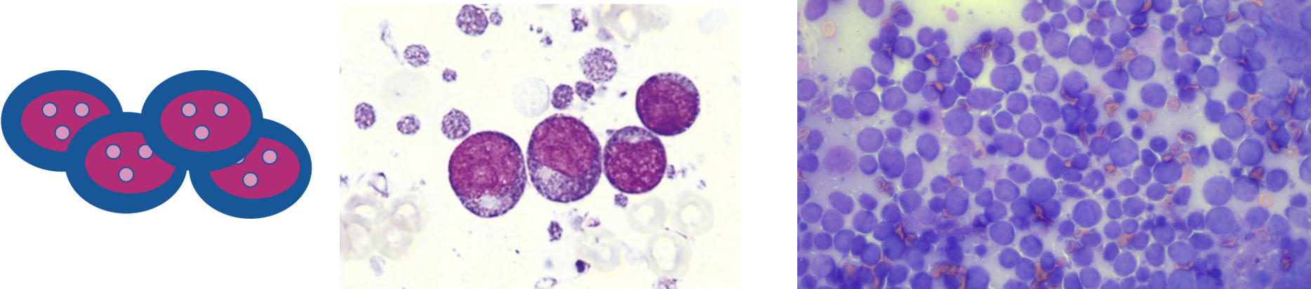

tingible body macrophage
phagocytosed debris of lymphocytes in macrophages

metastatic neoplasia
-lymph node maybe reactive
-presence of cells not normally in LN
-increased number of cells normally present often with atypical morphology


metastatic carcinoma

metastatic melanoma→ dark black brown pigment
approach to LN cytology

structure of spleen
-capsule
-red pulp
-white pulp

capsule of spleen
rich in elastic fibres and smooth muscle
red pulp of spleen
splenic sinusoids (rich in blood)
white pulp of spleen
splenic lymphoid tissue (antigen presenting cells, macrophages, b and t lymphocytes)
capsule of spleen function
contraction can release blood into circ (e.g. in response to acute blood loss)
red pulp of spleen function
reservoir of blood, filter for senescent or damaged erythrocytes, iron metabolism
white pulp of spleen function
immunity against bloodborne antigens (antigenic sensitisation)
when indicated to investigate spleen
-diffuse or symmetrical enlargement
-nodular or focal lesions
-abnormal US appearance
-evaluation of haematopoiesis
-diagnose neoplasia
-tumour staging
-often incidental
what is spleen good indication for
bone marrow→ easier to sample spleen than bm
key points for sampling spleen
-non suction technique to min blood contam
-spread gently
-keep cytologic prep away from formalin fumes
-avoid contam with lubricant gel→ bc usually US guided
use water/ saline

-ideally take sample before giving steroids
normal spleen look
-similar to LN but more blood
-neutrophils in blood
-90% small lymphocytes
-medium and large less than 5-10%
-rare plasma cells, macrophages, mast cells

splenomegaly causes
-reactive hyperplasia

-lymphoma

-splenitis

-metastatic neoplasia
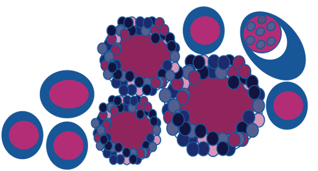
-extramedullary haematopoieis
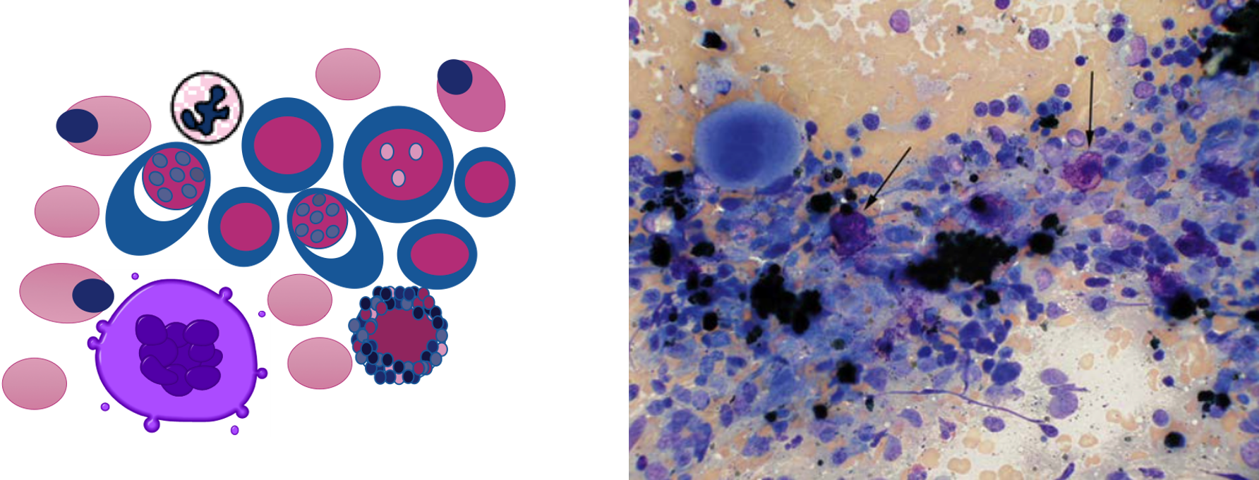
extramedullary haematopoiesis
if bone marrow having problems then spleen makes precursors (like in foetal life)

can see fe on splenic samples
erythroid island
erythrocyte precursors around macrophage to get iron to make haemoglobin

megakaryocyte
large platelet precursor cell found in the bone marrow or made in spleen when bone marrow not operating

infection or inflam of the spleen enlargement
-symmetrical or nodular enlargemenet
infection or inflam of spleen clinicopathological findings
-inflam leucogram
-hyperglobulinaemia
-infectious agent (e.g. leishmania, toxoplasma)
extramedullary haematopoiesis enlargement of spleen
symmetrical or nodular
extramedullary haematopoiesis clinicopathological findings
-anaemia
-other cytopaenias seen on CBC
haemolymphatic neoplasia type of enlargement
symmetrical or nodular
haemolymphatic neoplasia (e.g. lymphoma, multiple myeloma) clinicopathological findings
-lymphocytosis
-atypical cells in circulation
-cytopenias seen on CBC
-hyperglobulinaemia
-monoclonal gammopathy
-paraproteinuria
neoplasia like hystiocytic sarcoma or haemangiosarcoma enlargement
symmetrical or nodular
neoplasia like hystiocytic sarcoma or haemangiosarcoma clinicopathological findings
-regen anaemia +/- other cytopenias
-schistocytes
-acanthocytes
-thrombocytopenia
circulatory disturbances e.g. haematoma, torsion, splenic congestion enlargement
symmetrical or nodular
circulatory disturbances e.g. haematoma, torsion, splenic congestion clinicopathological findings
-regen anaemia
-abnormal liver values
-inflam/stress leucogram
infectious agents of spleen
-leishmania

-toxoplasma

neoplasia of spleen
-multiple myeloma

-haemangiosarcoma (mesenchymal)

multiple myeloma

tumour of plasma cells
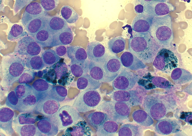
melanoma

mast cell tumour

carcinoma

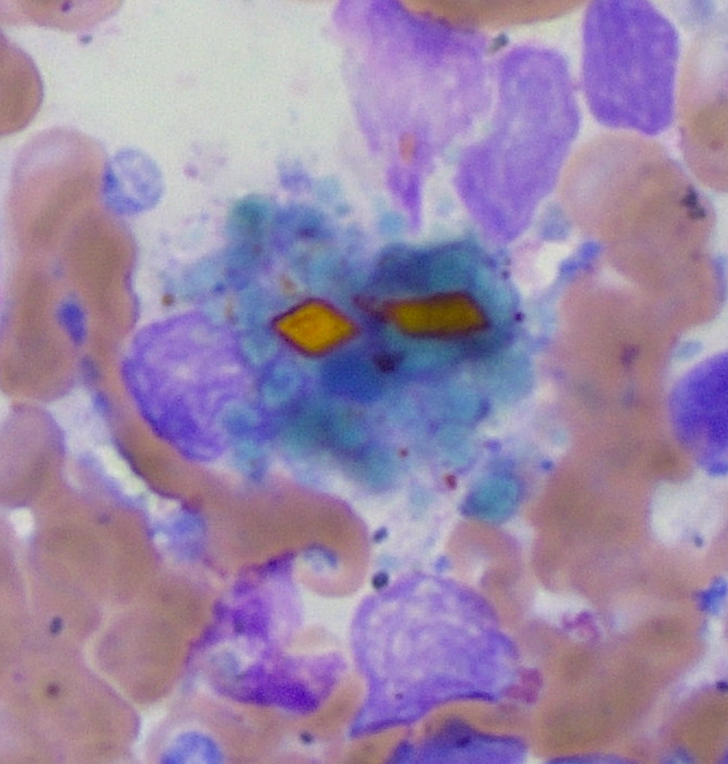
haematoma
-blood
-haemoglobin breakdown products (haemosiderin is blue, haematoidin is yellow and rhomboid)→ chronic haemorrhage
approach to spleen cytology and disease

structure of thymus
-primary lymphoid organ for development of t lymphocytes

where is thymus located
cranial mediastinum
-prominent before puberty then replaced by adipose tissue
2 most important components of thymus
epithelial cells and lymphocytes (CD4+ CD8+)
two pathologies of thymus
-thymoma
-lymphoma

thymoma origin
epithelial cells

lymphoma origin
lymphocytes
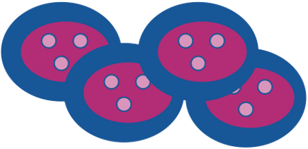
how do we differentiate between lymphoma and thymoma
differentiate between the cells
Thymoma → epithelial cells
Lymphoma → lymphocytes
treatment of thymoma
surgical
treatment of lymphoma
chemotherapy
how to investigate/ differentiate between thymoma and lymphoma
-imaging mass in cranial mediastinum
-cytology or/and flow cytometry
-histopathology for definitive
-paraneoplastic syndromes like hypercalcaemia, myasthenia gravis
what is myasthenia gravis most common in
thymoma
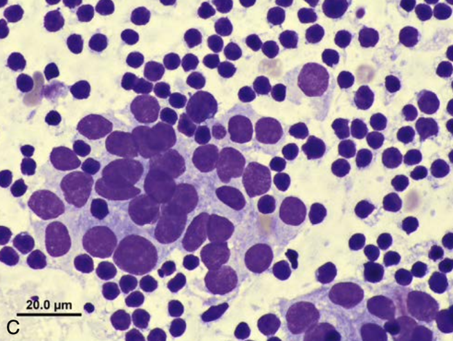
thymoma cytology
-epithelial cells
-small lymphocytes
-occasional mast cells

lymphoma cytology
-homogenous population of large atypical lymphocytes

