(D1) Ruminant Muscular Disorders
 Call Kai
 Learn
 Practice Test
 Spaced Repetition
 Match
 Flashcards
 Knowt Play
1/64

There's no tags or description
Looks like no tags are added yet.
65 Terms
clostridium is gram —
positive
what shape is clostridium
rod
how does clostridium spp. spread (4)
producing potent toxins
Form spores
Can be brought up in cultivated soils where they are then activated
Some spores are resistant to many disinfectants
what causes clostridium spp.
Ingest contaminated feed
Excess toxins produced by GIT when conditions predispose commensal clostridia to proliferate
A wound/injury
Bacterial translocation through edematous bowel
what clostridium spp. is in contaminated feed
Clostridium botulinum
Clostridium botulinum aka
botulism
how to diagnose clostridium spp. (4)
CK elevated
AST elevated
Impression smear sample of necrotic muscle is submitted for immunofluorescent detection
Clostridial toxemias
how to treat clostridium spp. (2)
Antibiotics
Surgery to remove necrotic muscle
how to prevent clostridium spp.
vaccinate
when to vaccinate for clostridium spp. (4)
boost calves at weaning
boost calves at one year
Adults vaccinated yearly as well
Annual vaccination is usually a standard protocol in the spring

whats wrong
blackleg
what bacteria causes blackleg
Clostridium chauvoei
clinical signs of blackleg (12)
Find animal dead
Bloating after death
putrefaction after death
Blood stained froth at nostrils
High fever
Increased HR
Very depressed
hot painful swelling Superficial muscle involvement initially
muscle painless later on
muscles cold later on
feel emphysema
Overlying skin can become cracked and dry
define putrefaction
the process of decaying or rotting
define emphysema
bubble wrap texture when palpating
how to diagnose blackleg (1)
infected muscles are dark/dry and emphysemic
If — — is isolated it is always significant as this bacteria never proliferates post-mortem
Clostridium chauvoei
where are malignant edemas found (2)
in a wound
in any muscle
what bacteria causes malignant edema (2)
Clostridium septicum
Clostridium novyi
what trauma causes malignant edema (4)
Penetrating trauma
Wound
umbilicus
iatrogenic via dirty needles
clinical signs of blackleg (4)
Fever
Increased HR
Depression
Soft doughy swellings
how to diagnose blackleg (2ish)
infected muscles are reddened/wet and emphysemic
— is a Component of enzyme glutathione peroxidase
selenium
— Protects internal cell membranes and organelles from oxidative damage
selenium
— protects tissues that are high in lipids such as cellular membranes, mitochondria, endoplasmic reticulum, and plasma membranes
vitamin E
— Deficiencies may also suppress the immune system
vitamin E
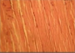
what kind of muscle
skeletal

what kind of muscle is it
smooth

what kind of muscle is it
cardiac
Muscular dystrophy show up in
rapidly growing animals
Cattle show increased retained placentas when they have deficiencies are — and —
selenium and vitamin E
clinical signs of the cardiac form of selenium and vitamin E (8)
Acute onset
Severe dyspnea
Frothy or blood stained discharge
pulmonary congestion
Tachycardia
+/- arrhythmia
Temp Normal
Recumbency
clinical signs of the skeletal form of selenium and vitamin E (14)
Lie down
try to stand
B/A/R
Stiffness
trembling when standing
tremors when standing
weakness when standing
Shoulder muscles swollen and firm
back muscles swollen and firm
hip muscles swollen and firm
Normal temp.
Myoglobinuria
Hunched appearance
Dysphagia
what is the most common form of selenium and viamin E defiecincy
skeletal muscle form
clinical signs of the smooth form of selenium and vitamin E (4)
Dystocia
Increased retained placentas
Chronic bloats
Increased LDA incidence
how to diagnosis selenium and vitamin E deficiency (8)
Elevated CK
elevated AST
Creatinine in urine increase
myoglobinuria in urine increase
glutathione peroxidase
Vitamin E levels
liver analyzed
muscles light
how to treat selenium deficiency and vitamin deficiency (5)
Parenteral selenium
Careful of overmedication
Cardiac form typically doesn’t respond as well
Give treatment a few days to work
Decrease stress and exertion
how to prevent selenium and vitamin E deficiency
Properly balanced diet
what are the 2 different types of Exertional Rhabdomyolysis
Sporadic Exertional Rhabdomyolysis
Chronic Exertional Rhabdomyolysis
what causes Sporadic Exertional Rhabdomyolysis (9)
Inadequate warm up
Over-exercise to fatigue
Exercise beyond level of fitness
Concurrent illness that contributes
low sodium
low potassium
low Vit E
low selenium
Ca:P imbalance
clinical signs of Sporadic Exertional Rhabdomyolysis (2)
Muscle cramping
Stiffness following exercise
how to diagnose Sporadic Exertional Rhabdomyolysis (4)
no previous history of Rhabdomyolysis
increased CK
increased AST
Myoglobinuria
how to treat Sporadic Exertional Rhabdomyolysis (7)
Stop exercise
Soft bedding
freshwater
Rest
+/- IV fluids
Acepromazine
Muscle relaxants
what is the goal of treatment of Sporadic Exertional Rhabdomyolysis
to minimize pain and reduce anxiety for the horse
what are the 2 types of Chronic Exertional Rhabdomyolysis (2)
Recurrent Exertional Rhabdomyolysis
Polysaccharide Storage Myopathy (PSSM) Type 1
Polysaccharide Storage Myopathy (PSSM) Type 2
what causes Recurrent Exertional Rhabdomyolysis (9)
Inherited muscle disorder
Abnormal intracellular Calcium regulation
stress
T. breds
Standardbreds
Arabians
fit
diets higher in nonstructural carbohydrates
young, fit nervous fillies
clinical signs of Recurrent Exertional Rhabdomyolysis (2)
Cramping
Stiffness post exercise
diagnosis of Recurrent Exertional Rhabdomyolysis (4)
History
Increased CK
increased AST
Muscle biopsy
how to treat Recurrent Exertional Rhabdomyolysis (3)
Management to decrease triggers
High fat diet
low starch diet
how to prevent exercise caused Recurrent Exertional Rhabdomyolysis (9)
Position the stall in a quiet area of the barn
Time of training
Turn-out
Avoid training regimes
Tranquilize before exercise to prevent excitement
treatment of lameness
Avoid stall rest or lay-up if possible
providing calm exercise if rested the day before
give medications that affect intracellular calcium regulation
how to prevent diet caused Recurrent Exertional Rhabdomyolysis (4)
high-quality grass
oat hay should form the basis of the diet.
a mixture (half alfalfa and half oat hay)
Vitamin and mineral supplements containing Vitamin E and Selenium are beneficial
PSSM stands for
Polysaccharide Storage Myopathy
define Polysaccharide Storage Myopathy type 1
excess storage of carbohydrates in the muscle
what breeds are typically affected by Polysaccharide Storage Myopathy type 1 (3)
quarter horses
morgans
draft breeds
clinical signs of Polysaccharide Storage Myopathy type 1
Tucked up
Wide stance
stiffness
Muscle fasciculation
muscle twitches
Sweating
Gait abnormality
colic
Draft horses - muscle atrophy and recumbency
clinical signs of Polysaccharide Storage Myopathy type 1 that are in draft horses specifically (2)
muscle atrophy
recumbency
Polysaccharide Storage Myopathy (PSSM) Type 2 affects what breeds (5)
Arabians
warmbloods
Morgans
T.breds
Q horses.
clinical signs of Polysaccharide Storage Myopathy type 2 that are in young horses specifically (3)
stiffness
difficulty rising
increased CK levels
clinical signs of Polysaccharide Storage Myopathy type 2 that are in warmbloods horses specifically (2)
Exercise intolerance
gait abnormalities
clinical signs of Polysaccharide Storage Myopathy type 2 (3)
Muscle atrophy
stiffness
soreness
how to diagnose Polysaccharide Storage Myopathy (2)
Increased CK levels
muscle biopsy
how to diagnose Polysaccharide Storage Myopathy type 1
genetic testing of hair or blood
how to diagnose Polysaccharide Storage Myopathy type 2 (2)
negative test for type 1
muscle biopsy for abnormal glycogen storage
how to treat Polysaccharide Storage Myopathy (6)
Rest
Feed forage at a rate of 1.5-2% of body weight
more than 15% of digestible energy as fat
<10% of digestible energy as starch
Gradual increase in exercise when horse is ready
Daily turnout
how to prevent Polysaccharide Storage Myopathy (2)
Feed more grass hays
limit richer legume hays such as alfalfa