BIPN 105 Midterm 2
 Call Kai
 Learn
 Practice Test
 Spaced Repetition
 Match
 Flashcards
 Knowt Play
1/31

Earn XP
Description and Tags
covers LE 7-10, Labs 7-10, problem sets 4-5, ALSO ANY complete names of Rs/their transduction pathways/cell types they're on/its effects, equations, setups of each Lab & why, any drugs used, physiology behind expected results
32 Terms
when comparing a STRIP of cardiac muscle (not intact heart) vs. skeletal muscle (SKM)
what is the difference in the passive (1) and active tension curves (~2)
try to draw both graphs out w/ 2 curves (cardiac, skeletal) on each graph
what are the labeled axes of the graphs
t/f: for (a strip of) cardiac muscle, the active tension curve is lower than the total tension curve
cardiac muscle is a bit more stiff than SKM, so passive tension curve is a bit above the SKM passive tension curve (look at pic)
cardiac muscle works on the ascending part of the active tension curve
cardiac muscle usually do NOT have the plateau or descending part of active tension curve that SKM has b/c cardiac muscle never stretches far enough to separate the myofilaments
…
x-axis is muscle length
y-axis is tension
__
true
for (a strip of) cardiac muscle, the active tension curve is lower than the total tension curve

for strip of cardiac muscle’s graph of tension vs. muscle length:
explain how you get contractions with different preloads (aka stretch lengths) with a constant total load using a transducer bar, support in 3 positions, and a transducer ← referring to the physical setup
& connect it to the tension vs. muscle length graph of (a strip of) cardiac muscle
(^ may help to draw it out to visualize it)
__
what type of transducer is used for looking at a frog’s heart in Lab 7: electrical and mechanical activity of the heart?
there is a muscle w/ a weight hanging from it (aka the constant total load)
there is a transducer that lets the muscle shorten & “support” that prevent the transducer bar from stretching any further than necessary (mimicking real physiology)
the transducer bar is pulled up when the muscle contracts
SO before a contraction (b/c talking about preload), moving the “support” furthest down/lowest position will get the least preload, so the cardiac muscle stretches the least before a contraction occurs → and if a muscle stretches more (before a contraction aka is an isometric contraction), then it will also shorten more (during a contraction) towards the total tension curve (referring to “support 3”)
aka least preload → least initial muscle stretch (before a contraction) → maximum muscle shortening
AND compared to the “support” being in the lowest position:
if the “support” doesn’t go as down/low as before, then that means more of the total load will be converted into preload (aka more preload) → SO have less afterload → SO when the cardiac muscle contracts, it doesn’t need to generate as much active tension to overcome the reduced afterload → SO the muscle shortens less than before when moving towards the total tension curve (referring to support 2 or 1 relative to support 3)
aka more preload (aka more initial stretch) → less afterload → overcome afterload faster b/c need to generate less active tension → muscle shortens less than before
_____
^ Lab 7: electrical and mechanical activity of the heart uses an isotonic (length) transducer
hint is that
lower position of the “support” = more preload/initial stretch → less afterload → less generated active tension → overcome afterload faster → more muscle shortening
(aka more preload = more muscle shortening)
(look at the red lines signifying the transducer bar positions depending on the support to know which has the most (least horizontal) vs least (horizontal) amount of stretch)

for strip of cardiac muscle’s graph of tension vs. muscle length:
explain how you get contractions with different afterloads(aka resistance) with a constant preload using only 1 transducer setting of a transducer bar, one support, and a transducer but 3 different afterloads ← referring to the physical setup
& connect it to the tension vs. muscle length graph of (a strip of) cardiac muscle
(^ may help to draw it out to visualize it)
__
t/f: increasing the afterload will increase the total load
the different afterloads are the different weights hanging off the muscle
IF you have a constant preload, then the initial stretch of the muscle is constant regardless of the afterload
WITH a lighter weight aka lower afterload will have a short isometric phase & then the isotonic phase will occur when moving towards the total tension curve
BUT a heavier weight aka higher afterload will have a longer isometric phase & its isotonic phase will be shorter than that of a lighter afterload, meaning that heavier afterload → causes more muscle shortening (in isotonic contraction)
& remember that a heavy enough afterload may cause no muscle shortening (isotonic contraction) due to it being unable to generate enough active tension to overload that very heavy afterload
____
true
increasing the afterload will increase the total load

def. inotropic state & positive and negative inotropic states
the inotropic state will shift the __ tension curve, but not the __ tension curve
SO for mixed contractions (having both isometric + isotonic contractions), changing the inotropic state will result in __/__ muscle shortening
inotropic state - contractility of muscle (contractility refers the force & speed of a contraction)
“+” inotropic state - increased contractility
“-” inotropic state - decreased contractility
__
the inotropic state will shift the active tension curve, but not the passive tension curve
SO for mixed contractions (having both isometric + isotonic contractions), changing the inotropic state will result in more or less muscle shortening

for an INTACT heart (aka cardiac chambers w/ volume):
name the axes for the total tension graph
if cardiac chambers fill up with MORE volume = __ stretch in cardiac muscle = __ length b/w sarcomeres (sarcomere is aka …)
x-axis is L ventricular volume (ml)
y-axis is L ventricular pressure (mmHg)
__
if cardiac chambers fill up with MORE volume = more stretch in cardiac muscle = increased length b/w sarcomeres (sarcomere is the contractile part of a muscle fiber/myofibril)

(remaining flashcards regarding cardiac stuff is going to be about the intact heart, not about strip of cardiac muscle)
in terms of LV anatomy:
def. diastole & systole (← for def. of diastole, what also happens during this time)
which valves open & close in terms of pressures of the cardiac chambers? ← group them in terms of diastole vs. systole
which of these represent the filling vs. ejection phase? which is to prevent backflow of blood?
def. preload & afterload
what signifies/represents preload and afterload?
which one of the “representations” equals the minimum aortic pressure aka minimum PAo?
diastole - ventricular relaxation
when the LV relaxes (IVR), the LA starts to contract/atrial systole (near the end of the filling phase) in order to push the last bit of blood into the LV before the LV contraction starts again
systole - ventricular contraction
…
diastole:
mitral valve opens when PLA > PLV
filling phase
aortic valve closes when PAo > PLV
to prevent blood from flowing from the aorta back into the LV
systole:
mitral valve closes when PLV > PLA
to prevent blood from flowing from the LV back into the LA
aortic valve opens when PLV > PAo
ejection phase
_____
preload - maximum filling before a contraction starts
EDV (end diastolic volume)
afterload - what the ventricle has to overcome before ejection (contraction/muscle shortening) can occur
DBP (diastolic blood pressure) = minimum PAo (← b/c minimum PAo is the minimum pressure threshold that the LV has to overcome before it can force open the aortic valve to begin ejecting blood/emptying itself)
explain flow of blood, including all cardiac chambers, valves, and blood vessels
name the 2 types of valves, which valves belong in which group?
…
state rule to help remember the “order” of blood vessels
LA pumps blood into the LV (via mitral aka bicuspid valve)
LV pumps blood into the aorta (via aortic valve)
aorta pumps blood throughout the body
via order of blood vessels: aorta → arteries → arterioles → capillaries
blood returns back to the heart
via order of blood vessels: capillaries → venules → veins (→ vena cavae)
blood enters the RA
RA pumps blood into the RV (via tricuspid valve)
RV pumps blood into the pulmonary artery (via pulmonary/pulmonic valve)
pulmonary artery pumps blood towards the lungs
__
atrioventricular valves (← aka b/w atria and ventricle)
mitral/bicuspid & tricuspid valves
semilunar valves
aortic & pulmonic valves
remember blood vessels in alphabetical order , so “Artery-arterioles-Capillaries-Venules-veins”
“MATPat” for remembering order of valves

for the cardiac cycle Pressure/Volume (P/V loop)
draw it out & label each phase, each corner, SBP, SV
^ describe filling phase (2 parts)
state at which points each of the valve closes or opens
state what area is systole vs. diastole
formula for Ejection Fraction
def. the term
ejection fraction %s for a healthy vs unhealthy heart
1 → 2: filling phase
(filling phase occurs when the mitral valve is open) most of filling phase is passive filling & near the end of filling phase, left atrial contraction/systole occurs to push the last bit of blood into the LV
2 → 3: isovolumetric contraction (IVC)
(similar to isometric contraction, where LV is at constant EDV so maintain the LV’s stretch while increasing PLV)
3 → 4: ejection phase
4 → 1: isovolumetric relaxation (IVR)
__
1: mitral valve opens (PLA > PLV)
2: mitral valve closes (PLV > PLA)
3: aortic valve opens (PLV > PAo)
4: aortic valve closes (PAo > PLV)
__
2 is when first reach EDV aka preload
4 is when first reach ESV & ESP (end systolic pressure, NOT SBP/systolic blood pressure)
3 is DBP aka afterload
the peak pressure b/w 3 → 4 is the SBP (systolic blood pressure)
in which there is maximum pressure in both the LV and aorta
the volume b/w 3 → 4 is the SV (stroke volume)
where stroke volume is the amount of blood ejected in 1 heartbeat
__
systole is IVC & ejection phase
diastole is IVR & filling phase
(atrial contraction is part of diastole b/c diastole is defined in terms of the LV)
_________________________
ejection fraction = SV / EDV
ejection fraction - the percent of blood pumping out from the heart’s main pumping chamber aka the LV for each heartbeat
healthy heart is 60-70%
unhealthy heart is <50%

for effects on the P/V loop:
if you increase preload (aka EDV)
draw the new P/V loop
what 2 things change?
this demonstrates what type of law? (← name, define, explain mech.)
…
what are 2 ways to increase the preload
increased SV
increased max pressure (aka SBP)
demonstrates the Frank-Starling Law of the Heart
where more filling (higher EDV) = more ejection (higher SV)
mech:
as the heart fills with more blood, the cardiac muscle fibers stretch more before the contraction occurs. So when the contraction occurs, these cardiac fibers will have stronger contractions
(basically, the heart pumps out (more or less) whatever volume of blood flows into it)
that extra filling is ejected when the next heartbeat occurs
^^ this is NOT a change in inotropy, so NO change in the total tension curve
…
2 ways to increase the preload:
constrict the veins or venules
venules and veins push blood into the heart, so constricting them will increase venous return / increase filling pressure
decrease HR
decrease HR means the LV will increase filling time
(& emptying time, but emptying time is not relevant to this case)

for effects on the P/V loop:
if you increase afterload (aka DBP)
draw the new P/V loop
what 2 things change? what about pulse pressure (← & def. term)?
…
what is 1 way to increase the afterload?
decreased SV
increased SBP (aka max pressure)
pulse pressure stays about the same (b/c both SBP and DBP increased by about the same amounts)
pulse pressure - the difference between SBP and DBP (aka “= SBP - DBP”)
…
1 way to increase afterload:
constrict the arterioles (NOT the arteries)
constricting the arterioles will increase resistance of blood flow from arteries into arterioles → SO more blood is trapped in the arteries at the end of diastole (diastole b/c blood flows out the heart into the arteries during LV relaxation), and this means that there is also resistance in the aorta when trying to push blood from the aorta into the arteries WHICH increases aortic pressure (aka higher PAo)
higher PAo means that the LV needs to contract more isovolumetrically to overcome the higher PAo that’s holding the aortic valve closed (aka need higher PLV in order to overcome the higher PAo)
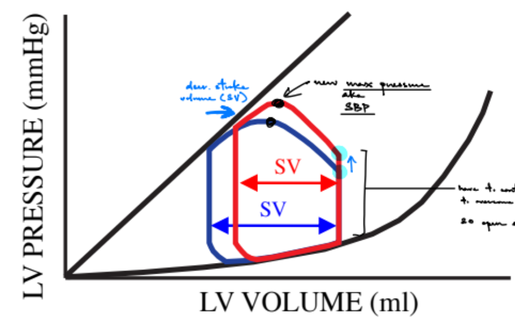
for effects on the P/V loop:
if you have a positive inotropic state w/ constant preload and afterload
draw the new P/V loop
what 2 things change? what about pulse pressure?
___________________________
what are some tricks to remember if the new P/V loop will move the R or L wall?
increased SV
increased SBP (aka max pressure)
increased pulse pressure
b/c SBP increased, but DBP stayed the same (aka constant afterload)
(also, constant preload means that the EDV stays the same)
___________________________
some tricks to remember if the new P/V loop will move the R or L wall:
if preload changed → filling changed (aka affects the filling phase)
SO move the R wall (EDV)
if afterload OR inotropic state changed → emptying changed (aka affects the ejection phase)
SO move the L wall (ESV)

in general for cardiac muscle fibers
describe the size & # of nucleus/nuclei
how are cardiac fibers connected (name spec. term)? this means that …
how is this similar to SM?
__
name the 3 types of cardiac fibers
cardiac muscle fibers
small, mononucleated
are electrically connected via intercalated disks, which contain gap junctions (← gap junctions are structures the connect the cytoplasm of two cells/fibers)
SO heart contracts as a “single unit”, where the heartrate is determined by the fastest firing cell (b/c the AP will spread from that fastest firing cell type to the rest of the cardiac cells)
^ both SM and cardiac muscle cells/fibers are myogenic (aka muscle fibers can trigger their own contractions)
__
contractile cells
nodal cells
conductive cells
for CONTRACTILE cardiac cells:
can also be called? (2)
they form the (1)
they generate __
organization (1) & have __ ATP use
…
state the conduction velocity, AP duration, and contraction duration of atrial & ventricular contractile cells
^ how does this relate to summation/tetanus? why?
…
__ contractile cells can beat faster than __ contractile cells (why?)
_______
name & describe the AP phases (← include the channels activated and their level of activity)
^ include automaticity (& def. this term)
^ what phases are similar to past muscle types or nerves that we learned & how are they similar?
→ SO the AP is “__” b/c of…
aka myocardium or myocytes
they form the chambers of the heart
contractile cells generate pressure
are striated & have high ATP use
…
contractile cells CAN’T summate or tetanize
b/c the electrical event of AP duration + refractory period is usually longer than the mechanical event of contraction duration
SO contractile cells can’t contract until they have completely relaxed, then filling can occur
…
atrial contractile cells can beat faster than ventricular contractile cells
b/c atrial contractile cells hit the refractory period sooner due to having shorter AP duration (so refractory period starts and ends much earlier) & shorter contraction duration, meaning the electrical and mechanical events both take less time than that of ventricular contractile cells
(i.e. 200 ms of atrial vs. 300 ms of ventricular)
_______
phase 0: fast de- (sim. to SKM b/c Na+)
Na+ influx
phase 1: small re- (sim. to SKM & nerves b/c VG-Na+ ch. quickly inactivate due to being time-dependent)
decreased Na+ influx b/c VG-Na+ channels are time-dependent & quickly inactivate
small K+ efflux
phase 2: Ca2+ plateau
balance b/w Ca2+ influx via LTCCs & K+ efflux
phase 3: fast re-
decreased Ca2+ influx ← b/c LTCCs will get inhibited/inactivated by Ca2+
(LTCCs are long-activated so long Ca2+ influx can result in this plateau)
increased K+ efflux
phase 4: stable Vrest/RMP
→ SO NO automaticity
automaticity - no spontaneous depolarization and AP / can’t generate its own electrical impulses aka APs
→ SO the AP is “fast” b/c of the fast de- in phase 0

for NODAL cardiac cells:
nodal cells don’t have ___
organization (1)
they __ contract, so…
__ conduction velocity that is much __ than contractile cells
nodal cells are found in the (2 ← describe where they are located)
for 1 of them, describe (1)
_______
name & describe the AP phases (← include the channels activated and their level of activity)
^ include automaticity (& explain the mech.)
^ what phase is similar to past muscle types that we learned & how are they similar?
^ include the name of a specific channel & what is another name for it?
→ SO the AP is “__” b/c of…
…
nodal cells usually DON’T have which phases?
__
what is the main pacemaker of the heart (for HR)?
__
3 ways you can increase AP frequency (and resulting increase in HR) in nodal cells
nodal cells don’t have T-tubules
are not striated
they do NOT contract, so nodal fibers don’t contribute to changes in pressure
slow conduction velocity that is much slower than contractile cells
nodal cells are found in the:
sinoatrial node (SAN) — near top of RA where vena cavae enter
atrioventricular node (AVN) — in the RA near the tricuspid valve
the AVN electrically connects the atrial & ventricular contractile cells b/c there is no electrical connection b/w the atria and ventricle → SO the electrical signal has to conduct through the AVN to get from the atria to ventricle in order to de-, fire AP, and cause mechanical event of contraction
_______
phase 0: slow de- (sim. to SM b/c Ca+)
Ca2+ influx via LTCCs
phase 2: shorter Ca2+ plateau
balance b/w Ca2+ influx via LTCCs & K+ efflux
phase 3: slow re-
decreased Ca2+ influx ← b/c LTCCs will get inhibited/inactivated by Ca2+
(LTCCs are long-activated so long Ca2+ influx can result in this plateau)
increased K+ efflux
phase 4: spontaneous de-
→ SO have automaticity
funny channels aka HCNA ch. (hyperpolarization cyclic nucleotide activated ch.) activate when re- occurs
these channels are mixed cation channels, so there is a lot of Na+ influx & some K+ efflux → resulting in If (aka current of funny ch.)
some Ca2+ ch. are also involved
OVERALL, will fire an AP when this spontaneous de- raises the Vm > VT
→ SO the AP is “slow” b/c of the slow de- in phase 0
…
nodal cells usually DON’T have phase 1 or phase 2
__
SAN (aka SA nodal fibers) is the main pacemaker of the heart (for HR)
__
3 ways you can increase AP frequency (and resulting increase in HR) in nodal cells
increase phase 4 slope
which increase the rate of spontaneous de-, so the Vm increases faster, so can reach VT faster to fire an AP faster
decrease VT
Vm reaches a lower VT faster
increase Vm
so that Vm is closer to VT

for CONDUCTIVE cardiac cells:
are specialized to do what? (1)
organization (1)
they __ contract, so…
__ conduction velocity that is much __ than contractile cells
conductive cells are found in the (2 ← where are they located)
__
for the AP:
the AP is “__” b/c …
very __ phase __, which means that conductive cells can act as __ __ when …
^in general:
phase 0 sets the (1)
phase 3 sets the (2)
phase 4 set the (1)
^^^ relate all of them to faster slope of that phase
are specialized to do spread depolarization (b/c are conductive)
are not striated
they do NOT contract, so nodal fibers don’t contribute to changes in pressure
fast conduction velocity that is much faster than contractile cells
conductive cells are found in the:
Bundle of His — in the interventricular septum (aka area b/w the L and R ventricles), where L branch goes to the LV & the R branch goes to the RV
Purkinje fibers — in the R and L ventricular walls
(where de- spreads from the BoH to Purkinje fibers aka fibers that make up the R & L ventricular walls)
__
for the AP:
the AP is “fast” b/c nodal cells have the FASTEST phase 0 slope
very slow phase 4 (spontaneous de-), which means that conductive cells can act as backup pacemakers when electrical impulses don’t get through the AVN
^ conductive fibers have slower phase 4 spontaneous de- than nodal fibers
(pacemaker cells are any cell types that can do spontaneous de-)
^in general:
phase 0 sets the conduction velocity
faster phase 0 slope = faster cond. vel.
phase 3 sets the AP duration & refractory period
faster phase 3 slope = shorter AP duration, so refractory period can start and end earlier
phase 4 set the AP firing frequency
faster phase 4 slope = faster AP frequency
differentiate b/w speed of conduction velocity VS. speed of phase 4 spontaneous de-
conduction velocity is how fast/slow an electrical signal can travel/be conducted (← the electrical signal/impulse is already generated)
how fast/slow spontaneous de- (aka automaticity) is = how fast/slow it takes for generate an electrical signal
for cardiac pacemakers in HUMANS
what is the order of primary to backup pacemaker cells that take over when higher-level ones fail to conduct their electrical signal
& state each of their approximate bpm
…
compared to humans, FROGS (like in lab):
have __ firing nodal cells (aka __ phase __ __ __)
do or don’t have conductive cells?
meaning…
for cardiac pacemakers in HUMANS
SAN — 80 bpm
AVN — 45 bpm
conductive — 35 bpm
…
compared to humans, FROGS (like in lab):
have slower firing nodal cells (aka frogs have slower phase 4 spontaneous de- than humans)
DON’T have conductive cells
meaning that there is no Bundle of His or Purkinje fibers, so frogs don’t have conductive backup pacemakers
explain pathway for spread of depolarization of cardiac muscle fibers/of the heart
^ for the AVN, explain the AVN delay (1, and what would happen if no delay?)
SAN starts de- (b/c is the main pacemaker cells)
→ spreads to de- in BOTH L & R atrial contractile cells (aka myocardium)
→ to de- in AVN
which electrically connects the L & R atria to the L & R ventricles, b/c there is no direct connection b/w atrial and ventricular contractile fibers
there is slow conduction velocity of nodal fibers, SO have a delay in spreading/conducting the signals to the ventricle (aka AVN delay)
this give the atria time to de-, fire an AP, contract, and push blood into the ventricle BEFORE the ventricle starts to contract
otherwise, the atria wouldn’t have the chance to push blood into the ventricle b/c the PLV being > PLA would close the atrioventricular valves (mitral and tricuspid valves)
→ to de- in Bundle of His
where the small branches de- the (interventricular) septum first
then, de- spreads to the tips of the L & R ventricles
→ to de- in Purkinje fibers
(de- spreads throughout the L & R ventricular walls)
→ to de- in the R & L ventricular contractile walls
then, de- travels up towards the valves (B/C the valves is where you want the blood to go out of the heart)

the MECHANICAL (not electrical) mech. of cardiac muscle fiber/heart contractions is similar to __ muscle contractions
explain the mech.
__
explain the electrical mech. for cardiac contractile cells (← in terms of how Ca2+ enters & leaves the cell)
^ state % of the Ca2+ that triggers contractions that is entering / leaving via each source / way of removal (4 total)
^ mention the name of the protein that is involved w/ SERCA-2 & its effect
…
how is this electrical mech. of cardiac contractile contraction diff. vs. similar from that of SKM (3 total)? from SM (1)?
the MECHANICAL (not electrical) mech. of cardiac muscle fiber/heart contractions is similar to skeletal muscle contractions:
Ca2+ binds to troponin in the muscle
to make the troponin-tropomyosin complex undergo conformational change, where tropomyosin pulls away from the myosin-binding sites on actin, in order to expose those myosin-binding sites on actin
myosin binds to actin using energy/ATP, where myosin ATPase converts ATP → ADP
crossbridge forms → powerstroke
__
electrical mech. for cardiac contractile cells:
the MAP (muscle AP) spreads along the sarcolemma & down T-tubules to activate LTCCs located on both the sarcolemma & in T-tubules
Ca2+ influx via LTCCs in the sarcolemma (is 70% of the Ca2+ that triggers contractions)
activation of LTCCs in the T-tubules will activate RyR-2 receptors, WHICH are Ca2+-dependent Ca2+ channels, to release Ca2+ from the SR into the cytosol of cardiac contractile cells (is 30% of the Ca2+ that triggers contractions)
Ca2+ will leave the contractile cell in 2 ways: via NCX & SERCA-2
the NCX pumps 1 Ca2+ out of the cell for every 3 Na+ into the cell (NCX removes 30% of the Ca2+ that triggers contractions)
SERCA-2 pumps Ca2+ back into the SR (SERCA-2 removes 70% of the Ca2+ that triggers contractions)
…
phospholamban (PLB) is a protein that inhibits the SERCA-2 pump, so that Ca2+ takes longer to be removed from the cytosol of a contractile cell & less Ca2+ is pumped into the SR
(b/c SERCA-2 regulates how fast & how much Ca2+ is going to be pumped into the SR)
…
contractile have LTCCs in the sarcolemma and T-tubules, while SKM only have LTCCs (i.e. DHPRs) in the T-tubules
contractile don’t have accessory proteins connecting the T-tubule LTCCs to the RyR-2 receptors, while SKM have accessory protein connecting T-tubule LTCCs to RyR-1 receptors
RyR-2 activation is DIRECTLY dependent on Ca2+, while RyR-1 activation is dependent on LTCCs (DHPRs) and accessory protein (← aka INDIRECTLY dependent on Ca2+)
-
both contractile and SM involve the NCX that behaves the same way in both types of muscle fibers
aka
2 sources of Ca2+ are Ca2+-induced SR release (via LTCC activation in T-tubules)(70%) & through LTCCs during AP (30%)
2 removals of Ca2+ are SERCA-2 (70%) & NCX (30%)
^ SO most of the Ca2+ is from the SR

in SYMPATHETIC system of the heart, what is the
main type of R
its G protein transduction pathway (← name only)
NTs/hormones that activate this R
what about the same questions regarding the PARASYMPATHETIC system of the heart?
sympathetic
beta-1 Rs (Gs transduction pathway) — activated by NE & E
(where NE is the NT, and E is the hormone)
(unlike beta-2 Rs that were also Gs & BUT activ. by only E)
para-
m2AChRs (Gi transduction pathway) — activated by ACh
(unlike m3AChRs that were Gq & BUT also activ. by ACh)
def. “inotropic, chronotropic, dromotropic” states/effects
inotropic - contractility
chronotropic - heartrate aka frequency
dromotropic - AVN conduction velocity
for sympathetic system, explain the mech. of beta-1 Rs in CONTRACTILE fibers
__
overall, you get:
increased (3), which all cause (1) → (1)
but ^ 1 of the 3 can also cause (1)
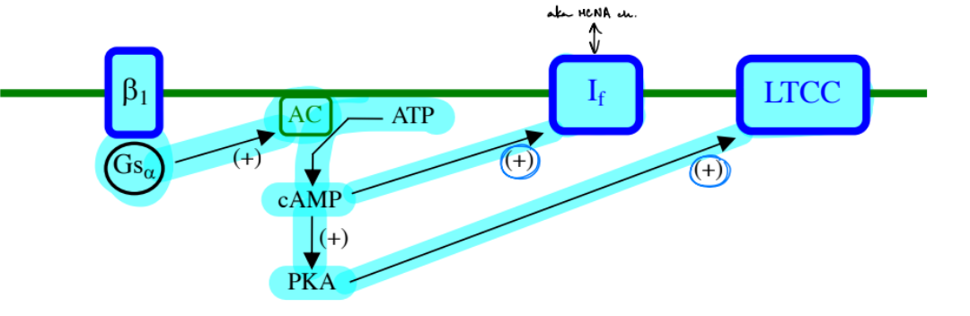
the beta-1 R is activated by NE or E, so get activated Gs alpha subunit, while the beta and gamma subunits split away
Gαs activates AC → AC converts ATP to cAMP → cAMP activates PKA
PKA has 3 effects:
PKA phosphorylates LTCCs on the sarcolemma & in T-tubules to make LTCCs more active, so get more Ca2+ influx
PKA phos. RyR-2 to make them more sensitive to Ca2+, which will increase its ability to release Ca2+ from the SR → so get more Ca2+ released from the SR into the cytosol
PKA phos. PLB, which inhibits PLB → so SERCA-2 activity increases → so more Ca2+ is pumped into the SR faster
__
overall, you get:
increased LTCC activity, RyR-2 activity, and SERCA-2 activity → which all cause increased Ca2+ in the cytosol → (get more crossbridges and powerstrokes) → increase contractility aka “+” inotropic effect
(increased SERCA-2 activity will increase the amount of Ca2+ in the SR, which means that more Ca2+ can be released into the cytosol)
but increased SERCA-2 activity can also cause decreased contraction duration

for sympathetic system, explain the mech. of beta-1 Rs in NODAL fibers
__
overall, you get:
increased (2), which all cause (3)
^ how does this translate into SAN and AVN effects?
the beta-1 Rs is activated by NE or E, so get activated Gs alpha subunit, while the beta and gamma subunits split away
Gαs activates AC → AC converts ATP to cAMP
→ cAMP activates PKA & cAMP also activates funny channels
PKA activates LTCCs ONLY on the sarcolemma (b/c nodal cells don’t have T-tubules)
__
overall, you get:
increased sarcolemma LTCC activity & funny channel activity → which all cause increased phase 0 slope, increased phase 4 slope, lower VT
b/c more Ca2+ influx causes faster de-
b/c more funny channel activity causes faster spontaneous phase 4 de- w/ Na+ influx
b/c Ca2+ can inhibit LTCCs to have 3 possible effects, where one of them is to lower the VT
translates into nodal effects:
for SAN:
increase HR aka “+” chronotropic effect
(b/c increased phase 4 slope & lower VT)
for AVN:
increase conduction velocity aka “+” dromotropic effect
(b/c increased phase 0 slope + phase 4 slope & lower VT)
for PARAsympathetic system, explain the mech. of m2AChRs in CONTRACTILE fibers
__
overall, you get what?
________________________________________________________
for PARAsympathetic system, explain the mech. of m2AChRs in NODAL fibers
__
overall, you get:
(3 effects), which all cause (4)
^ how does this translate into SAN and AVN effects?
para- system w/ contractile:
no mechanism b/c
there are no m2AChRs on contractile cells → SO have no effect
________________________________________________________
para- system w/ nodal:
the m2AChR is activated by ACh, so get activated Gi alpha subunit, while the beta-gamma subunit complex splits away
Gαi inhibits AC
SO get less cAMP & less PKA
WHILE the beta-gamma subunit complex will bind to KACh channels (aka ACh-activated K+ channels) → which cause K+ efflux for re- and hyper-
__
overall, you get:
decreased LTCC activity, decreased funny channel activity, and increased KACh channel activity → which all cause decreased phase 0 slope, decreased phase 4 slope, increased VT, decreased Vm
translates into nodal effects:
for SAN:
decrease HR aka “-” chronotropic effect
for AVN:
decrease conduction velocity aka “-” dromotropic effect

for Lab 7: Electrical & Mechanical Activity of the Frog Heart (frog ECG lab):
we are specifically using recording the ECG and contraction of an __ __ frog heart (←def.)
def. SV wave & SV-P wave
^ include what the sinus venosus is
the T wave may be recorded as __ wave where there are (2)
__
(remember this is regarding frog hearts)
t/f: the SV wave precedes the P wave (why?)
t/f: the SV-P wave may occur before the T wave (why?)
we are specifically using recording the ECG and contraction of an in situ frog heart (← frog hear that is surgically exposed to be studied, but is still in its original position within the chest)
SV wave - the electrical deflection of sinus venosus de-, where the main pacemaker cells in the frog heart are located in the sinus venosus chamber
SV-P wave - the ECG/electrical deflection of sinus venosus de- (SV wave) AND the immediately following R & L atrial de- (P wave) occur so fast that overlap and are seen as a combined SV-P wave (which would just be the P wave in a mammalian, i.e. human, ECG)
the T wave may be recorded as double wave where there are Tstart & Tend waves
____
true: the SV wave precedes the P wave
b/c the main pacemaker cells in the frog heart is within the sinus venous chamber, so the sinus venosus will de- before the ventricle depolarizes
true: the SV-P wave may occur before the T wave
frog ventricular cells have a very long Ca2+ plateau (long phase 2 aka ST segment), so it takes a long time for ventricular re- to start after ventricular de- occurred
SO the sinus venosus + atria de- of the NEW heartbeat may already start before ventricular cells of the OLD heartbeat have begun to re-
ALSO B/C frog hearts don’t have conductive fibers (aka no BoH/PF conductive fibers), the electrical signals from the sinus venous & both atria (seen as SV-P wave) will travel very slowly across the AV junction
→ SO the T wave will finish right before QRS complex starts (aka SO that re- of the old heartbeat will finish right before ventr. de- of the new heartbeat begins)

for Lab 7: Electrical & Mechanical Activity of the Frog Heart (frog ECG lab):
what type of transducer is used?
why do we need to hold the transducer bar in the middle of its range of motion & then release/depress the centering switch?
how is the calibration data collected?
what type of stimulator, transducer, preamplifier input setting, and filters are used for this lab?
use an isoTONIC (length) transducer
(b/c want to look at change in muscle length when the tension(?) is constant)
we need to hold the transducer bar in the middle of its range of motion & then release/depress the centering switch
b/c that middle range of motion will be set as the baseline aka zero reference point
get calibration data by:
starting w/ the transducer bar all the way down & then moving it up 5 mm (using a ruler) and hold it there briefly & repeat at least 3 times
…
use Grass stimulator, isotonic (length) transducer, bipolar AND “+” monopolar inputs, AC & DC couple
Grass stim. b/c it delivers higher voltages & can better control the frequency, duration, and voltage which is important for cardiac muscle
(not Powerlab stim. b/c it delivers low voltages that are good for sensitive isolated nerves, like sciatic nerve, but not for cardiac muscle)
remember:
a transducer will convert mechanical signals (in this case, the amount of muscle shortening during a contraction) into voltage that can be measured by the computer
transducer output data is the x-axis, the thing we want to know is the y-axis
for Lab 7: Electrical & Mechanical Activity of the Frog Heart (frog ECG lab):
how to calculate HR
def. systolic contraction amplitude (SCA)
what can be inferred about SCA when compared relative to what?
how do you calculate SCA in this lab?
NOTE to remember that the “-” r.e. is on the ventricle, and “+” r.e. is on the atria
HR is (1 beat/RR interval in s) * (60 s/1 min)
aka (60s/min) / (RR interval in beat/s)
systolic contraction amplitude (SCA) - the amp. of the mechanical contraction
can be inferred from the graph that if SBP increases, then the SCA increases
calculate SCA by converting transducer output into SCA (in mm) using the calibration equation
NOTE to remember that the “-” r.e. is on the ventricle, and “+” r.e. is on the atria
for Lab 7: Electrical & Mechanical Activity of the Frog Heart (frog ECG lab):
what 3 drug treatments were used & how were they administered? (← include Ringer’s solution)
^ what are the effect of these 3 treatments on HR and SCA? explain
__
remember: atropine does what?
record control ECG and contractions → squirt epinephrine (E) in Ringer’s sln onto heart → squirt Ringer’s sln on heart to wash the E off
record control ECG and contractions → squirt ACh in Ringer’s sln onto heart → squirt Ringer’s sln on heart to wash the ACh off
record control ECG and contractions → squirt atropine in Ringer’s sln onto heart → THEN squirt ACh in Ringer’s sln onto heart → squirt Ringer’s sln on heart to wash the atropine + ACh off
__
E will increase HR, and can either increase or decrease SCA (all relative to the sinus venous chamber)
E activates beta-1 Rs in sympathetic system on contractile cells & nodal cells → will increase LTCC, funny channel, SERCA-2, and RyR-2 activity
→ faster phase 4 slope → faster HR
SCA can increase b/c higher contractility
SCA can decrease b/c ventricle fills up w/ less blood, so weaker contractility
ACh decrease HR, and decrease SCA
ACh activates KACh ch. in parasympathetic system in nodal cells, which decreases Vm, so slower to de- to reach VT
will inhibit cAMP and PKA activation, so less LTCC activation, so less Ca2+ influx, so less contractility
atropine increases HR, and no change on SCA
atropine inhibits/blocks ACh from binding to m2AChRs, which reduces the effects of m2AChRs → meaning that the sinus venous can de- faster to increase HR frequency
In both frogs and mammals, vagal (parasympathetic) nerve fibers heavily innervate the sinus venosus / SA node and the atria, but sparsely innervate the ventricle (the muscular chamber responsible for generating the main systolic contraction trace). Because the ventricle receives almost no tonic parasympathetic input at rest, there is virtually no baseline ACh sitting on ventricular m2AChRs for atropine to block.
for Lab 7: Electrical & Mechanical Activity of the Frog Heart (frog ECG lab):
(for part D: external cardiac pacing & refractory period determinants)
what stimulator was used in this part?
how do you know when/at what point the control contractions start to have refractory patterns ← based on graph?
based on concepts, why does SCA decrease as the stimulus frequency is increased?
refractory pattern has alternating bigger and smaller contractions. which chambers of the heart are contracting during during bigger/smaller contractions?
when refractory period starts, which cardiac cells are entering their refractory period? b/c of large stim. voltage, it is probably their absolute refractory period
what causes refractory period in these cells & why is it much longer than the refr. period of skeletal muscle cells? how does this long refr. period affect the ability of cardiac cell contractions to have summation
used Grass stimulator
control contractions start to have refractory patterns when same size contr. → become big-small-big-small patterns
_______________
SCA decr. as stim. frequency incr.
b/c higher frequency means less time for filling & IVR (aka less time for diastole), while systole (times for IVC & ejecting) stays ~constant
less time for ventricle to relax means that there is less blood being pumped into the ventricle during diastole → so lower EDV
less blood volume in LV = less stretch of the LV wall = less actin-myosin crossbridge formations and powerstrokes → so lower systolic/contractile force, which is visualized as lower SCA (aka amp. of a mechanical contraction)
__
during smaller contractions:
2 atria b/c is smaller, so generates not as much contractile/systolic force as the ventricle (aka small SCA than that of the ventricle)
during bigger contractions:
ventricle (remember frog hearts only have 1 ventricle) b/c ventricle has a much thicker myocardial wall, so generates more contractile/systolic force (aka higher SCA)
^ not sinus venosus for bigger b/c it has very little muscle mass, so its contraction is too weak to show up as peaks
__
ventricular contractile cells are entering refractory period @ the start of the overall refractory period
b/c these cells makeup the majority of the heat muscle & drive the big contraction peak (that we see happens before the smaller contr. peak), which will then be followed by refractory period. This means that ventr. contr. cells enter refr. period before the atrial contr. cells do
__
^ ARP of ventricular contr. cells are caused by VG-Na+ channels rapdily closing their inactivation gates (become inactivated state) right after phase 0 de- occurs in ventr. contractile cells
(remember contractile cell phase 0 de- involves Na+, not Ca2+)
VG-Na+ channel inactivation can’t be removed until repolarization occurs
…
ARP is much longer in cardiac muscle than in SKM
b/c cardiac muscle have a phase 2 Ca2+ plateau, while SKM don’t have this Ca2+ plateau
the Ca2+ plateau makes it so that re- doesn’t occur immediately after de-, so there is a delay in re- aka delay in removing inactivation of VG-Na+ channels
^ a longer refractory period (part of electrical event) makes it so that cardiac muscle can’t summate or tetanize, unlike SKM
important b/c if the heart could tetanize, it would remain in a sustained contraction state aka a sustained heartbeat → meaning that the heart would stop pumping blood
for Lab 7: Electrical & Mechanical Activity of the Frog Heart (frog ECG lab):
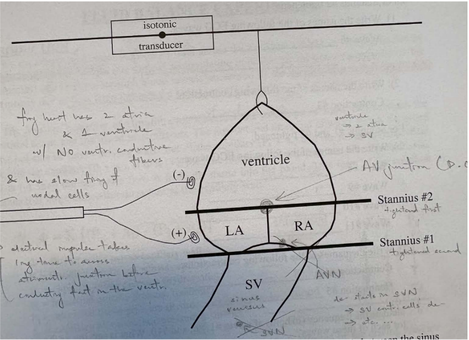
def. Stannius ligatures
their purpose
where were the Stannius ligatures placed? (2)
what would change from the control vs. when:
stannius #2 is tightened
stannius #1 is tightened while #1 is still tight
Stannius ligatures - sutures placed b/w chambers of the heart
when these ligatures are tightened, they prevent the spread of electrical activity past the ligature
…
Stannius #1 b/w sinus venous(SV) and the atria (aka both atria)
Stannius #2 b/w the atria (aka both atria) and the ventricle
^ both are loosely placed & are only tightened right before recording ECG and contraction data
…
control vs. when stannius #2 is tightened
control vs.
stannius #1 is tightened while #1 is still tight
ignore annotations on pic; just know that how the setup looks (i.e. where the Stannius ligatures are) & how the frog’s heart chambers are organized/connected